Impact Factor




Theranostics 2026; 16(15):8827-8843. doi:10.7150/thno.134232 This issue Cite
Research Paper
Feedback-regulated 3D-printed hydrogel micropatch for integrated therapy of fungal keratitis
Yujie Liu1,2,*, Li Ma2,*, Guodong Fang3, Tan Li1, Hongran Zhao1, Ting Wu1,2, Jiajia Zhang1,2, Xia Qi1, Xiangyue Hu1, Hongwei Wang1, Chao Yang3,  , Depeng Shi1,
1. Eye Institute of Shandong First Medical University, State Key Laboratory Cultivation Base, Shandong Provincial Key Laboratory of Ophthalmology, Qingdao Eye Hospital of Shandong First Medical University, Qingdao 266071, China.
2. Department of Ophthalmology, First Affiliated Hospital of Dalian Medical University, Dalian 116011, China.
3. College of Materials Science and Engineering, Qingdao University, Qingdao, Shandong, 266071, China.
* These authors contributed equally to this work.
Received 2026-3-11; Accepted 2026-8-1; Published 2026-8-24
Abstract
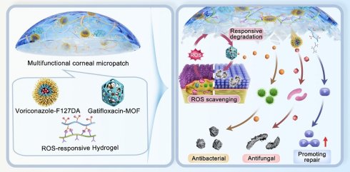
Rationale: Fungal keratitis remains clinically challenging due to limited drug bioavailability, frequent dosing, reactive oxygen species (ROS)-mediated stromal damage, perforation risk, and bacterial coinfection. In the study, we developed a feedback-regulated multifunctional corneal micropatch (MFCP) via 3D printing for the treatment of fungal keratitis.
Methods: The dual-network hydrogel micropatch comprises an ROS-responsive hydrogel (RRH), voriconazole-loaded F127DA micelles (VCZ-F127DA), and gatifloxacin-loaded cerium metal-organic frameworks (GAT-MOFs). We characterized its physicochemical properties and evaluated its biocompatibility, corneal healing capacity, ROS scavenging activity, and antimicrobial performance. Histological staining, cytokine assays, and transcriptomic sequencing were used to assess its therapeutic efficacy in a mouse model of fungal keratitis.
Results: MFCP exhibits tunable curvature, high transparency, and mechanical properties matching those of the cornea, along with good biocompatibility. High ROS level can accelerate breakdown of the micropatch and promote drug release, generating strong synergistic antifungal and antibacterial activities. The released GAT-MOFs scavenge excessive ROS, forming a negative feedback loop. In the mouse model of fungal keratitis, MFCP mitigates corneal infection, lowers proinflammatory cytokines, and accelerates tissue repair, with therapeutic efficacy superior to that of voriconazole eye drops. Transcriptomic analysis reveals suppression of NF-κB-mediated inflammatory signaling and upregulation of corneal regenerative pathways after treatment with MFCP.
Conclusion: These findings demonstrate that MFCP represents a viable therapeutic option for fungal keratitis.
Keywords: fungal keratitis, dual-network hydrogel, multifunctional corneal micropatch, cerium metal-organic frameworks, 3D printing
Introduction
Fungal keratitis, mainly caused by Fusarium solani (F. solani) and Aspergillus fumigatus, is a sight-threatening corneal infectious disease [1, 2]. It is a leading cause of corneal blindness worldwide, with a much higher burden in developing regions [3, 4]. Multiple clinical and behavioral factors have driven its steadily climbing incidence. Several factors have been linked to fungal keratitis and its rising global incidence, including ocular trauma, contact lens wear, ophthalmic surgery, and prolonged glucocorticoid use [5-9].
The early treatment of fungal keratitis mainly relies on topical antifungal eye drops [10-12]. Treatment outcomes are often compromised by rapid precorneal clearance and limited bioavailability [13-15]. Advanced carriers, such as hydrogels and nanomedicines, improve ocular drug retention and bioavailability [16-18]. However, most existing platforms offer only passive sustained release and cannot adapt to progressive pathological changes [19]. This passive release strategy increases the risk of antifungal resistance. A feedback-regulated delivery platform that adjusts drug release to disease dynamics could overcome these limitations [20].
Reactive oxygen species (ROS) generated by host immune activation can damage the corneal stroma, potentially causing thinning, perforation, and vision loss [21-24]. Bacterial coinfection worsens the prognosis of fungal keratitis, with enucleation rates up to 21% in some reports [25-27]. Antifungal monotherapy alone is insufficient [16, 28, 29]. Even after infection and inflammation subside, residual stromal defects can still perforate. 3D-printed hydrogel patches offer a way to fill ulcerated areas, stabilize the cornea, and support healing [30, 31]. A 3D-printed patch combining ROS scavenging, antibacterial capacity, healing promotion, and stromal reinforcement could meet multiple needs in fungal keratitis management [32].
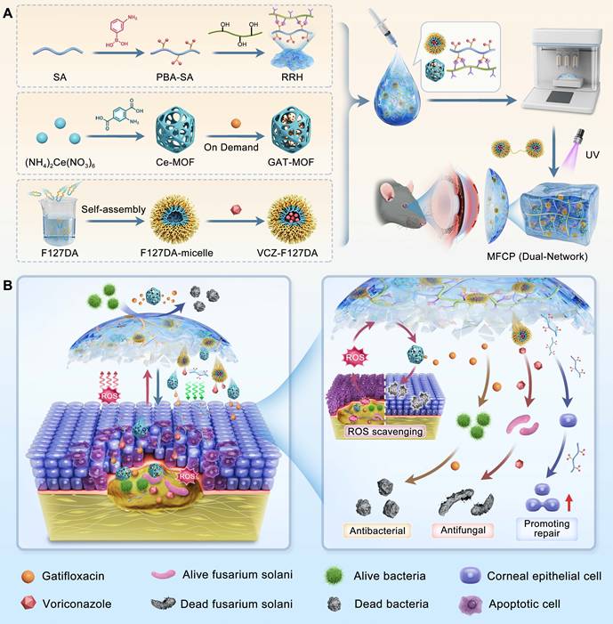
In this study, a multifunctional corneal micropatch (MFCP) was developed for feedback-regulated treatment of fungal keratitis (Scheme 1). The patch was fabricated via 3D printing using a bioink, which was composed of an ROS-responsive hydrogel (RRH), voriconazole-loaded F127DA micelles (VCZ-F127DA), and gatifloxacin-loaded cerium metal-organic frameworks (GAT-MOFs, prepared by facile stirring and enabling sustained and tunable release). High ROS levels cleave boronate ester bonds within the patch, triggering degradation and a burst of drug release to fight acute infection. The released GAT-MOFs scavenge ROS and curb inflammation; as ROS decline, degradation slows and release tapers, forming a self-regulating cycle. In addition, ROS-responsive release, antimicrobial activity, antioxidant effects, and healing promotion of MFCP were evaluated, and transcriptomics was used to investigate its mechanism against fungal keratitis.
Fabrication and mechanisms of multifunctional corneal micropatch (MFCP) for fungal keratitis. (A) Fabrication of MFCP from ROS-responsive hydrogel (RRH), voriconazole-loaded F127DA micelles (VCZ-F127DA), and gatifloxacin-loaded metal-organic frameworks (GAT-MOFs) by 3D printing. (B) Proposed mechanisms of action of the MFCP in fungal keratitis: ROS-responsive degradation, ROS scavenging by MFCP (forming a negative feedback loop), and antibacterial, antifungal, and promoting repair.
Methods
Preparation of GAT-MOFs
Cerium metal-organic frameworks (Ce-MOFs) were prepared by reacting a solution of (NH4)2Ce(NO3)6 (822 mg in 3 mL deionized water) with acetic acid (3 mL), anhydrous ethanol (20 mL), and 2-aminoterephthalic acid (272 mg) [33]. The reaction mixture was maintained under continuous stirring at 25 °C for 2 h. The solid was pelleted by centrifugation, rinsed with ethanol and H₂O, followed by drying in vacuo at 60 °C overnight. These synthesized Ce-MOFs (10 mg) were then used for drug loading by codispersing with GAT (30 mg) in deionized water (10 mL) and stirring at 4 °C for 24 h. After centrifugation and removal of the unloaded drug in the supernatant, the solid was dried under vacuum at low temperature to yield the GAT-MOFs.
Fabrication of MFCP
Sodium alginate (SA, 1.0 g) was dissolved in deionized water (100 mL), followed by sequential addition of EDC (0.96 g) and 3-aminophenylboronic acid (0.39 g). The mixture was kept under agitation at 25 °C for 24 h. It was dialyzed against deionized water (MWCO: 3500 Da) over 3 days, followed by lyophilization to yield phenylboronic acid-grafted alginate (PBA-SA). An RRH was formed by mixing 4% (w/v) PBA-SA solution with 4% (w/v) polyvinyl alcohol solution. VCZ-F127DA were prepared by dissolving F127DA at 15% (w/v) in a 0.25% (w/v) lithium phenyl-2,4,6-trimethylbenzoylphosphinate solution at 4 °C [34], adding 1% VCZ solution (w/v), sonicating at 300 W for 5 min under cooling, and incubating at 37 °C for 2 h to facilitate micellization. The bioink was formulated by adding GAT-MOFs (125 µg/mL) to a 1:1 mixture of the RRH and VCZ-F127DA. This bioink was loaded into a 3D bioprinter syringe and extruded according to a G-code file designed based on corneal curvature. The printed structure was subsequently crosslinked under 405 nm UV light for 30 s to yield the MFCP. A dual-network corneal patch (DNCP) was fabricated identically but using the bioink without GAT-MOFs.
Physicochemical characterization
Determination of GAT loading capacity
The content of GAT was quantified via a UV-near-visible light spectrophotometer (UV-3600Plus, Shimadzu, Japan), with a detection wavelength of 405 nm. The unbound GAT in the supernatant was calculated based on the established linear calibration curve A = 0.069C + 0.0416 (R2 = 0.9997). The drug loading capacity (DL%) was determined using the formula: DL% = (WT - WS) / Wnp × 100%, where WT, WS and Wnp denote total drug, the drug in the supernatant, and the dry particle weight, respectively.
Drug release curve of MFCP
Swollen MFCPs were immersed in 1 mL of PBS or H2O2 solutions (50, 100, or 200 μM) at 37 °C with shaking (60 rpm). At different time points, the release medium was sampled (1 mL) and replaced with the same volume of fresh medium. UV-vis spectrophotometry was used to quantify the cumulative release of GAT and VCZ from each sample.
ROS scavenging activity of MFCP
Commercial kits were used to assess ROS scavenging activity of MFCP against H2O2, hydroxyl radicals (·OH), and superoxide anion (O2⁻˙). We suspended MFCP samples in 3 mL of 100 mM H2O2 or PBS and recorded oxygen levels every 5 min over 30 min using a portable meter to monitor dissolved oxygen.
Cell experiments
In vitro biocompatibility study
Human corneal epithelial cells (HCECs) and human corneal fibroblasts (HCFs) were adopted to investigate the biocompatibility of MFCP. Live/dead staining: cells in 6-well plates (1 × 10⁵/well) were incubated with MFCP or its extracts (20–100 mg/mL) for 24 h, then stained. CCK-8: cells in 96-well plates (1 × 10⁴/well) were cultured for 24 h, treated with extracts (same concentrations) for 24 h, and assayed per kit instructions. Profibrotic risk was assessed in HCFs after 24 h exposure to extracts, with α-SMA and TGF-β1 mRNA expression determined via qRT-PCR (normalized to GAPDH).
Intracellular ROS detection
Cells were treated for 2 h with PBS, H2O2 (100 µM), H2O2 (100 µM) + Ce-MOFs (12.5 µg/mL), or H2O2 (100 µM) + MFCP extract (100 mg/mL). They were then stained with 10 µM DCFH-DA (10 min, 37 °C), washed, counterstained with Hoechst 33342, and examined by fluorescence microscopy. The ROS fluorescence intensity was quantified using ImageJ software.
Cell migration and proliferation experiment
HCECs were seeded in 6-well plates and grown until they reached 90% confluence. We created a linear scratch in each well with a sterile 200-µL pipette tip. After washing off the detached cells, we added fresh medium containing PBS, VCZ (1%), GAT-MOFs (12.5 µg/mL), DNCP extract (100 mg/mL), or MFCP extract (100 mg/mL) to the cultures. At the indicated time points (0, 12, and 24 h), wound images were taken. The wound area was quantified with ImageJ, and the percentage of closure was determined.
In vitro antimicrobial efficacy
Antibacterial activity
The antibacterial activity of MFCP against Staphylococcus epidermidis (S. epidermidis) and Staphylococcus aureus (S. aureus) was assessed using disk diffusion, colony counting, and live/dead staining [18]. In the disk diffusion test, 7-mm-diameter wells were punched in Mueller-Hinton agar and loaded with PBS, GAT, GAT-MOFs, DNCP, or MFCP. The plates were then inoculated with bacteria and incubated for 24 h, after which the inhibition zones were measured. To quantify bacterial survival, suspensions (1 × 10⁷ CFU/mL) were treated with the same agents for 12 h, diluted, plated, and counted after 24 h. Live/dead staining with Calcein-AM/PI allowed visualization of bacterial viability under fluorescence microscopy.
Antifungal activity study
F. solani (AS 3.1829) was purchased from the China General Microbial Strain Preservation Center (Beijing, China). A spore suspension (1 × 10⁷ CFU/mL) was treated with PBS, 100 µM H2O2, MFCP (100 mg/mL), or MFCP (100 mg/mL) + 100 µM H2O2. Disk diffusion, colony counting, and live/dead staining were performed as described above for the antibacterial evaluation.
Animal experiments
C57BL/6 mice (female, 6-8 weeks old) were purchased from Spafu (Beijing, China). All animal experiments were conducted following ARVO guidelines and received approval from the Ethics Committee of Shandong Eye Institute (SDSYKYJS No. 20250107).
Biocompatibility study in vivo
The in vivo biocompatibility of MFCP was examined in normal mice. All animals were divided into three experimental groups (n = 3). The test group had the MFCP placed on the cornea; the control groups were given topical 1% VCZ drops or PBS, with treatments applied three times daily. On day 7, ocular tolerance was assessed by slit-lamp, optical coherence tomography (OCT), and fluorescein staining, followed by eye enucleation for H&E staining.
Corneal wound healing
Corneal wounds were generated in normal mice as described previously [35]. After pentobarbital anesthesia, a 2.5 mm epithelial defect was made with an Algerbrush® II rust ring remover. The mice were randomized into five groups (n = 3) and administered with DNCP, MFCP, GAT-MOFs (12.5 µg/mL), 1% VCZ eye drops, or PBS. The DNCP and MFCP were applied once, whereas all other treatments were given topically three times daily. Corneal wound healing was monitored by fluorescein staining and bright-field imaging at 0, 24 and 48 h post-injury.
In vivo antifungal activity study
Fungal keratitis model was induced with mice using the previously reported procedures [36, 37]. Corneal wounds were created as described above. Sterile filter disks (2 mm) soaked with F. solani (1 × 10⁷ CFU/mL) were placed on the epithelial defect, and the eyelids were sutured closed for 24 h. Model validity was assessed using a standardized scoring system. Mice with established fungal keratitis were randomly divided into five groups: PBS, 1% VCZ eye drops (three times daily), 1% VCZ + 0.3% GAT eye drops (each instilled three times daily), DNCP and MFCP. Clinical severity scores based on the scoring system were recorded on days 1, 3, 5, and 7 (n = 6).
On day 3, corneas were harvested for fungal burden assessment via calcofluor white staining, with ImageJ measuring mycelial invasion depth (n = 3). On day 7, corneas were collected for H&E staining (n = 3), cytokine quantification (IL-1β, IL-6, and TNF-α) (n = 3), and transcriptomic analysis (n = 3). The antifungal effect of MFCP on corneal tissue was assessed by colony counting. Treated eyes were sampled on days 1, 3, 5, and 7 (n = 3 each). The collected corneal tissues were homogenized, serially diluted, and plated on Sabouraud dextrose agar; fungal colonies were enumerated after incubation to determine the residual fungal burden.
Statistical analysis
Data are presented as mean ± standard deviation. All experiments were repeated at least three times. Statistical analysis was performed using one-way analysis of variance or Student's t-test (GraphPad Prism 8). P < 0.05 was considered statistically significant (*P < 0.05, **P < 0.01, ***P < 0.001).
Results and Discussion
Characterization of GAT-MOFs
Ce-MOFs were prepared at room temperature from ammonium cerium nitrate and 2-aminoterephthalic acid, followed by GAT loading (Figure 1A). SEM showed uniform particles with tens of nanometers in size, and EDS confirmed the presence of C, N, O, and Ce in the nanoparticles (Figure S1). TEM revealed well-dispersed nanoparticles with an average size of 26.5 ± 5.1 nm (Figures 1B-C). XRD exhibited characteristic peaks at 6.9° and 8.0° (Figure 1D), consistent with the previously reported Ce-UiO-66 [33]. XPS survey spectra revealed Ce, O, C, and N signals, and high-resolution Ce 3d spectra showed both Ce3+ and Ce4+ in a mixed-valence state (Figures 1E-F). This redox-active couple is expected to confer ROS-scavenging activity, as reported for analogous Ce-based systems [38, 39].
Characterization of GAT-MOFs. (A) Synthesis process of GAT-MOFs. (B) TEM micrograph, (C) particle size analysis, (D) XRD patterns, (E) XPS spectrum, and (F) Ce 3d XPS spectrum of Ce-MOFs. (G) FTIR, (H) BET surface area, (I) pore-size distribution of Ce-MOFs and GAT-MOFs. (J) Drug loading capacity of GAT-MOFs.
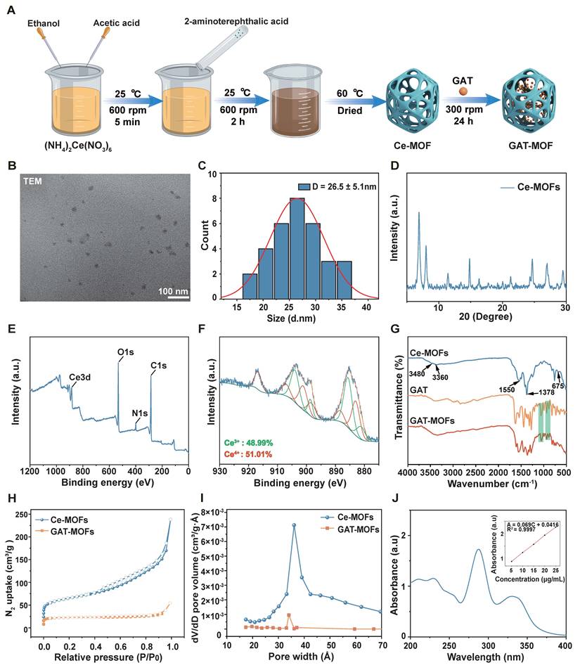
FTIR confirmed the incorporation of amino groups into Ce-MOFs (Figure 1G). The spectrum exhibited Ce-O stretching at 675 cm-1 and typical carboxylate bands at 1550 and 1378 cm-1. The peaks at 3360 and 3480 cm-1 confirmed the N-H stretching of the amino groups. These amino groups may form hydrogen bonds with carboxyl/hydroxyl groups on the ocular surface [40]. The positive zeta potential of +1.71 mV (Figure S2) should promote adhesion to the negatively charged cornea via electrostatic attraction [41]. These properties should prolong ocular retention and improve drug utilization.
FTIR confirmed GAT loading into Ce-MOFs (Figure 1G). BET showed a specific surface area of 215.48 m2/g for pristine Ce-MOFs, with pore sizes predominantly in the 2.3–4.2 nm range (Figure 1H). After GAT loading, the surface area decreased to 69.76 m2/g, and the pore-size distribution narrowed markedly (Figure 1I), consistent with pore filling by the drug. UV-vis spectroscopy quantified the loading capacity at 29.6% ± 2.1% (Figure 1J).
Characterization of MFCP
The MFCP was fabricated using extrusion-based 3D printing with a bioink formulated from RRH, GAT-MOFs, and VCZ-F127DA (Figure 2A). PBA-SA was the key component conferring ROS-responsiveness to the system. Compared with unmodified SA, PBA-SA showed new FTIR peaks at 1480, 1340, and 700 cm-1 (Figure 2B). The bands at 1480, 1340, and 700 cm-1 are assigned to benzene C=C stretching, boronic acid B-O stretching, and aromatic C-H out-of-plane bending, respectively. The 1H NMR spectrum revealed aromatic proton signals at 7.1–7.7 ppm (Figure 2C), confirming conjugation of phenylboronic acid groups.
Characterization of MFCP. (A) Synthesis routine of MFCP. (B) FTIR and (C) 1H NMR spectra of PBA-SA. (D) Shear-thinning test, (E) SEM, (F) breaking elongation, (G) tensile strength, (H) light transmittance, (I) swelling ratio of light-crosslinked F127DA hydrogel (F127DA-Gel), double-network hydrogel corneal micropatch without GAT-MOFs (DNCP), and MFCP. (J) In vitro release curves of voriconazole (VCZ) and (K) gatifloxacin (GAT) from MFCP. *P < 0.05, **P < 0.01, ***P < 0.001.
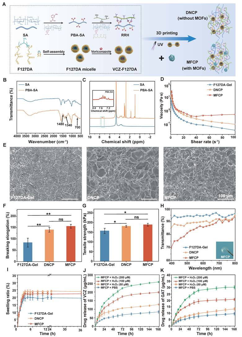
F127DA, a biocompatible hydrophobic drug carrier, forms a stable hydrogel (F127DA-Gel) via UV-triggered photo-crosslinking in the presence of a photoinitiator [34]. Rheological analysis revealed solid-like elastic behavior of all hydrogels, including F127DA-Gel, the DNCP, and the MFCP. As shown in Figure S3, the storage modulus (G′) of these hydrogels consistently exceeded their loss modulus (G″) throughout the tested frequency range. All three hydrogels were shear-thinning, while MFCP showed higher viscosity than F127DA-Gel and DNCP (Figure 2D). SEM revealed the morphology of F127DA-Gel, DNCP and MFCP (Figure 2E).
Beyond conventional casting and molding methods, 3D printing provides structural versatility, and allows precise fabrication of tunable porosity, layer-specific mechanical gradients, and anisotropic architectures [42-44]. These features have been associated with therapeutic benefits, including mechanical support, controlled drug release, and cell infiltration. The micropatch can be fabricated by 3D printing to match the corneal curvature for surface fit (Figure S4). Tensile testing revealed that MFCP exhibits an elongation at break of 155 ± 10% and a fracture strength of 189 ± 15 kPa, exceeding those of the F127DA-Gel (Figures 2F-G) [44]. These features are essential for temporary support and perforation prevention in infected corneas [45]. MFCP exhibited high transparency in the visible range of 400–800 nm, ensuring no visual obstruction after attachment to the ocular surface (Figure 2H). Its equilibrium water content of 88.8 ± 1.4% (Figure S5) and swelling ratio of 22.2 ± 0.5% (Figure 2I) in PBS closely resemble those of the natural corneas. This allows the micropatch to maintain corneal surface moisture and promote epithelial repair [46].
We measured VCZ and GAT release from MFCP under PBS and H2O2 (50, 100, 200 µM) conditions (Figures 2J-K). The results demonstrate a slow and sustained release profile in PBS. Higher release rates were observed under H2O2 conditions mimicking severe infection, with a burst observed within 24 h. The release rate decelerated over time, as the H2O2 concentration was progressively reduced due to the scavenging of Ce-MOFs. The on-demand release behavior can be attributed to the ROS-sensitive cleavage of dynamic borate ester bonds in the PBA-SA network, and is subsequently downmodulated as the scavenging of ROS by Ce-MOFs. These findings suggest that MFCP functions as a ROS-responsive delivery system rather than a passive drug depot.
Biocompatibility evaluation
We evaluated the safety of MFCP through in vitro and in vivo biocompatibility tests. The cytocompatibility of MFCP was assessed using 3D and 2D cocultures of HCECs and HCFs (Figure 3A). Both cell types adhered to and proliferated on the MFCP surface (Figure 3B). Live/dead staining with MFCP extracts at different concentrations showed no significant increase in cell death for either cell type (Figure 3C). Fluorescence-based live/dead staining (Figure S6) and CCK-8 assays (Figure 3D) confirmed that MFCP extracts did not reduce HCECs or HCFs viability. Instead, a dose-dependent increase in cell proliferation was observed. α-SMA and TGF-β1 mRNA levels were also measured in HCFs treated with MFCP extracts (Figure S7). MFCP did not upregulate either gene at any tested concentration. This indicates it does not drive HCFs toward a myofibroblast phenotype, suggesting a low risk of corneal scar formation.
Biocompatibility evaluation of MFCP. (A) Schematic illustration of the cell culture experiment. (B) Coculture model of HCECs and HCFs on MFCP. (C) Viability assessment of corneal cells exposed to MFCP extracts via live/dead and (D) CCK-8 assays. (E) C57BL/6 mouse corneas and (F) H&E-stained corneal/retinal sections following treatment with PBS, 1% VCZ, or MFCP (n = 3). *P < 0.05, **P < 0.01, ***P < 0.001
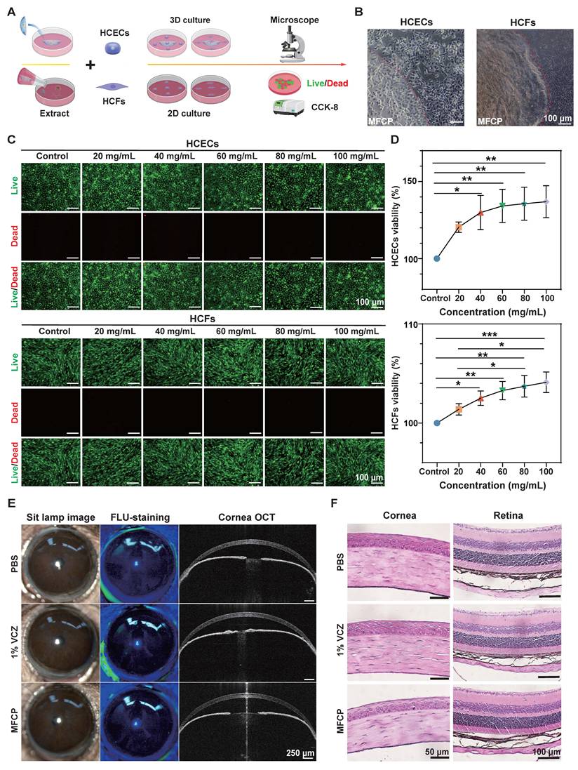
We placed MFCP to the corneal surface of healthy C57BL/6 mice over a 7-day period, with PBS and 1% VCZ eye drops as controls. There was no conjunctival hyperemia, edema, or corneal opacity in any group by slit-lamp and OCT examinations (Figure 3E), and no obvious inflammatory cell infiltration in the corneal stroma of all groups by H&E staining (Figure 3F). No obvious toxicity or inflammation was observed in vivo, suggesting that MFCP is well tolerated and suitable for further evaluation in fungal keratitis.
Promotion of corneal wound healing
We assessed cell migration and proliferation using in vitro scratch assays (Figure 4A). Quantitative analysis showed that DNCP and MFCP extracts accelerated scratch closure compared with the other groups at 12 and 24 h (Figure 4B). DNCP had a slightly higher closure rate than MFCP at 12 h, possibly due to the cytotoxic effect of GAT released from MFCP [47]. By 24 h, the closure rates of DNCP and MFCP were similar. The superior scratch closure in the DNCP and MFCP groups likely arises from alginate resulting from degradation of the micropatch, which promotes cell migration and proliferation [48].
Promotion of corneal wound healing by MFCP. (A) Representative images and (B) quantification of cell scratch assay. (C) Schematic of the mouse corneal wound model. (D) Slit-lamp images and (E) corneal wound healing rates at 0, 24, and 48 h (n = 3). *P < 0.05, **P < 0.01, ***P < 0.001.
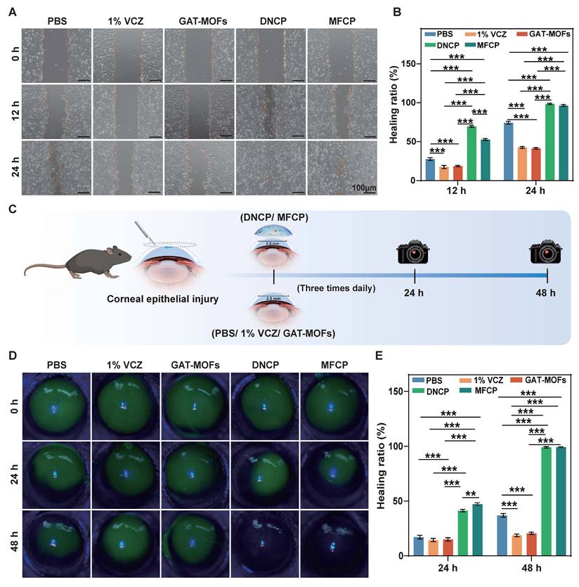
Corneal wound healing was assessed in vivo, and wound closure was tracked by slit-lamp imaging at 0, 24, and 48 h (Figure 4C). Nearly complete re-epithelialization occurred within 48 h in the DNCP and MFCP groups, faster than in the PBS group (Figures 4D and S8). MFCP promoted faster closure than DNCP at 24 h (Figure 4E). This enhanced in vivo performance likely reflects the antioxidant capacity of Ce-MOFs, which scavenge ROS and alleviate oxidative stress, accelerating healing [49].
ROS scavenging activity
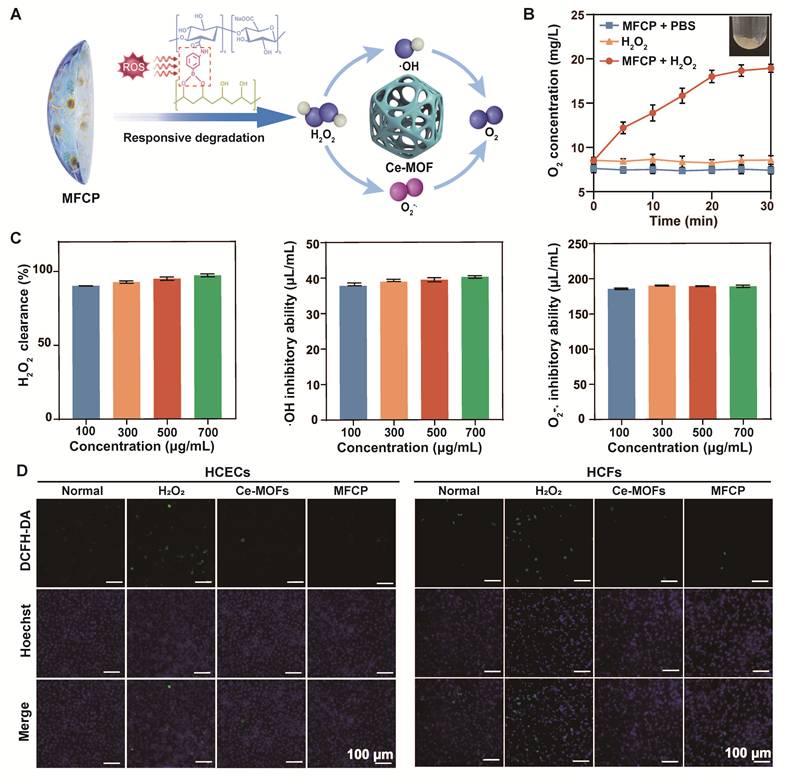
The antioxidant mechanism of MFCP involves ROS-responsive cleavage of borate ester bonds and subsequent release of Ce-MOFs, which catalyze the conversion of H2O2, O2⁻˙, and ·OH into molecular oxygen (Figure 5A). Incubation of MFCP with H2O2 generated oxygen in a time-dependent manner, confirming its catalytic decomposition of H2O2 (Figure 5B). The ROS-elimination activity of Ce-MOFs was quantified using chemical assay kits (Figure 5C). At 500 μg/mL, Ce-MOFs showed 95.68 ± 1.26% H2O2 elimination in a peroxidase-mimetic assay, along with superoxide dismutase-like (189.12 ± 0.64 μL/mL) and hydroxyl radical scavenging (39.72 ± 0.63 μL/mL) activities. A cellular oxidative stress model was established by exposing HCECs and HCFs to 100 μM H2O2. Fluorescence microscopy showed strong green fluorescence in H2O2-treated cells, indicating elevated ROS levels (Figure 5D). Pretreatment with Ce-MOFs or MFCP extract reduced fluorescence to near-baseline levels (Figure S9). These results confirm the ROS-scavenging ability of Ce-MOFs, thus mitigating oxidative stress.
ROS-scavenging activity of MFCP. (A) Schematic of ROS-responsive MFCP degradation and ROS elimination by Ce-MOF. (B) Oxygen generation from MFCP upon H2O2 exposure. (C) ROS scavenging activities of Ce-MOFs against H2O2, ·OH, and O2⁻˙. (D) Intracellular ROS in cells exposed to Ce-MOFs or MFCP.
In vitro antibacterial activity
We evaluated the antibacterial activity of MFCP using zone of inhibition (ZOI), colony counting, and live/dead staining assays (Figure 6A) [18, 50, 51]. GAT-MOFs and MFCP produced distinct inhibition zones against S. epidermidis and S. aureus, but the PBS and DNCP groups showed none (Figure 6B). Against S. epidermidis, the ZOIs were 39.53 ± 0.77 mm for GAT-MOFs and 39.01 ± 0.31 mm for MFCP. Against S. aureus, the ZOIs were 43.27 ± 2.51 mm for GAT-MOFs and 42.96 ± 1.69 mm for MFCP (Figure 6C). All values exceeded those of the controls. Free GAT solution and GAT-MOFs were compared at equivalent GAT concentrations (Figure S10). No significant difference was found, indicating that GAT loading preserved its antibacterial activity.
In vitro antibacterial activity of MFCP. (A) Schematic of the antibacterial evaluation methods. (B) ZOI images and (C) inhibition zone diameters. (D) Colony images and (E) CFU counts. (F) Live/dead staining, and (G) fluorescence intensity quantification. All groups were respectively treated with PBS, GAT-MOFs, DNCP, or MFCP. *P < 0.05, **P < 0.01, ***P < 0.001.
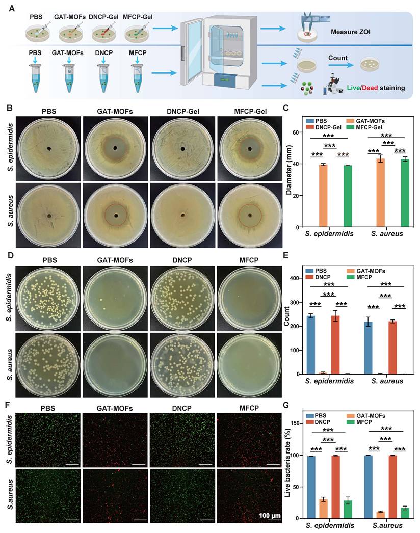
Colony counting assays showed reduced CFU numbers and sizes after GAT-MOF or MFCP treatment (Figure 6D). The PBS and DNCP groups exhibited dense bacterial growth, confirming the bactericidal activity of the drug-loaded formulations (Figure 6E). Live/dead staining further visualized bacterial viability. The PBS and DNCP groups showed predominantly green fluorescence, indicating high survival. The GAT-MOFs and MFCP groups exhibited strong red fluorescence, corresponding to extensive bacterial death (Figure 6F). Quantitative fluorescence analysis confirmed higher antibacterial rates for both materials against both strains (Figure 6G).
In vitro antifungal activity
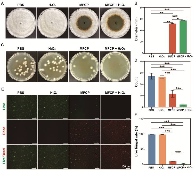
We evaluated the antifungal activity of MFCP in the absence and presence of H2O2 (Figure 7A). H2O2 alone showed no antifungal activity. MFCP alone and MFCP + H2O2 (100 µM) produced inhibition zones of 51.83 ± 0.47 mm and 57.30 ± 0.50 mm, respectively (Figure 7B). Colony counting assays showed reduced fungal growth with MFCP alone, and nearly abolished colony formation with MFCP + H2O2 (Figures 7C-D). Live/dead staining showed a shift from green (live) to red (dead) fluorescence after MFCP treatment, with further increase upon H2O2 cotreatment (Figures 7E-F). The enhanced antifungal activity of MFCP + H2O2 likely arises from exogenous H2O2 accelerating MFCP degradation, leading to increased VCZ release and stronger antifungal efficacy.
In vitro antifungal activity of MFCP. (A) ZOI images and (B) inhibition zone diameters. (C) Colony images and (D) CFU counts. (E) Live/dead staining and (F) fluorescence intensity quantification. All groups were treated with PBS, H2O2, MFCP, or MFCP + H2O2. *P < 0.05, **P < 0.01, ***P < 0.001.
In vivo therapeutic efficacy of MFCP against fungal keratitis
In vivo antifungal efficacy of MFCP was evaluated in a mouse model of fungal keratitis (Figure 8A). We monitored corneal changes in different treatment groups by slit-lamp imaging on days 1, 3, 5, and 7 after infection (Figure 8B). MFCP was the only treatment that kept corneas near-normal throughout the 7-day period, with only mild opacity at day 3 and minimal residual changes by day 7. In the PBS group, marked opacity and edema appeared by day 3 and worsened to hypopyon, neovascularization, and ulceration by day 7. 1% VCZ and 1% VCZ + 0.3% GAT exhibited modest antifungal effects, with little difference between them. DNCP reduced edema and neovascularization more effectively than the drug treatments, though it still didn't match MFCP. MFCP maintained the lowest clinical severity scores throughout the 7-day treatment period (Figure 8C).
In vivo therapeutic efficacy of MFCP against fungal keratitis. (A) Treatment timeline. (B) Representative corneal images and (C) clinical scores during the follow-up period after infection. (n = 6). (D) Calcofluor white staining of corneas on day 3 (blue indicates fungal hyphae) (n = 3). (E) H&E staining of corneas and (F) corneal inflammatory cytokine levels on day 7 (n = 3). *P < 0.05, **P < 0.01, ***P < 0.001.
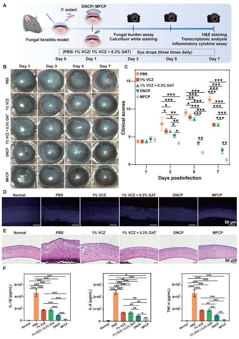
We assessed corneal fungal burden by CFU counting after MFCP treatment. Compared with day 1, fungal colonies were almost undetectable by day 3 (Figure S11). Calcofluor white staining on day 3 revealed that hyphae were rarely detected in the MFCP and DNCP groups (Figure 8D). VCZ and VCZ + GAT groups showed some reduction compared with the PBS group, but hyphae were still present. MFCP and DNCP showed comparable efficacy in reducing hyphal invasion, with depths of 21.2 ± 3.1 μm and 23.4 ± 4.2 μm, respectively (Figure S12). By contrast, hyphal invasion was deeper in the other groups: PBS (146.4 ± 3.7 μm), VCZ (118.6 ± 1.7 μm), and VCZ + GAT (123.1 ± 1.4 μm) (Figure S12).
We evaluated the impact of different treatments on corneal structure and inflammation using H&E staining (Figure 8E). MFCP-treated corneas retained complete tissue structure and barely any infiltrating immune cells. VCZ and VCZ + GAT groups had less edema and fewer infiltrates than the PBS group, but considerable pathology persisted. These changes were milder in the DNCP group, though MFCP group remained the closest to normal corneal morphology. These findings indicate that MFCP effectively alleviated inflammation, supported by molecular analysis of proinflammatory cytokines in corneal tissues (Figure 8F).
Transcriptomic profiling of MFCP-treated corneas
We performed RNA sequencing on corneal tissues harvested on day 7 (Figures 9A-B). The MFCP group yielded 917 DEGs relative to the PBS group. Among these, 156 genes were upregulated and 761 downregulated. Genes involved in extracellular matrix remodeling (e.g., Mmp13) and biological rhythms (e.g., Bmal1) were upregulated. Inflammatory chemotaxis genes such as Ccl11 were downregulated [52-54].
Transcriptomic profiling of MFCP-treated corneas (n = 3). (A) Volcano plot of DEGs between PBS and MFCP groups. (B) Heatmap of DEGs. (C, D) GO enrichment analysis of the upregulated and downregulated DEGs. (E, F) KEGG pathway enrichment analysis of the upregulated and downregulated DEGs.
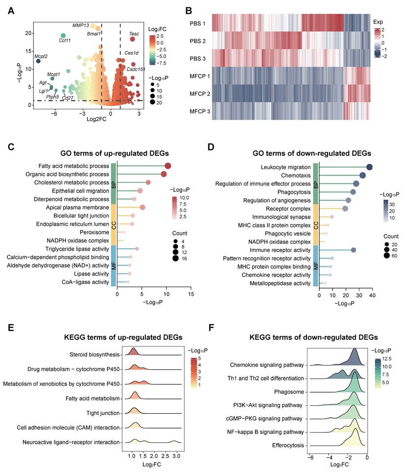
GO enrichment analysis of DEGs showed that upregulated genes were associated with metabolic and structural repair pathways, including fatty acid metabolism, cholesterol metabolism, epithelial cell migration, and tight junction (Figure 9C). Downregulated genes were linked to immune and inflammatory pathways, such as leukocyte migration, chemotaxis, immune effector regulation, and phagocytosis (Figure 9D). KEGG pathway analysis gave similar results (Figures 9E-F). Upregulated pathways included steroid biosynthesis, fatty acid metabolism, and tight junction. Downregulated pathways included chemokine, NF-κB, and PI3K-Akt signaling [54, 55].
MFCP treatment inhibited key proinflammatory pathways and genes, likely reflecting its dual anti-inflammatory design. Ce-MOFs scavenge ROS and limit NF-κB activation; voriconazole controls infection and eliminates persistent immune stimulation. These actions may promote inflammation resolution. MFCP also upregulated genes involved in lipid metabolism, cell migration, and cell junctions, suggesting a role in tissue repair. Sodium alginate from MFCP degradation may provide repair signals and promote epithelial regeneration and barrier restoration.
Conclusions
In this study, we developed a feedback-regulated hydrogel corneal micropatch via 3D printing for the integrated therapy of fungal keratitis. The micropatch was printed using a bioink containing dynamic boronate ester bonds, enabling closed-loop feedback regulation of on-demand drug release and hydrogel degradation in response to ROS levels in the infected microenvironment. By integrating antifungal, antibacterial, antioxidant, and mechanical support functions, the MFCP demonstrated superior therapeutic efficacy compared to conventional eye drops, offering a novel feedback-regulated and multifunctional platform for managing fungal keratitis.
Supplementary Material
Supplementary materials and figures.

Acknowledgements
We acknowledge the financial support of the Shandong Provincial Natural Science Foundation (No. ZR2026MS1353), Qingdao Natural Science Foundation (No. 25-1-1-258-zyyd-jch) and National Natural Science Foundation of China (No. 82000898, to LM).
Author contributions
Y.L. and L.M. contributed equally to this work. Y.L. and G.F. performed most experiments including physicochemical characterization, cell experiments, antibacterial experiments, and animal experiments. T.L., X.Q. and T.W. participated in performing the animal experiments. H.Z. participated in analyzing the transcriptome data. J.Z. and X.H. participated in antibacterial experiments. The manuscript was written by D.S. and revised by H.W., C.Y., and L.M.
Data availability
All data needed to evaluate the conclusions in the paper are present in the paper and the Supplementary Materials. Additional data related to this paper may be requested from the authors.
Competing Interests
The authors have declared that no competing interest exists.
References
1. Donovan C, Arenas E, Ayyala RS, Margo CE, Espana EM. Fungal keratitis: Mechanisms of infection and management strategies. Surv Ophthalmol. 2022;67(3):758-69
2. Sharma N, Bagga B, Singhal D, Nagpal R, Kate A, Saluja G. et al. Fungal keratitis: A review of clinical presentations, treatment strategies and outcomes. Ocul Surf. 2022;24:22-30
3. Brown L, Leck AK, Gichangi M, Burton MJ, Denning DW. The global incidence and diagnosis of fungal keratitis. Lancet Infect Dis. 2021;21(3):e49-57
4. Duran-Ospina JP, de la Hoz MJ, Maddela NR, Lapo-Talledo GJ, Siteneski A, Montes-Escobar K. Global research on keratomycosis: New insights from latent Dirichlet allocation and HJ-Biplot-driven knowledge mapping study. Diagn Microbiol Infect Dis. 2024;110(1):116442
5. Niu L, Liu X, Ma Z, Yin Y, Sun L, Yang L. et al. Fungal keratitis: Pathogenesis, diagnosis and prevention. Microb Pathog. 2020;138:103802
6. Ling JYM, Yeung SN, Chan CC, Trinh T, Antaki F, Harissi-Dagher M. et al. Trends and clinical outcomes of fungal keratitis in Canada: A 20-year retrospective multicentre study. Am J Ophthalmol. 2024;265:147-55
7. Bisen AC, Sanap SN, Agrawal S, Biswas A, Mishra A, Verma SK. et al. Etiopathology, epidemiology, diagnosis, and treatment of fungal keratitis. ACS Infect Dis. 2024;10(7):2356-80
8. Ung L, Bispo PJM, Shanbhag SS, Gilmore MS, Chodosh J. The persistent dilemma of microbial keratitis: Global burden, diagnosis, and antimicrobial resistance. Surv Ophthalmol. 2019;64(3):255-71
9. Mohd-Tahir F, Norhayati A, Siti-Raihan I, Ibrahim M. A 5-year retrospective review of fungal keratitis at hospital universiti sains malaysia. Interdiscip Perspect Infect Dis. 2012;2012:851563
10. Bock R, Roth M, Geerling G. Mycotic keratitis - signs, diagnosis, therapy. Klin Monbl Augenheilkd. 2025;242(6):636-42
11. Cai J, Yang C, Wei Q, Lian H, An L, Zhang R. Natamycin versus natamycin combined with voriconazole in the treatment of fungal keratitis. Pak J Med Sci. 2023;39(3):775-9
12. Reddy DC, Asif MI, Bari A, Velpandian T, Agarwal T, Maharana PK. et al. Comparative evaluation of topical monotherapy (natamycin or natasol) and combination therapy (natamycin and voriconazole) in mild-moderate fungal keratitis. Cornea. 2024;44(8):970-5
13. Mascarenhas M, Chaudhari P, Lewis SA. Natamycin ocular delivery: Challenges and advancements in ocular therapeutics. Adv Ther. 2023;40(8):3332-59
14. Liu S, Bai Q, Jiang Y, Gao Y, Chen Z, Shang L. et al. Multienzyme-like nanozyme encapsulated ocular microneedles for keratitis treatment. Small. 2024;20(21):e2308403
15. Ruan M, Wang R, He Y. Novel drug delivery systems for the management of fungal keratitis. J Ocul Pharmacol Ther. 2024;40(3):160-72
16. Yu M, Li L, Liu Y, Wang T, Li H, Shi C. et al. Pathogenesis and treatment strategies for infectious keratitis: Exploring antibiotics, antimicrobial peptides, nanotechnology, and emerging therapies. J Pharm Anal. 2025;15(9):101250
17. Kumar G, Kalita AH, Ahmed J, Saxena S, Sharma S, Mehta A. et al. Advancing fungal keratitis treatment: transitioning from conventional amphotericin B therapy to nanocarrier-based delivery systems. Naunyn Schmiedebergs Arch Pharmacol. 2025;398(11):14617-35
18. Shi DP, Qi X, Ma L, Zhao LH, Dou SQ, Wang Y. et al. Fabrication of nanozyme-thixotropic anionic hydrogel coating with multi-enzyme-mimicking activity for the treatment of fungal keratitis. Chem Eng J. 2024;486:150264
19. Fisher MC, Alastruey-Izquierdo A, Berman J, Bicanic T, Bignell EM, Bowyer P. et al. Tackling the emerging threat of antifungal resistance to human health. Nat Rev Microbiol. 2022;20(9):557-71
20. Wu S, Song R, Liu T, Li C. Antifungal therapy: Novel drug delivery strategies driven by new targets. Adv Drug Deliv Rev. 2023;199:114967
21. Hua X, Chi W, Su L, Li J, Zhang Z, Yuan X. ROS-induced oxidative injury involved in pathogenesis of fungal keratitis via p38 MAPK activation. Sci Rep. 2017;7(1):10421
22. Hoffman JJ, Burton MJ, Leck A. Mycotic keratitis-A global threat from the filamentous fungi. J Fungi (Basel). 2021;7(4):273
23. Awad R, Ghaith AA, Awad K, Mamdouh Saad M, Elmassry AA. Fungal keratitis: Diagnosis, management, and recent advances. Clin Ophthalmol. 2024;18:85-106
24. Fernandes M, Vira D, Dey M, Tanzin T, Kumar N, Sharma S. Comparison between polymicrobial and fungal keratitis: Clinical features, risk factors, and outcome. Am J Ophthalmol. 2015;160(5):873-81 e2
25. Devarajan B, Sharma S, Mills B, Prajna L, Venkatesh PN, Dharmalingam K. Dysbiosis of bacterial and fungal microbiomes affects the disease process and treatment outcome in fungal keratitis. Exp Eye Res. 2026;262:110745
26. Ahn M, Yoon KC, Ryu SK, Cho NC, You IC. Clinical aspects and prognosis of mixed microbial (bacterial and fungal) keratitis. Cornea. 2011;30(4):409-13
27. Muni I, Behera HS, Sahu SK, Priyadarshini SR, Das S. Microbiological profile of culture-positive fungal keratitis. Eye Contact Lens. 2024;50(6):265-9
28. Kim YE, Kim J. ROS-scavenging therapeutic hydrogels for modulation of the inflammatory response. ACS Appl Mater Interfaces. 2022;14(20):23002-21
29. Dunster E, Johnson WL, Wozniak RAF. Antimicrobial drug-drug interactions in the treatment of infectious keratitis. Cornea. 2023;42(12):1555-61
30. Jia S, Bu Y, Lau DA, Lin Z, Sun T, Lu WW. et al. Advances in 3D bioprinting technology for functional corneal reconstruction and regeneration. Front Bioeng Biotechnol. 2022;10:1065460
31. Namli I, Gupta D, Singh YP, Datta P, Rizwan M, Baykara M. et al. Progressive insights into 3D bioprinting for corneal tissue restoration. Adv Healthc Mater. 2026;15(3):e03372
32. Cheung Ma W, Liang Goh G, Meera Priyadarshini B, Yee Yeong W. 3D printing and 3D-printed electronics: Applications and future trends in smart drug delivery devices. Int J Bioprint. 2023;9(4):725
33. Dai S, Montero-Lanzuela E, Tissot A, Baldoví HG, García H, Navalón S. et al. Room temperature design of Ce(iv)-MOFs: From photocatalytic HER and OER to overall water splitting under simulated sunlight irradiation. Chem Sci. 2023;14(13):3451-61
34. Li M, Wei R, Liu C, Fang H, Yang W, Wang Y. et al. A "T.E.S.T." hydrogel bioadhesive assisted by corneal cross-linking for in situ sutureless corneal repair. Bioact Mater. 2023;25:333-46
35. Shah D, Aakalu VK. Murine corneal epithelial wound modeling. Methods Mol Biol. 2021;2193:175-81
36. Montgomery ML, Fuller KK. Experimental models for fungal keratitis: An overview of principles and protocols. Cells. 2020;9(7):1713
37. Wu TG, Wilhelmus KR, Mitchell BM. Experimental keratomycosis in a mouse model. Invest Ophthalmol Vis Sci. 2003;44(1):210-6
38. Cai Y, Ren W, Du C, Zhang J, Zhao Y, Yu J. et al. Cerium oxide nanozymes: Exploring synthesis, catalytic activity, and biomedical potential. Small. 2025;21(34):e2504436
39. Chen J, Wan J, Li C, Wei Y, Shi H. Synthesis of novel Fe0-Fe3O4/CeO2/C composite cathode for efficient heterogeneous electro-Fenton degradation of ceftriaxone sodium. J Hazard Mater. 2022;437:129393
40. Lei M, Ji Y, Zhang S, Hou B, Lu Y, Sun J. et al. Engineering a functionality-integrated artificial cornea stromal substitute: Janus bio-adhesive implant with a collagen-based multi-scale biomimetic skeleton. Bioact Mater. 2025;51:740-57
41. Wei Y, Mao Y, Zhu R, Xu Y, Qi Q, Wen X. et al. Intraocular fate of surface charge-dependent nanomicelles via topical administration: Posterior delivery and transport pathway. J Control Release. 2025;388(Pt 2):114398
42. Dufaud M, Marquette C, Jorgensen C, Perrier-Groult E, Noël D. A natural composite hydrogel laden with mesenchymal stromal cells for osteochondral repair: Comparison between casting and 3D bioprinting. Bioprinting. 2024;43:e00366
43. Damiri F, Fatimi A, Liu Y, Musuc AM, Fajardo AR, Gowda BHJ. et al. Recent advances in 3D bioprinted polysaccharide hydrogels for biomedical applications: A comprehensive review. Carbohydr Polym. 2025;348(Pt B):122845
44. Sanchez-Rubio A, Hope L, Barcelona-Estaje E, Jayawarna V, Williams J, Salmeron-Sanchez M. Gelatin vs GelMA in alginate-based bioinks as a platform for versatile 3D bioprintable in vitro systems. Biomater Adv. 2025;177:214408
45. García-López C, Rodríguez-Calvo-de-Mora M, Borroni D, Sánchez-González JM, Romano V, Rocha-de-Lossada C. The role of matrix metalloproteinases in infectious corneal ulcers. Surv Ophthalmol. 2023;68(5):929-39
46. M A, Saxena A, Mishra D, Singh K, George SD. Microfluidic contact lens: Fabrication approaches and applications. Microsyst Nanoeng. 2025;11(1):59
47. Kowalska J, Banach K, Rzepka Z, Rok J, Karkoszka M, Wrześniok D. Changes in the oxidation-reduction state of human dermal fibroblasts as an effect of lomefloxacin phototoxic action. Cells. 2022;11(12):1971
48. Mazurek Ł, Kuś M, Jurak J, Rybka M, Kuczeriszka M, Stradczuk-Mazurek M. et al. Biomedical potential of alginate wound dressings - From preclinical studies to clinical applications: A review. Int J Biol Macromol. 2025;309(Pt 2):142908
49. Na Y, Woo J, Choi WI, Lee JH, Hong J, Sung D. α-Tocopherol-loaded reactive oxygen species-scavenging ferrocene nanocapsules with high antioxidant efficacy for wound healing. Int J Pharm. 2021;596:120205
50. McMahon S, Kennedy R, Duffy P, Vasquez JM, Wall JG, Tai H. et al. Poly(ethylene glycol)-based hyperbranched polymer from RAFT and its application as a silver-sulfadiazine-loaded antibacterial hydrogel in wound care. ACS Appl Mater Interfaces. 2016;8(40):26648-56
51. Liu Z, Zhao X, Yu B, Zhao N, Zhang C, Xu FJ. Rough carbon-iron oxide nanohybrids for near-infrared-II light-responsive synergistic antibacterial therapy. ACS Nano. 2021;15(4):7482-90
52. Yan Z, Kavanagh T, Harrabi RDS, Lust ST, Tang C, Beavil R. et al. FRET sensor-modified synthetic hydrogels for real-time monitoring of cell-derived matrix metalloproteinase activity using fluorescence lifetime imaging. Adv Funct Mater. 2024;34(21):adfm 202309711
53. Ze Y, Wu Y, Tan Z, Li R, Li R, Gao W. et al. Signaling pathway mechanisms of circadian clock gene Bmal1 regulating bone and cartilage metabolism: A review. Bone Res. 2025;13(1):19
54. Polosukhina D, Singh K, Asim M, Barry DP, Allaman MM, Hardbower DM. et al. CCL11 exacerbates colitis and inflammation-associated colon tumorigenesis. Oncogene. 2021;40(47):6540-6
55. Li D, Guo YY, Cen XF, Qiu HL, Chen S, Zeng XF. et al. Lupeol protects against cardiac hypertrophy via TLR4-PI3K-Akt-NF-κB pathways. Acta Pharmacol Sin. 2022;43(8):1989-2002
Author contact
Corresponding authors: Depeng Shi, e-mail: dpshisdfmu.edu.cn; Chao Yang, e-mail: yangchaoedu.cn.