Impact Factor

- Issue 1; 2027
- Issue 16; 2026
- Issue 15; 2026
- Issue 14; 2026
- Issue 13; 2026
- Volume 17; 2027
- Volume 16; 2026
- Advance Articles
- Past Issues
- Cover Images
- Cover Suggestion
- Index & Coverage
- Special Issues
1. Introduction
2. Different types and...
3. CRISPR epigenome editing
4. CRISPR screening and drug...
5. CRISPR/Cas nucleic acid...
6. Disease model construction
7. CRISPR technology in various...
8. Progress in delivery systems
9. Current challenges in CRISPR...
10. Conclusions and perspectives
Acknowledgements
References



Theranostics 2026; 16(16):9384-9415. doi:10.7150/thno.136062 This issue Cite
Review
Recent advances in CRISPR technologies for biomedical applications and disease treatment
Xue Wu1*, Xiaorong Kou2*, Liping Bai2, Chao Liu3, Qinjie Wu2, Ming Zhang1  , Ning Wang2 , Changyang Gong2
1. Department of Ophthalmology, West China Hospital, Sichuan University, Chengdu, 610041, China.
2. Department of Biotherapy, Cancer Center and State Key Laboratory of Biotherapy, West China Hospital, Sichuan University, Chengdu, 610041, China.
3. Department of Otolaryngology-Head &Neck Surgery, West China Hospital, Sichuan University, Chengdu, 610041, China.
*These authors contributed equally to this work.
Received 2026-4-12; Accepted 2026-8-28; Published 2026-9-24
Abstract
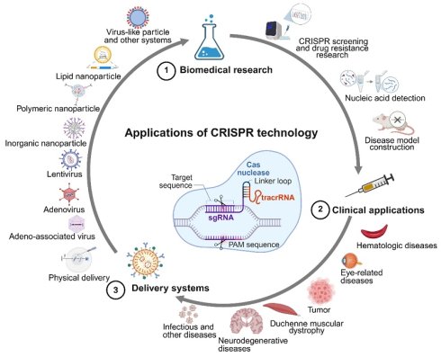
Clustered regularly interspaced short palindromic repeats (CRISPR), a precise genome-editing platform, has revolutionized genetic engineering. The highly selective and programmable characteristics of CRISPR have enabled its broad application. Recent advances in CRISPR biotechnology have promoted its widespread use in various biomedical fields, such as CRISPR screening. Ongoing preclinical and clinical trials will help determine the full capabilities of CRISPR in disease therapy. The efficacy of CRISPR-based applications is fundamentally dependent on optimized delivery strategies. Multiple approaches for delivering the CRISPR components have been investigated, primarily relying on physical, viral and non-viral delivery methods. Each method has specific strengths and limitations. Notably, non-viral delivery systems hold significant promise for CRISPR applications, offering distinct advantages such as transient expression, low immunogenicity, and fewer size constraints for cargo delivery. Various biomaterials demonstrate strong potential for delivering CRISPR components to achieve gene editing, including inorganic nanoparticles, polymeric nanoparticles, lipid nanoparticles, metal–organic frameworks, DNA origami, and virus-like particles. Therefore, this review provides an overview of recent research on the molecular principles of CRISPR and its biomedical applications. Recent advances in CRISPR-based treatments and their delivery vectors are also reviewed. Finally, the challenges faced by CRISPR technology are discussed.
Keywords: CRISPR, genome editing, biomedical research, disease treatment, delivery system
1. Introduction
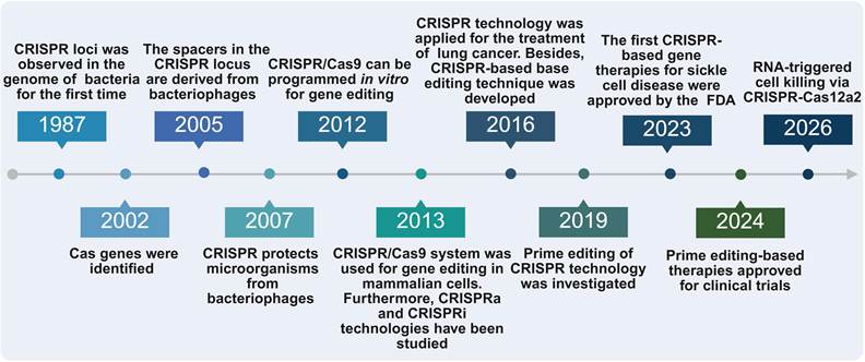
Genome editing has been widely exploited not only to prevent or alleviate disorders resulting from gene mutations but also to explore pathogenic biomolecular processes. The major available genome-editing platforms encompass zinc finger nuclease (ZFN), transcription activator-like effector nuclease (TALEN), and clustered regularly interspaced short palindromic repeats (CRISPR) systems [1]. The CRISPR technology is a novel nuclease system originally discovered in bacteria that protects microorganisms from bacteriophages by cleaving foreign nucleic acid sequences [2]. Furthermore, CRISPR system has been optimized for widespread application in fundamental biological research (Figure 1) [3-16]. CRISPR components include a CRISPR-associated protein system (Cas) effector for nuclease activity and single-guide RNA (sgRNA) for specific targeting [17]. Cas effectors can be more easily customized for specific targets than conventional gene editing systems such as TALEN and ZFN [18]. Diverse CRISPR effector proteins with various sizes, structures, compositions and targeting mechanisms have been discovered. These discoveries have greatly broadened the utilization of CRISPR for genome editing and laid the foundation for its clinical application.
Brief timeline for the development of CRISPR technology [3-16]. Created with BioRender.com.
Recently, the highly selective and programmable mechanisms of CRISPR have facilitated widespread utilization across diverse biological and medical applications. Beyond classical application, base editing systems can effectively correct single-base mutations [17]. Unlike nuclease-based methods, base editing does not introduce double-strand breaks (DSBs). In addition, the newly developed CRISPR prime editing can theoretically produce all possible base substitutions [13]. Prime editing also introduces targeted mutations without DSBs and may result in insertions of up to 44 bp via complementary prime editing guide RNA (pegRNA) [13]. Furthermore, CRISPR-based genetic tools, such as CRISPR activation (CRISPRa) and CRISPR interference (CRISPRi), also exhibit remarkable specificity [19]. Overall, the flexibility and simplicity of CRISPR system have enabled its wide employment in various biomedical fields, including CRISPR screening and drug resistance research, CRISPR/Cas nucleic acid detection, and disease model construction [20-23]. These advancements have accelerated basic research and spurred innovation across the life sciences.
Progress in CRISPR technology has driven innovations in biological research and illustrated enormous therapeutic capacity [14,24]. Notably, CRISPR has demonstrated remarkable utilization across various diseases, including hematological, eye-related, metabolic, neurodegenerative, and autoimmune diseases, as well as other intractable disorders. Ongoing preclinical and clinical trials will help determine the full capabilities of CRISPR [24]. The first-in-human phase I clinical trial of editing PD-1 in T cells to treat lung cancer demonstrated the clinical application of CRISPR technology [11]. Moreover, Casgevy (exagamglogene autotemcel) has been authorized for managing patients with either sickle cell disease (SCD) or transfusion dependent β-thalassemia (TDT) [14,25-27]. Furthermore, CRISPR delivery strategies play an important role in clinical application. The main methods for delivering CRISPR components include physical, viral and non-viral delivery approaches [2,20,28-33]. Each approach presents a unique profile of strengths and limitations. Physical strategies can deliver CRISPR components by penetrating cellular barriers [20,28]. Viral vectors, particularly adeno-associated viruses (AAVs), exhibit superior delivery efficiency and tissue-specific tropism, but encounter obstacles including immunogenicity and restricted cargo capacity. In contrast, non-viral delivery systems provide enhanced biosafety by circumventing immune responses and can be modified to improve delivery precision [29-33]. Optimizing these delivery systems is pivotal for advancing CRISPR toward clinical translation.
While its advantages are undeniable, several challenges remain in CRISPR-based treatments, such as off-target effects and ethical issues [34,35]. Current research focuses on refining CRISPR specificity and developing novel strategies to overcome its limitations. Therefore, this review presents an overview of recent research on the molecular principles and biomedical applications of CRISPR. Recent advances in CRISPR-based treatments and their delivery vectors are also reviewed. Finally, the challenges encountered by CRISPR technology are discussed.
2. Different types and mechanisms of CRISPR technology
2.1. CRISPR/Cas effector proteins
CRISPR/Cas effector proteins can be classified into six types (I-VI), each with a characteristic Cas nuclease component, which are grouped into two kinds (class I or class II). Specifically, Class I systems (types I, III, and IV) rely on multi-subunit Cas effector proteins and can be employed for eukaryotic gene editing, but their complexity has restricted their widespread application [36-38]. Nevertheless, Class II systems, including type II (Cas9), type V (Cas12), and type VI (Cas13), utilize a single Cas effector protein to accomplish targeted RNA or DNA cleavage, thus exhibiting enormous promise for gene editing [38]. Among them, Cas9 derived from Streptococcus pyogenes has been broadly utilized and can generate DSBs at precise genomic loci using programmable gRNA molecules [39]. Meanwhile, Cas12a utilizes a single crRNA with a defined protospacer-adjacent motif (PAM), exhibiting high specificity, low off-target effects and strong potential for nucleic acid detection [40-46]. In contrast, Cas13a/b targets RNA without a strict PAM requirement, primarily facilitating effective post-transcriptional knockdown [46-50]. The main features of these class II systems have been summarized in Table 1.
Distinctive features of representative Class II Cas effectors in genome editing.
| Type | Cas protein | gRNA | PAM requirement | Target type | Collateral cleavage | Characteristics | Refs |
|---|---|---|---|---|---|---|---|
| Type II | Cas9 | crRNA and tracrRNA | Yes, NGG | DNA | No | Most commonly used for gene editing | [7,46,51] |
| Type V | Cas12a | crRNA | Yes, TTTV | DNA | ssDNA | High specificity, low off-target effects | [40-46] |
| Type VI | Cas13a/b | crRNA | No, PFS | RNA | ssRNA | Post-transcriptional knockdown | [46-50] |
Notably, the miniaturization of enzymes in CRISPR technology has emerged as a pivotal optimization strategy. The Cas12 family has several compact nucleases, such as CasX (Cas12e, <1000 amino acids) and Cas12f (Cas14, 400–700 amino acids) [52,53]. The DNA cleavage properties, compact size, and non-pathogenic origin of CasX enzyme confer significant advantages over other CRISPR/Cas genome-editing enzymes [52]. Cas12f nuclease is among the smallest genome-editing tools currently available and has advantages in efficient delivery via size-limited delivery vectors such as AAV [53]. Similar to other Cas12 nucleases, Cas12f can cleave nonspecific ssDNA upon binding to complementary target DNA, making it a promising tool for nucleic acid detection [42,54]. In addition, compared with Cas9 and Cas12a nucleases, Cas12f nuclease markedly reduces large deletions and chromosomal translocations [55]. However, the gene-editing activity of Cas12f is inferior to that of the widely used Cas9 and Cas12a [56]. Notably, following Cas effector-mediated cleavage, the resulting DSB is repaired by the endogenous DNA repair mechanisms, which ultimately dictate the final outcome of genome editing.
2.2. DNA repair mechanisms for genome editing
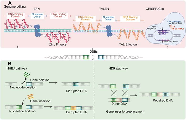
The repair of DSBs is mediated predominantly by two distinct pathways, specifically non-homologous end joining (NHEJ) and homology-directed repair (HDR). As a versatile genome engineering tool, CRISPR technology leverages these mechanisms to achieve targeted genetic modifications. Specifically, three major genome-editing platforms, namely ZFN, TALEN, and CRISPR/Cas system, all of which function by generating targeted DSBs at predefined genomic loci, are depicted in Figure 2A. Notably, unlike ZFN and TALEN that rely on direct protein‒DNA interactions for target recognition, CRISPR/Cas system achieves sequence specificity primarily via programmable base pairing between sgRNA and target DNA, thereby introducing targeted DSBs [9,57]. With respect to the downstream repair processes, the two major DSB repair pathways are depicted in Figure 2B, where NHEJ pathway ligates the two ends of a DSB and predominates as an efficient repair mechanism, whereas HDR pathway utilizes a homologous donor template to achieve precise repair. Consequently, the NHEJ pathway introduces insertions or deletions (indels) without requiring a homologous donor, which disrupts protein-coding sequences to achieve functional gene knockout [2,17,58,59], which remains the most widely utilized CRISPR application and serves as the foundation for loss-of-function studies. In contrast, as an alternative pathway to NHEJ, the HDR pathway allows cells to repair DSBs by introducing exogenous template DNA [17,60]. Specifically, this procedure is significantly more effective for inserting foreign gene sequences at a site when the target DSB is introduced using Cas effectors. With this approach, disease-causing mutations can be repaired. Nevertheless, although HDR can precisely facilitate targeted gene modification, it is generally less efficient than NHEJ which does not require a DNA template to generate insertions or deletions [20].
DNA repair mechanisms for genome editing. (A) ZFN, TALEN, and CRISPR/Cas technologies can induce DSBs at targeted sites. (B) Endogenous DNA repair can be completed via the NHEJ or HDR pathway. Created with BioRender.com.
2.3. Base editing
Owing to the inefficiency of HDR and the indeterminacy of NHEJ, base editing systems possess a considerable ability to correct single-base mutations [17]. Unlike nuclease-based methods, base editing does not introduce DSBs. Thus, base editing system provides a targeted and significantly effective method for correcting SNPs related to specific disorders [17]. The base editing system primarily comprises two types: adenine base editor (ABE) and cytosine base editor (CBE). The ABE system comprises sgRNA and adenosine deaminase-fused nickase Cas9 (nCas9), resulting in effective base transitions with a lower occurrence of indels [17]. In contrast, CBE system utilizes a cytidine deaminase-fused nCas9 to cause C·G-to-T·A editing at target gene loci [12]. With the development of basic research, base editing systems have become more flexible and effective [61]. However, the mutagenic properties of base editors have raised concerns about off-target effects. Therefore, improvements in base editing system need to focus on providing accurate single nucleotide editing. While base editing achieves precise single nucleotide transition with high efficiency, it is constrained by the limited base transitions, which cannot meet the repair needs of all single nucleotide variants. These limitations motivated the development of prime editing, which utilizes an engineered nCas9 fused to reverse transcriptase, enabling small insertions, deletions and base conversions without inducing DSBs or requiring exogenous donor DNA templates [62].
2.4. Prime editing
Notably, recently developed CRISPR tools, known as prime editing tools, can theoretically produce all possible base substitutions [13]. Table 2 summarizes the key characteristics of base editing and prime editing systems. Mechanistically, prime editing variants generally exploit nCas9 fused with reverse transcriptase and utilize pegRNA [24]. Specifically, pegRNAs are composed of sgRNA scaffold with a 3' extension that provides a reverse transcription template, thereby creating specific alterations at the target site [63]. Prime editing introduces targeted mutations in the absence of DSBs and may result in insertions of up to 44 bp via complementary pegRNAs [13]. Third-generation prime editing systems, represented by PE3 and PE3b, utilize a supplementary sgRNA to nick the non-edited strand [24], which greatly improves editing efficacy [13]. Building on these advances, a prime editing therapy for chronic granulomatous disease was cleared for clinical trials in 2024, representing a pivotal milestone in the clinical translation of prime editing [15].
Main characteristics of base editing and prime editing.
| Type | DSB requirement | Editing scope | Main advantages | Limitations | Refs |
|---|---|---|---|---|---|
| Base editing | No, single-strand break | Single nucleotide transition | High editing efficiency; low indel rate | Dependent on fixed distance to PAM; bystander edits | [12,62,64] |
| Prime editing | No, single-strand break | Small insertions, deletions or base conversions | No exogenous donor DNA template required; low off-target effect | Lower efficiency; reverse transcriptase lacks proofreading activity | [13,14,62] |
2.5. Chemical and physical control
Recently, chemical and physical methods for modulating the CRISPR system have achieved promising outcomes in enhancing spatiotemporal controllability. Commonly used chemical and physical strategies include small-molecule induction, thermal modulation, ultrasonic activation and optical regulation of the CRISPR system [65-68]. Doxycycline and rapamycin are representative small molecules for chemical regulation [69-71]; however, they tend to diffuse freely and may have toxic side effects. Moreover, thermal modulation affects intracellular biological processes and is difficult to control precisely, while ultrasound displays limited penetration capacity in cells and tissues [72]. In contrast to these alternative approaches, optical regulation of Cas9 function exhibits distinct advantages in terms of non-invasiveness, spatiotemporal specificity, and reversibility [73]. Nevertheless, a prevalent drawback of such gene regulatory systems is potential leakage, characterized by residual activity under non-illuminated conditions. This issue can be alleviated by optimizing the interaction interfaces between effector proteins and photosensitive elements [74]. Additionally, inefficient penetration of deep tissue during in vivo applications remains a key challenge for current photoactivation-based strategies [75]. Collectively, each chemical and physical modulation strategy involves unique benefits and drawbacks. Therefore, the selection of an optimal modulation strategy should be tailored to the specific demands of the experimental setting or therapeutic application.
3. CRISPR epigenome editing
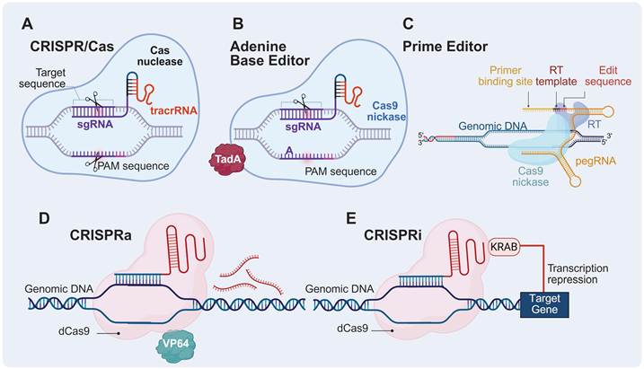
Based on evolved CRISPR platforms, significant progress has been made in the development of epigenome editing tools, including CRISPRa and CRISPRi, DNA methylation and histone modification editing, as well as integrated systems such as CRISPRon and CRISPRoff. Notably, unlike the genome editing tools shown in Figure 3A-C, epigenome editing tools typically utilize catalytically dead Cas9 (dCas9) fused to diverse effectors to precisely control gene transcription and epigenetic marks, leaving the DNA sequence unchanged [76].
Extended CRISPR technology. (A) Conventional CRISPR/Cas system: The sgRNA directs the Cas nuclease to the target genomic locus adjacent to the protospacer adjacent motif sequence. The Cas nuclease introduces DSBs at the target site, which trigger DNA repair mechanisms such as NHEJ or HDR. (B) Adenine base editor: The nCas9 is fused to an adenine deaminase domain (e.g., TadA). This system mediates precise single-base transition at the target site without generating DSBs. (C) Prime editor: The system generally exploits nCas9 fused to reverse transcriptase and utilizes pegRNA. Specifically, pegRNAs comprise sgRNA scaffold with a 3' extension that provides a reverse transcription template, thereby creating specific alterations at the target site. (D) CRISPRa: A catalytically inactive dCas9 is fused to transcriptional activator domains (e.g., VP64). The complex is recruited to the promoter region of the target gene to upregulate endogenous gene expression at the transcriptional level without altering genomic sequences. (E) CRISPRi: Catalytically inactive dCas9 is fused to the transcriptional repressor domain (e.g., KRAB). Upon binding of the dCas9-repressor complex to the promoter region of the target gene, the KRAB domain recruits repressive chromatin modifiers to suppress target gene transcription, achieving sequence-specific gene silencing. Created with BioRender.com.
3.1. CRISPR activation and interference
Apart from the typical CRISPR application of targeted gene knockout, CRISPRa and CRISPRi represent the most widely used epigenome editing tools, which enable targeted transcriptional activation and repression, respectively (Figure 3D-E) [77]. Specifically, Table 3 presents representative examples of gene activation and inhibition via CRISPRa and CRISPRi technology. Moreover, transcriptional regulation by CRISPRa and CRISPRi is utilized in a broad spectrum of biomedical applications, such as genetic screening, gene function studies, and preclinical research on hereditary diseases [77,78].
Gene activation and inhibition via CRISPRa and CRISPRi technology.
| Type | Function | Domain | Target cell | Delivery vector | Target | Efficiency | Refs |
|---|---|---|---|---|---|---|---|
| CRISPRa | Reversible gene activation | VP64 | MDA-MB-231 | Polymeric nanoparticle | miR-524 | 3-fold in vivo | [94] |
| VP64 | Astrocytes | Lentivirus, AAV | Ngn2 | 20-fold in vitro | [87] | ||
| VP64 | Astrocytes | Lentivirus, AAV | Isl1 | 10-fold in vitro | [87] | ||
| VP64 | A2780 | Polymeric nanoparticle | CT45 | 4-fold in vitro | [95] | ||
| VP160 | C2C12 | Plasmid electroporation | Lama1 | Not applicable | [96] | ||
| CRISPRi | Reversible inhibition | KRAB | Rod cells | AAV | Nrl | Up to 80% in vivo | [93] |
| KRAB | AML-12 | AAV | PCSK9 | 30% in vivo | [97] |
3.1.1. CRISPRa
Gene activation offers crucial phenotypic knowledge and may be influenced by multiple aspects, including cell type, basic level of gene expression, sgRNA efficiency, and epigenetic characteristics [79,80]. Notably, a threefold increase in basal gene expression levels can be effective for certain phenotypes, while some phenotypes require higher expression levels. Whether a universal dCas9 effector can achieve general and effective activation in different cell types and gene targets remains to be explored [81]. The single activation domain commonly used during early research was VP64 [10,82]. On the basis of existing technologies, other alternative single-activation domain systems, including those involving VP160, p300, and CREB-binding protein (CBP), continue to be developed [83,84]. VP160 and VP64 similarly promote the upregulation of target genes [85]. However, the p300 domain substantially increased gene activation by up to two times that of VP64 in HEK293T cells [83]. Similarly, the CBP domain can induce more than twofold activation of some targets relative to the combinatorial synergistic activation mediator system [86].
Furthermore, CRISPRa is a promising approach that can reveal the complex role of target genes in cell differentiation and has a major influence on effective disease therapy. For example, CRISPRa targeting the Ngn2 and Isl1 genes resulted in specialized differentiation of mouse embryonic fibroblasts or astrocytes into motor neurons [87]. With respect to the cardiac abnormalities caused by dystrophin deficiency, mortality in mice with heart failure was effectively reduced by tail-vein injection of dCas9-VP64 packaged with AAV9 to activate dystrophin [88]. However, the CRISPRa system may be unsuitable for a single viral delivery approach because of its larger size relative to that of Cas9 [89]. As alternatives to AAVs, nanoparticles are capable of delivering the dCas9 system to targeted sites, activating tumor suppressor factors in situ to effectively inhibit tumor progression [90].
3.1.2. CRISPRi
The CRISPRi system originally utilized the KRAB domain as a transcriptional repressor to achieve powerful transcriptional inhibition [91]. Owing to its greater specificity, CRISPRi can exhibit more efficient suppression when it targets miRNA clusters than short hairpin RNA (shRNA) and short interfering RNA (siRNA) [92]. The inhibition of miRNAs is promising for treating specific malignant tumors with high miRNA expression levels [77]. The development of novel drug discovery methods can be enhanced and accelerated by the CRISPRi system, facilitating the evolution of innovative treatments. For example, CRISPRi is currently being used in preclinical studies of retinal diseases. Retinitis pigmentosa, which is a hereditary disease that results in severe photoreceptor degeneration, can be treated in animal models by using KRAB-dCas9 [93]. In this therapy, CRISPRi components were delivered by subretinal injection of AAV vectors and inhibited Nrl gene expression, which reprogrammed the rods into cone-like cells. These results demonstrated that inhibiting Nrl gene expression rescued the degradation of photoreceptors and demonstrated significant potential for the treatment of retinitis pigmentosa. However, much remains to be further explored in terms of the fundamental characteristics and innovative applications of the CRISPRi system.
3.2. DNA methylation and histone modification editing
DNA methylation and histone modification constitute the two predominant mechanisms of epigenetic regulation. DNA methylation at promoter CpG dinucleotides generally leads to transcriptional silencing by hindering transcription factor binding and promoting a compact chromatin structure, which is predominantly mediated by the DNA methyltransferase (DNMT) family [98]. Specifically, DNMT1 maintains established methylation patterns and DNMT3A/3B catalyze de novo methylation, whereas ten-eleven translocation (TET) enzymes mediate active demethylation via 5-methylcytosine oxidation [99-102]. The effect of histone modifications on gene expression is either activating or repressive, with the functional outcome dictated by the modification type [103]. For instance, histone methyltransferases and demethylases dynamically regulate methylation, while acetyltransferases and histone deacetylases (HDACs) similarly control acetylation. Notably, the removal of acetyl groups by HDACs typically induces chromatin compaction, thereby leading to transcriptional repression [104]. Leveraging these regulatory mechanisms, recent studies have successfully achieved histone modifications by fusing dCas9 to various effector enzymes, notably LSD1, G9A, EZH2, and P300 [14,83]. Collectively, these advances highlight the therapeutic potential of epigenetic modifiers to correct aberrant gene expression patterns in intractable diseases, such as cancer, neurodegenerative disorders, and metabolic syndromes [76].
3.3. CRISPRon and CRISPRoff
Epigenome editing systems integrating multiple epigenetic effectors exhibit superior efficacy in achieving stable and robust gene regulation. Notably, the development of CRISPRon and CRISPRoff platforms, which integrate multiple epigenetic effector domains, represents a landmark advance [105,106]. CRISPRoff system establishes heritable long-term gene silencing via targeted DNA methylation that persists across cell divisions. Correspondingly, CRISPRon can reverse this silencing through targeted DNA demethylation, thereby offering unprecedented reversibility and tunable control over epigenetic landscapes [105]. Unlike single-component effectors, such as dCas9-DNMT3A-DNMT3L and dCas9-KRAB, CRISPRoff combines the DNMT3A and DNMT3L effector domains with the KRAB repression domain, thereby achieving epigenetic silencing with significantly enhanced durability and stability [105,106]. In contrast, CRISPRon system achieves reversal of CRISPRoff-mediated epigenetic silencing by fusing dCas9 to the TET1 catalytic domain paired with engineered sgRNAs, thereby restoring transcriptional activity [105]. Using a dCas9-TET demethylation tool, researchers reversed the durable PCSK9 silencing imposed by epigenetic editors, effectively reactivating transcription [107]. This inherent tunability could theoretically mitigate the long-term risks associated with irreversible genome editing, representing a promising strategy for managing chronic diseases that demand adjustable therapeutic interventions.
4. CRISPR screening and drug resistance research
CRISPR-based high-throughput screening has emerged as a cornerstone of functional genomics due to its programmability and flexibility. Unlike RNA interference (RNAi) screening, which relies on partial post-transcriptional silencing and is characterized by variable knockdown efficiency and frequent off-target binding to partially complementary transcripts, CRISPR-based screening generates defined genetic alterations at the DNA level with superior consistency and lower off-target effects [108,109]. Specifically, CRISPR off-target activity is inherently constrained by PAM recognition and can be further mitigated by using high-fidelity Cas variants and optimized sgRNA design. CRISPR screening has been deployed across a continuum of experimental models. In vitro screening offers unparalleled capacity and experimental controllability but fails to recapitulate the complex tissue architecture. Indirect in vivo screening partially addresses this limitation by introducing sgRNA libraries into cultured cells prior to murine transplantation, thereby recapitulating in vivo physiological and pathological processes and enabling the investigation of oncogenes [110], tumor suppressors [111-113], synthetic lethal genes [114], and immunotherapy regulators [115]. However, transplanted cells remain constrained by organ microenvironment [116]. Direct in vivo screening circumvents these constraints by inducing mutagenesis directly in target tissues while preserving the microenvironment and immune function [116], as exemplified by intracranial injection of AAV carrying sgRNA achieving direct mutagenesis in the animal brain [117]. Nevertheless, the capacity of direct in vivo CRISPR screening to faithfully recapitulate the pathogenesis of diverse disorders warrants further systematic evaluation.
By utilizing customized sgRNA libraries, CRISPR screening can simultaneously assess multiple genomic components, including protein-coding genes, noncoding RNAs and enhancers [118], facilitating unbiased identification of regulators driving specific phenotypes such as tumorigenesis [116]. Notably, this approach has revealed therapeutic targets, including CMTM6 as a candidate antitumor target [119] and POR as a regulator of ferroptosis [120]. Furthermore, a customized miRKOv2 library was developed to identify miRNAs essential for prostate cancer cell survival through CRISPR screening (Figure 4A) [121]. Beyond target discovery, CRISPR-based findings can guide drug development by directing therapeutic agents against pathogenic genes to disrupt critical biological pathways and selectively inhibit malignant cells [21,122,123]. Key antioncogenes (TP53 and PTEN) and oncogenes (MYC, KRAS, and EGFR) have been identified via CRISPR technology as promising drug targets [124]. However, translating CRISPR-based discoveries into clinically efficacious therapeutics remains challenging, owing to off-target effects, inefficient gene editing, and the necessity for rigorous preclinical and clinical validation [124,125].
CRISPR screening and drug resistance research. (A) Schematic diagram illustrating the identification of essential miRNAs in prostate cancer cells via miRNA-focused CRISPR screening. Adapted with permission from [121], copyright 2025 The Authors. (B) Flowchart of a genome-wide CRISPR screening to identify genes linked to anti-HER2 resistance using a pooled human GeCKOv2.0 lentiviral sgRNA library. Adapted with permission from [127], copyright 2022 The Authors. (C) Schematic overview of a CRISPR/Cas9-based screening system for cisplatin resistance research. Adapted with permission from [129], copyright 2022 The Authors.
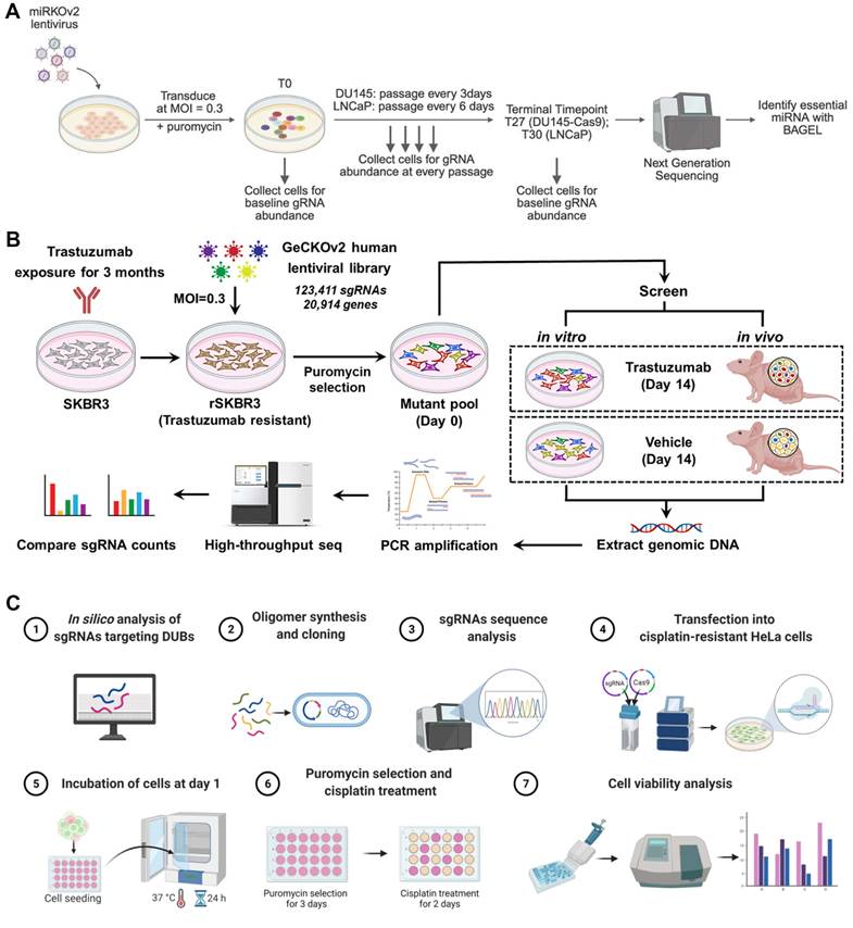
Drug resistance research represents another major application of CRISPR screening. Gene mutations are important drivers of acquired drug resistance [126], and CRISPR screening enables systematic identification of resistance-associated genes. For example, a genome-wide CRISPR screening in rSKBR3 cells employing a lentiviral library of 123411 sgRNAs successfully identified anti-HER2 resistance genes through integrated in vitro and in vivo approaches (Figure 4B) [127]. Similarly, CRISPR screening in chronic myeloid leukemia cells uncovered gene perturbation profiles that mediate drug resistance and sensitization to tyrosine kinase inhibitors and other therapeutic agents, as reflected by relative sgRNA enrichment or depletion [128]. Furthermore, CRISPR/Cas9 screening demonstrated that USP1 conferred resistance to cisplatin treatment (Figure 4C) [129]. Collectively, these studies illustrate the utility of CRISPR screening in identifying resistance mutations to facilitate targeted drug development. Overall, candidate genes and drug targets for disease treatment can be identified by CRISPR screening. However, downstream regulatory mechanisms require further characterization, and the development of efficacious medications directed at these targets remains challenging, as the comprehensive functions of the identified genes cannot be fully elucidated owing to the inherent stochasticity of experimental procedures [130].
5. CRISPR/Cas nucleic acid detection
Pathogen nucleic acid detection based on genetic elements enables timely diagnosis and therapy. Nevertheless, such diagnostics rely on technologies that can detect trace concentrations of nucleic acids with excellent specificity and sensitivity [131]. In this review, current advancements in CRISPR/Cas nucleic acid detection are discussed.
Approaches that facilitate the specific, sensitive, and rapid analysis of nucleic acid sequences contribute greatly to accurate disease diagnosis and efficient clinical therapy [132]. Established genomic diagnostic approaches, including polymerase chain reaction (PCR), demonstrate advantages in amplifying genomic sequences. However, they have drawbacks in terms of practical utility and cost [133]. Most existing nucleic acid detection methods require complicated and cumbersome instruments, in addition to skilled staff. Owing to multiple manual steps that take time and materials, the cost of detection is high [39]. Isothermal amplification methods are quicker than PCR. They avoid the need for complex devices such as thermocyclers and can be performed at a stable temperature [134]. Nevertheless, these emerging benefits entail the sacrifice of specificity and sensitivity. Despite efforts toward optimization, SNPs are not always able to be accurately distinguished [135], although the ability to detect changes in small nucleotides is critical in the identification of pathogens and diseases. Currently, the demand for time-efficient and cost-effective nucleic acid testing methods is still growing in fields such as genotyping and pathogen detection. Consequently, nucleic acid-based diagnostics must combine the flexibility, specificity, and sensitivity of existing genomic diagnostic methods with the usability, speed, and cost efficiency of isothermal amplification approaches [136]. Compared with existing nucleic acid detection methods, CRISPR/Cas-based systems offer promising alternatives (Table 4).
Characteristics of CRISPR/Cas nucleic acid detection.
| Category | Traditional detection | CRISPR/Cas detection | Refs |
|---|---|---|---|
| Properties | PCR amplification and detection | Targeted identification and cleavage by CRISPR/Cas system | [132,133,139] |
| Equipment requirement | Sophisticated thermocycler and fluorescence detection equipment | Constant temperature condition | [134,136] |
| Cost | High | Cost-effective | [39] |
| Specificity | High specificity is achieved through primer and probe design | The target sequence is identified by gRNA with high specificity and can be distinguished from single base mutations | [135,137,138] |
| Efficiency | Time-consuming and amplification is required | Fast and no complicated thermal cycle steps are required | [134,137] |
| Application scenarios | Laboratory testing, high throughput screening | Field testing, resource limited environment | [39,137-139] |
In general, CRISPR-based biosensing with isothermal signal amplification and high base resolution promises to be a remarkable approach to meet the demands of ultrasensitive nucleotide detection, including SNP identification, cancer mutation testing, human genotyping and pathogen detection, especially in point-of-care diagnostic applications [137-139]. In recent decades, the unique properties of Cas proteins, such as Cas12 and Cas13, have enabled excellent sensitivity and selectivity for nucleotide detection [140]. In addition to DNA and RNA, CRISPR-based sensing can also contribute to the detection of small molecules and proteins [138,140]. Research has indicated that CRISPR biosensing methods may be adjusted and modified within a few days to identify specific viruses, thus enabling diagnostic preparation for future pandemics [39]. Owing to their increased accuracy and sensitivity, optimal quick diagnostic methods can be used in multiple clinical applications. As a result, CRISPR technology is highly important for the rapid and specific diagnosis and treatment of infectious diseases [141].
6. Disease model construction
6.1. Human stem cells and organoids
Integrating stem cell treatment with CRISPR-based genome editing is highly promising for managing multiple genetic disorders. Disorders resulting from monogenic mutation, including SCD, muscular dystrophy and cystic fibrosis, may benefit from this strategy [142]. For example, transplanting the functional stem cells produced via CRISPR technology into patients has demonstrated the potential to restore normal blood cells and reduce the clinical manifestations of SCD [143]. Human pluripotent stem cells (hPSCs) can also be treated using CRISPR technology for applications in multiple fields such as cell therapy and drug screening, thereby addressing the limitations of hPSCs (Figure 5) [144-146]. The application of precise gene modification of hPSCs can contribute to regenerative medicine [144,146] and the analysis of the genetic differences between different types of cells and tissues. In addition, the CRISPR/Cas9 technique seems to be significantly effective and precise for hPSC gene editing and has various benefits over previous approaches, including ZFN and TALEN [147,148]. Nevertheless, each disease is associated with unique challenges that require careful assessment to determine the appropriateness of genome editing via CRISPR and subsequent stem cell treatment [149]. Moreover, ethical concerns must also be considered carefully. Therefore, rigorous ethical principles to confirm that genome editing is performed conscientiously are necessary [142].
The applications of human stem cells and organoids. Stem cell sources include somatic stem cells derived from adult human or rodent tissues, induced pluripotent stem cells (iPSCs) reprogrammed from somatic cells, and embryonic stem cells isolated from the blastocyst. All of these stem cell types can be genetically engineered via CRISPR technology to introduce specific mutations. Pluripotent stem cells differentiate into ectoderm-derived organoids (brain, retina, skin), mesoderm-derived organoids (heart, kidney), and endoderm-derived organoids (stomach, intestine, pancreas), which can also be directly engineered via CRISPR systems. These platforms, particularly CRISPR-modified models, facilitate disease modeling, cancer research, drug discovery, cell therapy, and personalized medicine. Created with BioRender.com.
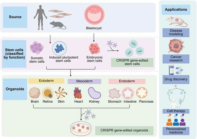
Organoid technology allows the three-dimensional reconstruction of multiple tissues from stem cells that recapitulate important tissue phenotypes [150] and can be used as disease models. CRISPR technology works well in genetically engineering organoids for the study of single-gene diseases and cancers [151]. The use of organoid culture for tumor modeling represents the latest advance in drug discovery. As an alternative strategy for optimizing treatment regimens, researchers have leveraged the ability of organoids to expand tissues directly and use them for drug screening (Figure 5) [152-154]. In one study, cancer organoids with different genotypes presented different drug sensitivities and reproduced predicted responses [155]. Drug screening based on organoids may contribute to clinical decision-making in terms of choosing effective therapies and avoiding unnecessary administration of ineffective drugs.
6.2. Engineered animal model
The most prominent application of CRISPR technology involves the development of engineered animal models to investigate pathogenic mechanisms and evaluate treatment strategies. Previously preferred method for genome modification, gene targeting of homologous recombinant embryonic stem cells, seems to be high-cost and requires the establishment of cell lines [156]. CRISPR system overcomes these obstacles and is currently routinely applied to produce gene mutations in mice [157,158]. The modification of zygotes by CRISPR is particularly powerful for editing multiple targets simultaneously or for introducing novel alterations into current animal models with various transgenic alleles [158]. Provided that appropriate gRNAs are selected, CRISPR/Cas9 gene targeting may be accomplished in different murine backgrounds. In addition, Cas9 ribonucleoprotein complex (RNP) electroporation of mouse zygotes has been achieved, eliminating the need for laborious procedures [159,160]. Although this gene editing method is extremely effective, more investigations are needed to optimize the HDR pathway for larger templates. In summary, innovative strategies promote the development of engineered animal models. In addition to its use in germline editing, CRISPR technology may also be utilized for modifying somatic cells. To promote the use of CRISPR technology in somatic cells and mouse models, transgenic mice with constitutive, conditional, or inducible Cas9 expression have been produced [161-163]. Besides, sgRNA sequences can be transduced in pools or individually to modify cells in transgenic mice expressing Cas9. In a recent study, Cas9 RNP electroporation was effectively utilized to directly modify primary immune cells in mice [164]. This strategy facilitates the assessment of phenotypes caused by specific perturbations in immune cells [164].
7. CRISPR technology in various disease treatments
CRISPR technology has contributed greatly to disease treatment (Table 5), including hematological, eye-related, metabolic, neurodegenerative, and autoimmune diseases, as well as other intractable disorders [165-168]. Ongoing preclinical and clinical trials will help determine the full capabilities of CRISPR [24]. The pace of progress in CRISPR-based disease therapy is determined by multiple factors. Specifically, compared with polygenic conditions (e.g., autoimmune diseases), in which pathogenic mechanisms remain incompletely elucidated and therapeutic outcomes are less predictable, monogenic disorders with well-characterized causal mutations (e.g., SCD and TDT) are more amenable to CRISPR-mediated gene correction [26,169]. Another critical determinant is targeting cell accessibility. Diseases affecting easily accessible or ex vivo manipulable cells (e.g., hematopoietic stem cells in β-hemoglo-binopathies) have progressed more rapidly than those requiring delivery across the blood–brain barrier (e.g., Huntington’s disease) [26,170]. Moreover, the availability of effective delivery systems plays a critical role. Organs with clinically validated delivery platforms (e.g., the liver via lipid nanoparticle, the retina and choroid via subretinal AAV) have facilitated significantly faster clinical translation compared to organs lacking efficient and safe delivery strategies (e.g., the lung and pancreas) [171,172]. Building on this framework, the following discussion systematically elaborates on the research progress of CRISPR-based therapies for each disease category.
Overview of gene therapy based on CRISPR technology in clinical trials.
| Target locus | ClinicalTrials.gov identifier | Year | Phase | Disease indication | Intervention | Sponsor |
|---|---|---|---|---|---|---|
| PD-1 | NCT02793856 | 2016 | Phase 1 | Lung cancer | Infusion of edited cells | Sichuan University, China |
| PD-1 | NCT03747965 | 2018 | Phase 1 | Solid tumors | Infusion of edited cells | Chinese PLA General Hospital, China |
| PD-1 | NCT04417764 | 2019 | Phase 1 | Advanced hepatocellular carcinoma | PD-1 knockout engineered T-cell infusion | Central South University, China |
| PD-1 | NCT03081715 | 2017 | Not applicable | Advanced esophageal cancer | Infusion of PD-1 knockout T cells | Hangzhou Cancer Hospital, China |
| PD-1 | NCT03044743 | 2017 | Phase 1/2 | Advanced stage Epstein‒Barr virus associated malignancies | Infusion of edited cytotoxic T lymphocytes with PD-1 knockout | The Affiliated Nanjing Drum Tower Hospital of Nanjing University Medical School, China |
| PD-1 and TCR | NCT03545815 | 2018 | Phase 1 | Solid tumors | CRISPR/Cas9 treated CAR-T cell infusions | Chinese PLA General Hospital, China |
| TCR and PD-1 | NCT03399448 | 2018 | Phase 1 | Myeloma, sarcoma, melanoma | Infusion of edited cells | University of Pennsylvania, USA |
| HPV E6/E7 | NCT03057912 | 2018 | Phase 1 | HPV-related cervical intraepithelial neoplasia | Plasmid in gel | First Affiliated Hospital, Sun Yat-Sen University, China |
| CLL-1 | NCT06128044 | 2024 | Phase 1 | Relapsed/refractory acute myeloid leukemia | Allogeneic CAR-T cell therapy | Caribou Biosciences, Inc., USA |
| TGFβR | NCT04976218 | 2022 | Phase 1 | Advanced EGFR positive solid tumors | Infusion of edited cells | Chinese PLA General Hospital, China |
| CD5 | NCT04767308 | 2021 | Phase 1 | Hematopoietic malignancies | Infusion of edited cells | Huazhong University of Science and Technology, China |
| CD33 | NCT04849910 | 2021 | Phase 1/2 | Acute myeloid leukemia and myelodysplastic syndromes | Genome edited hematopoietic stem and progenitor cell therapy | Vor Biopharma, USA |
| CD33 | NCT05662904 | 2023 | Phase 1 | Acute myeloid leukemia | CD33-deleted CD34+ hematopoietic stem cells | German Cancer Research Center, Germany |
| CD52, TRAC | NCT04557436 | 2020 | Phase 1 | B-cell acute lymphoblastic leukemia | Infusion of edited allogenic T cells | Great Ormond Street Hospital for Children NHS Foundation Trust, UK |
| TRAC, PD1 | NCT04637763 | 2021 | Phase 1 | Relapsed/refractory B cell non-Hodgkin lymphoma | CRISPR-edited allogeneic CAR-T cell therapy | Caribou Biosciences, Inc., USA |
| TRAC, B2M | NCT04502446 | 2020 | Phase 1 | Relapsed or refractory T-cell or B-cell malignancies | Allogeneic CRISPR/Cas9-engineered T cells | CRISPR Therapeutics AG, Switzerland |
| TRAC, B2M | NCT04244656 | 2020 | Phase 1 | Multiple myeloma | Edited allogeneic T cells | CRISPR Therapeutics AG, Switzerland |
| TRAC, B2M | NCT05795595 | 2023 | Phase 1/2 | Relapsed or refractory solid tumors | Edited allogeneic T cells | CRISPR Therapeutics AG, Switzerland |
| TRAC, B2M | NCT05722418 | 2023 | Phase 1 | Relapsed/refractory multiple myeloma | Allogeneic CAR-T cell therapy | Caribou Biosciences, Inc., USA |
| TRAC, B2M | NCT05643742 | 2023 | Phase 1/2 | Relapsed or refractory B-cell malignancies | Edited allogeneic T cells | CRISPR Therapeutics AG, Switzerland |
| CISH | NCT04426669 | 2020 | Phase 1/2 | Metastatic gastrointestinal cancers | Autologous CISH inactivated tumor infiltrating lymphocytes | Intima Bioscience, Inc., USA |
| CISH | NCT05566223 | 2023 | Phase 1/2 | Metastatic non-small cell lung cancer | Autologous CISH inactivated tumor infiltrating lymphocytes | Intima Bioscience, Inc., USA |
| BCL11A | NCT03655678 | 2018 | Phase 2/3 | TDT | Infusion of edited cells (CTX001) | Vertex Pharmaceuticals Incorporated, USA |
| BCL11A | NCT03745287 | 2018 | Phase 2/3 | Severe SCD | Infusion of edited cells (CTX001) | Vertex Pharmaceuticals Incorporated, USA |
| BCL11A | NCT05356195 | 2022 | Phase 3 | Pediatric participants with TDT | Infusion of edited cells (CTX001) | Vertex Pharmaceuticals Incorporated, USA |
| BCL11A | NCT05329649 | 2022 | Phase 3 | Pediatric participants with severe SCD | Infusion of edited cells (CTX001) | Vertex Pharmaceuticals Incorporated, USA |
| BCL11A | NCT05477563 | 2022 | Phase 3 | TDT or severe SCD | Infusion of edited cells (CTX001) | Vertex Pharmaceuticals Incorporated, USA |
| BCL11A | NCT06300723 | 2024 | Not applicable | SCD | CD34+ autologous hematopoietic stem and progenitor cells edited at the BCL11A gene | Bioray Laboratories, USA |
| BCL11A | NCT05951205 | 2024 | Phase 3 | Adolescent and adult participants with severe SCD, βS/βC genotype | Infusion of edited cells (CTX001) | Vertex Pharmaceuticals Incorporated, USA |
| HBB | NCT03728322 | 2018 | Phase 1 | Thalassemia | Infusion of edited cells | Allife Medical Science and Technology Co., Ltd., China |
| HBG1/2 | NCT06041620 | 2023 | Not applicable | TDT | Autologous CRISPR/Cas12b edited hematopoietic stem cells | Institute of Hematology & Blood Diseases Hospital, China |
| F9 | NCT06379789 | 2024 | Phase 1/2 | Hemophilia B | CRISPR/Cas9 based F9 gene insertion therapy administered via intravenous infusion | Regeneron Pharmaceuticals, USA |
| OTOF | NCT06025032 | 2024 | Phase 1 | Hearing loss | CRISPR/Cas13 RNA base editing therapy administered via intracochlear injection | HuidaGene Therapeutics Co., Ltd., China |
| DMD | NCT05514249 | 2022 | Phase 1 | DMD | CRD-TMH-001 administered via intravenous injection | Cure Rare Disease, Inc, USA |
| PCSK9 | NCT05398029 | 2022 | Phase 1 | Heterozygous familial hypercholesterolemia, atherosclerotic cardiovascular disease, and uncontrolled hypercholesterolemia | Disrupted expression of the PCSK9 gene by base editing technology via intravenous infusion | Verve Therapeutics, Inc., USA |
| CEP290 | NCT03872479 | 2019 | Phase 1/2 | Leber congenital amaurosis 10 | EDIT-101 administered via subretinal injection | Editas Medicine, Inc., USA |
| VEGFA | NCT06031727 | 2023 | Phase 1 | Neovascular age-related macular degeneration | Cas13 technology using one single AAV vector upon subretinal injection | HuidaGene Therapeutics Co., Ltd., China |
| MYOC | NCT06465537 | 2024 | Not applicable | Primary open-angle glaucoma | Intracameral injection of virus-like particle | Shanghai BDgene Co., Ltd., China |
| CCR5 | NCT03164135 | 2017 | Not applicable | HIV-1 | Infusion of edited cells | Affiliated Hospital of the Academy of Military Medical Sciences, China |
| HIV-1 | NCT05144386 | 2022 | Phase 1 | Aviremic HIV-1 infected adults | CRISPR/Cas9 gene editing system delivered by AAV9 | Excision BioTherapeutics, USA |
7.1. Hematologic diseases
β-Hemoglobinopathy
β-Hemoglobinopathy, which affects the β chain of hemoglobin, represents the most prevalent monogenic disorder and manifests mainly in the form of SCD and TDT [173]. SCD is a single-gene disease resulting from β-globin gene mutation that causes ischemic tissue injury and organ failure [174]. β-Thalassemia, by contrast, can be attributable to multiple mutations resulting in decreased production of functional β-globin, which in turn causes impaired erythropoiesis and hemolysis. Regardless of the differences in underlying pathology, mutations that increase the level of fetal hemoglobin (HbF) can ameliorate both SCD and TDT because γ-globin has antisickle characteristics and is capable of replacing the function of β-globin [173]. Therefore, several strategies involving CRISPR technology for treating β-hemoglobinopathies are dependent on the reactivation of γ-globin [63].
Recently, many clinical trials have aimed to address β-hemoglobinopathies using different CRISPR-based tools. For example, CTX001 is a CRISPR-mediated autologous progenitor cell therapy for TDT and SCD (ClinicalTrials.gov Identifiers: NCT03655678 and NCT03745287) [26]. Exagam-glogene autotemcel (Casgevy) for treating SCD and TDT is a major milestone for CRISPR in the clinic [14,25-27]. In general, CRISPR-based gene therapy can function as a promising strategy for treating β-hemoglobinopathy. In addition, the great progress involving CRISPR has laid the foundation for this breakthrough technology to be applied more widely to other genetic diseases.
Hemophilia
Hemophilia A is attributable to FVIII gene mutations, and hemophilia B derives from FIX gene mutations [175]. Gene therapy is regarded as the most promising approach for hemophilia therapy, inducing endogenous synthesis of FVIII or FIX [175]. A promising gene therapy approach utilizes CRISPR/Cas technology [176]. In an ex vivo study, the germline cells of hemophilia B mice were injected with three different Cas9 variants to evaluate the effectiveness and safety of genetic correction [177]. In addition, Tang et al. integrated the active FIX variant into human iPSCs in vitro using CRISPR technology [178]. The results revealed that the FIX activity was increased by 4.2-fold [178]. Gene therapy by CRISPR technology provides valuable perspectives for research on hemophilia treatment.
7.2. Eye-related diseases
Leber congenital amaurosis 10 represents a hereditary retinal disorder induced by CEP290 gene mutations that cause severe dystrophy of photoreceptor cells and poor vision in early infancy [179]. A single-dose escalation clinical trial of EDIT-101 to treat Leber congenital amaurosis 10 was performed in 2019 (ClinicalTrials.gov identifier: NCT03872479) [180]. EDIT-101 represents a potential genome editing treatment in which Cas9 and sgRNA are delivered by the AAV5 vector via subretinal injection [180]. The treatment can target photoreceptor cells to correct CEP290 gene mutations and restore the functional expression of CEP290 [181]. Although anti-VEGF drugs are beneficial for decreasing ocular neovascularization, multiple challenges still need to be considered [182]. Genome editing of VEGFR2 in vivo through CRISPR/Cas9 system delivered by AAV vectors inhibited neovascularization in animal models of laser-induced choroidal neovascularization and oxygen-induced retinopathy upon intravitreal injection [183].
7.3. Metabolic diseases
Familial hypercholesterolemia
The common genes responsible for more than 90% of familial hypercholesterolemia involve LDLR as the primary gene, alongside APOB and PCSK9. Familial hypercholesterolemia can be divided into two types in accordance with whether only one allele is affected or whether both alleles are affected simultaneously, which are respectively denoted as heterozygous familial hypercholesterolemia or homozygous familial hypercholesterolemia [184]. Through the use of CRISPR technology, such as CRISPR/Cas nuclease and CRISPR base editor, in vivo modification of individual genomes can be achieved. In animal models with familial hypercholesterolemia variations, RNA-guided CRISPR/Cas9 nucleases repaired LDLR point mutations by disrupting the DNA double strands of targeted genes [167]. Therefore, LDLR expression partly recovered and the level of LDL-C decreased. Furthermore, targeting PCSK9 may be ineffective for homozygous familial hypercholesterolemia patients with negligible or absent LDLR function. However, further research on off-target effects and safety in humans is still needed.
Phenylketonuria
Phenylketonuria can be caused by disease-causing variations in the phenylalanine hydroxylase (PAH) gene, inducing an increase in blood phenylalanine, which causes neurotoxicity [185]. Current drug administration and diet control approaches are not immediately effective and decrease instead of normalizing blood phenylalanine levels [186]. The most common PAH variant in phenylketonuria is the P281L variant. Adenine base editing successfully restored the P281L variant in cell lines and humanized phenylketonuria model mice. In addition, after correcting PAH in the livers of humanized phenylketonuria mice, persistent normalization of the blood phenylalanine concentration within 48 hours after intervention was observed. These results illustrate that CRISPR technology holds great potential for treating phenylketonuria, but further exploration is essential.
Cystic fibrosis
Cystic fibrosis represents an archetypal inherited disease caused by inactivating mutations in the CFTR gene [187], predominantly influencing the reproductive system, digestive system and respiratory system. Gene therapy targeting cystic fibrosis has been challenging. The lungs are chronically exposed to harsh external environments and are endowed with defense systems to prevent inhaled pathogens and particles from entering lung cells. By utilizing lung-targeting lipid nanoparticles (LNPs) that contain CRISPR components for genome editing, intravenous airway targeting was achieved in the animal model of cystic fibrosis [188]. LNPs carrying CRISPR base editor and guide RNA were able to accurately correct the CFTR gene mutations that resulted in cystic fibrosis [187].
7.4. Tumor
Rapid progress in CRISPR technology can be leveraged to address multiple challenging and fundamental issues related to malignant tumors. Targeting a range of tumors is currently among the most common clinical applications of CRISPR editing [14]. This section focuses on preclinical and clinical studies, including CAR-T and TCR-T based therapeutic research. Multiple CRISPR-based treatments for cancer are currently undergoing clinical trials (Table 5). For example, in one clinical trial, cervical cancer-associated HPV E6/E7 oncogenes were targeted using CRISPR technology (ClinicalTrials.gov Identifier: NCT03057912) [189].
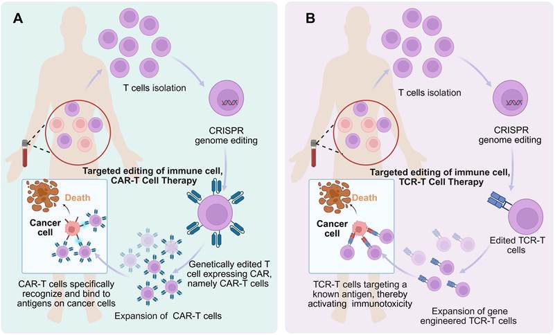
Recent investigations have focused on enhancing CRISPR editing efficacy in immune cells to correct pathogenic mutations [190,191]. For example, genome targeting via CRISPR technology can correct disease-causing IL2RA mutations in T cells and partly salvage IL2RA expression [190]. With the continuous development of adoptive immune cell therapy, there are also increasing opportunities for exploring novel targeted therapies based on CRISPR-engineered cells. Chimeric antigen receptor T-cell immunotherapy (CAR-T) allows genetically edited T cells from patients to express a chimeric antigen receptor (CAR) on their surface, resulting in sustained antitumor effects (Figure 6A) [192]. These engineered CARs help T cells specifically identify and combine with unique antigens on tumor cells, thereby activating antitumor cytotoxicity. CAR-T cell therapy may significantly relieve manifestations in patients with various kinds of blood cancers and solid tumors [193]. In addition, immunological therapy associated with gene correction via CRISPR is rapidly moving toward clinical practice. One clinical trial utilized Cas9 nucleases to modify autologous T cells ex vivo, knocking out the immune checkpoint PD-1 and endogenous T-cell receptor (TCR) to avoid mismatches or abnormal signaling, facilitating specific edited TCRs that could target tumor antigens [194]. The transfer of edited T cells back into patients presents significant prospects for the immunotherapy of malignant tumors (Figure 6B). Nevertheless, these applications are still under continuous development, with sustained efforts to realize efficacious and safe targeted gene therapy tailored to immune cells.
Genome editing of immune cells using CRISPR. (A) CAR-T cell therapy; (B) TCR-T cell therapy. Created with BioRender.com.
Importantly, the crosstalk between malignant cells and the tumor microenvironment (TME) also regulates tumor development [195-198]. CRISPR/Cas methods can precisely target genes that regulate the immune TME and metabolic pathways within tumors, thereby inhibiting tumor growth and metastasis [199]. Modulating the TME via CRISPR/Cas technology disrupts the homeostasis that supports tumor cells, thereby suppressing the abnormal behavior of tumors [200,201]. In summary, by targeting the genetic basis of tumors, immune cells and TME, CRISPR-based gene editing paves the way for fundamentally altering the behavior of tumors and their interactions with adjacent tissues, revolutionizing cancer therapy.
7.5. Duchenne muscular dystrophy
Duchenne muscular dystrophy (DMD) represents a hereditary muscle disorder [202]. CRISPR/Cas9 technology works at the DNA level, offering innovative prospects for DMD gene therapy. Furthermore, considerable progress in the field of DMD has been achieved since the emergence of genome editing via CRISPR [63]. AAV9 delivery of CRISPR/Cas9 via intraperitoneal, intramuscular, or systemic routes resulted in the expression of corrected dystrophin in animal models of DMD, partly restoring myocardial or skeletal function [202-204]. The general approach of these therapies is to correct point mutations in mouse models of DMD [202-204]. In addition, in vitro genome editing of iPSCs represents a viable strategy for DMD therapy [205]. Different types of mutations require different strategies. Exon knock-in by the CRISPR/Cas9 system via iPSC electroporation seems to be a beneficial approach for rescuing DMD with dystrophin gene mutations [205]. After iPSCs are modified and differentiated, functional dystrophin can be expressed [205]. Similar results were obtained when the reading frames of iPSCs were recovered by CRISPR technology in DMD with frameshift mutations [206].
7.6. Hereditary hearing loss
Hereditary hearing loss is a typical illustration of the application of gene therapy in inherited diseases [207]. Recently, more than 150 pathogenic genes related to hereditary hearing loss have been identified [208]. Owing to the increasing concern about hearing impairment, increasing numbers of studies are endeavoring to treat hereditary hearing loss by gene therapy, which is a significant single-gene disorder and the most prevalent category of congenital deafness. Currently, the prospective curative efficacy of the smallest Cas13 protein, CasRx, has been explored in neonatal Beethoven mice [209]. The Tmc1 mRNA level was downregulated by 70% via the CRISPR/CasRx system, and this treatment increased the survival rate of hair cells and alleviated hearing impairment [209]. This experimental verification illustrated the effectiveness and safety of CRISPR technology in the management of inherited hearing loss [210].
7.7. Neurodegenerative diseases
Alzheimer's disease
Causative gene mutations have been found in approximately 1% of familial Alzheimer's disease cases [211]. The CRISPR/Cas9 tool has attracted extensive attention considering treatment strategies for Alzheimer's disease involving the correction of genetic defects [212]. Utilization of the CRISPR system in Alzheimer's disease has been described, which remains a continuously growing research field [165]. Previous research has reported the utilization of dual AAV vector system to target the mutant gene that causes Alzheimer's disease, one packaging APPsw-specific gRNA and the other containing Cas9. The virus was assessed in Tg2576 mice by intrahippocampal injection and was also evaluated in neuronal cells derived from Tg2576 mouse embryos, which exhibited decreased Aβ production [213]. Furthermore, brain abnormality and cell death in animal models of Alzheimer's disease were rescued by targeting the methylation of APP through a dCas9–Dnmt3a approach [165].
Parkinson's disease
Currently, no strategy has been developed to completely cure Parkinson's disease, but genome editing techniques, particularly CRISPR/Cas9, are expected to achieve perpetual correction in hereditary Parkinson's disease with mutated genes [214]. Gordon et al. reported that augmenting PK2 signalling exerts a neuroprotective effect, while blocking its receptor accelerates dopaminergic neuron loss in animal models of Parkinson's disease [215]. Furthermore, CRISPR/Cas9-mediated knockout of PK2 was shown to exacerbate neuronal susceptibility to neurotoxin-induced cell death, an effect reversed by recombinant PK2 [214,215]. In addition, Chen et al. applied CRISPR system to downregulate SNCA in human embryonic stem cells, slowing the progression of SNCA-related pathological results [216].
Huntington's disease
Huntington's disease, an autosomal dominant disorder, arises from CAG repeat expansion in the huntingtin (HTT) gene, leading to the production of mutated HTT protein (mHTT) [211]. CRISPR gene editing can selectively inactivate mHTT in patient-derived fibroblasts [217], Huntington's disease model mice [218] and human-differentiated iPSCs [219]. It was first used for the permanent and selective inactivation of mHTT alleles in patient-derived fibroblasts by specialized allele-specific CRISPR/Cas9 to remove a broad region of HTT DNA, leading to the almost complete elimination of mRNA and mHTT proteins [217]. In addition, Kolli et al. studied the inhibitory effects of CRISPR/Cas9 systems on mHTT. The results demonstrated that mHTT silencing by CRISPR technology reduced the expression of mHTT in mesenchymal cells [220].
7.8. Autoimmune diseases
Systemic lupus erythematosus
Systemic lupus erythematosus can be featured by the destruction of self-antigen tolerance and manifests as the production of autoantibodies, inflammation and organ damage [221]. Although the pathogenesis of systemic lupus erythematosus has not yet been fully clarified, genetic traits constitute the etiology of this disorder [222]. The CXorf21 gene has been proposed as a suitable candidate gene for CRISPR/Cas9 treatment in systemic lupus erythematosus. The CXorf21 knockdown in vitro by CRISPR/Cas9 led to lower expression levels of IL-6 and TNF-alpha [168].
Type 1 diabetes
Type 1 diabetes can result in damage to pancreatic β cells, which mediate insulin secretion. Transplantation of insulin-secreting β cells may be an efficient and definitive treatment for type 1 diabetes [223]. Although the supply of primary β cells is incredibly limited, methods for generating β cells from pluripotent stem cells are being widely studied. Pluripotent stem cells can undergo gene editing through CRISPR/Cas to correct mutations that cause diabetes and eventually obtain effective β cells [224]. For instance, CRISPR technology was utilized to correct mutations of INS genes or WFS1 genes in iPSCs obtained from diabetes patients. Corrected iPSCs subsequently differentiated into stem cell-derived β cells with normal insulin folding and function, demonstrating considerable value for clinical application [225,226].
7.9. Infectious diseases
Infectious diseases are a serious challenge for public health and require new strategies for treatment [227]. CRISPR-based gene therapy targeting pathogen-specific genes provides a new strategy to fight infectious agents. CRISPR can also be used to identify the key host properties that pathogens exploit in infectious diseases, facilitating the development of creative approaches to reduce their virulence [228]. In addition, CRISPR can be used to develop diagnostic methods that contribute to the prompt detection of distinctive genetic markers of pathogens. One clinical trial (ClinicalTrials.gov identifier: NCT03164135) demonstrated that CRISPR gene editing targeting CCR5 could suppress human immunodeficiency virus type 1 (HIV-1) [229]. Furthermore, viral genomes may be removed from human cells by CRISPR [230]. One study using eight different sgRNAs that target hepatitis B virus revealed that integration of the viral genome into hepatocellular carcinoma cells was suppressed by CRISPR technology, consequently decreasing the expression of viral antigens.
8. Progress in delivery systems
Multiple methods for delivering CRISPR/Cas components to promote optimal gene editing activity have been explored [2]. CRISPR systems can be delivered primarily via physical, viral and non-viral approaches. Each delivery platform exhibits unique strengths and inherent limitations that must be carefully evaluated depending on the specific therapeutic application [231]. Physical methods, notably microinjection and electroporation, are particularly well-suited for in vitro and ex vivo applications due to their high efficiency and lack of cargo size constraints, but they are unsuitable for systemic in vivo delivery [20]. Viral vectors, most notably AAV, remain unparalleled in terms of efficiency and tissue-specific tropism, but they are associated with several concerns, including immunogenicity and limited cargo capacity [232-234]. Non-viral delivery approaches, particularly LNPs and emerging platforms such as engineered exosomes and virus-like particles, are progressively narrowing the efficiency gap while offering superior safety and targeting specificity [235-238]. The convergence of these technologies exhibits considerable potential to broaden the applications of CRISPR system.
8.1. Physical delivery methods
Physical delivery methods can deliver CRISPR components by penetrating cellular barriers [20,28]. The predominant physical delivery approaches include microinjection, electroporation, and hydrodynamic delivery. Microinjection involves a needle (0.5–5.0 μm in diameter) to deliver small quantities of substances into specific intracellular regions, such as the cytoplasm or nucleus [28]. Additionally, unlike size-limited delivery vectors, microinjection is not constrained by cargo size [20]. Moreover, microinjection is most appropriate for in vitro as well as ex vivo applications. As a visually observable and real-time traceable method, it is widely applied in fertilized mouse eggs and zebrafish embryos because of its low lethality and high delivery efficiency [239]. However, the primary limitations of microinjection are its manual operation and incompatibility with in vivo applications [20].
Compared with other delivery methods, electroporation is less cell type dependent and highly efficient at transfecting traditionally refractory cells. Similar to microinjection, electroporation remains effective for ex vivo or in vitro applications. Ex vivo electroporation has been extensively utilized in clinical trials, most notably in the realms of cancer immunotherapy and the treatment of inherited hematological disorders [11,26,194]. However, electroporation for CRISPR delivery has challenges, such as cytotoxicity, high costs, and operational complexity [240].
The efficiency of hydrodynamic delivery depends primarily on the anatomical structure of the target organ and its expansion rate post-intravascular injection [241]. When a substantial amount of solution is abruptly infused into the capillaries, it causes rapid expansion of the cell membrane. This leads to invagination formation, through which the solution gains access to the cellular interior without exogenous delivery vehicles [242]. This method is commonly used in gene therapy research for rodents. However, it is not considered suitable for application in large animals or humans, primarily due to its impracticality and insufficient safety profile [241]. For example, acute overload of the systemic circulation may lead to severe circulatory dysfunction [243].
Taken together, physical delivery methods, notably microinjection and electroporation, are particularly well suited for in vitro and ex vivo applications. Nevertheless, their insufficient safety profile and operational complexity limit their utility for systemic in vivo delivery, highlighting the need for complementary viral or non-viral strategies when tissue-specific editing is required.
8.2. Viral delivery methods
Viral delivery approaches have been identified as the most widely utilized strategies for efficiently delivering CRISPR components in vivo [77]. High-capacity viral vectors, including lentivirus (LV) and adenovirus (AdV) vectors, can encapsulate every component of the CRISPR system [77]. Nevertheless, AAV vectors with favorable efficiency have received much attention (Table 6). In addition, different organs can be targeted by specific AAV serotypes [233]. For example, AAV5 was utilized to deliver CRISPR system for treating Leber congenital amaurosis 10 [24]. The advantages of AAV include high transduction efficiency, low carcinogenic risk, serotype-related target cell specificity, long-lasting therapeutic effects, and remarkable clinical success [244-246]. CRISPR system delivery via AAV has performed well, but AAV can encapsulate only a limited amount of cargo [234]. Therefore, compared with relatively large SpCas9 (4.2 kb), smaller Cas9 orthologs might be more appropriate for delivery by AAV [24]. Although dual AAV vectors can be utilized, an all-in-one construct with a truncated Cas9 nickase represents a more appealing solution that has recently been investigated [247]. Moreover, although the efficiency of the virus vector deserves serious consideration, safety issues should not be ignored. In addition to the limited AAV package size, DNA encapsulated in AAV may unintentionally be integrated into the induced on-target DSB [248]. Because of the AAV vector DNA and AAV capsid, gene therapy delivered by AAV has the risk of activating the host immune system [233]. For this reason, many studies have focused on regulating AAV-related immunogenicity [249]. For example, optimized AAV serotypes with increased efficiency at low doses can partially overcome these challenges [250].
Common characteristics of CRISPR viral delivery methods.
| Delivery vector | Type | Most common cargo | Advantages | Limitations | Refs |
|---|---|---|---|---|---|
| AAV | Non-enveloped | ~4.7 kb, ssDNA | Efficient delivery; different serotypes can target specific tissues | Low packaging capacity; preexisting immunity | [117,204,251,252] |
| AdV | Non-enveloped | 8 kb-36 kb, dsDNA | No genomic integration; good genetic capacity | Inflammatory response; nonpersistent gene-editing effects | [252-260] |
| LV | Enveloped | ~10 kb, ssRNA | Persistent genetic effects; low immunogenicity | Genomic integration; low efficiency in vivo | [161,252,261-267] |
8.3. Non-viral delivery methods
Effective and safe non-viral delivery strategies for gene editing are urgently needed and have unique benefits in delivering gene cargo, including enabling instantaneous expression, low immunogenicity, fewer cargo size restrictions, and convenient mass production [268]. Many biomaterials, such as inorganic nanoparticles (NPs), polymeric NPs, LNPs, metal–organic frameworks, DNA origami, engineered exosomes, and virus-like particles, exhibit a significant ability to deliver CRISPR components for robust gene editing [2,269,270].
8.3.1. Inorganic NPs
Gold nanoparticles
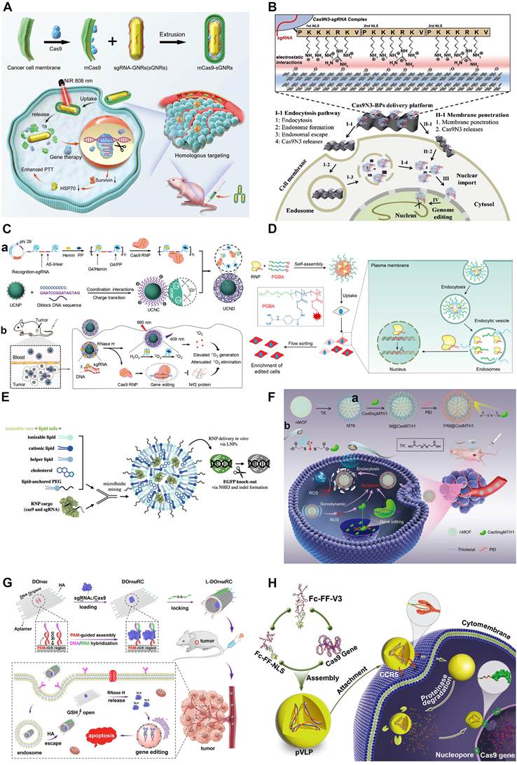
Gold nanoparticles (AuNPs), which exhibit distinct physical and chemical characteristics, are considered excellent non-viral vectors [271]. The physicochemical characteristics of AuNPs enable regulation of the surface charge by combining with cationic molecules to facilitate DNA binding via electrostatic interactions [272]. The effectiveness of AuNPs as non-viral delivery vectors has been illustrated in numerous studies [273,274]. Furthermore, arginine-modified AuNPs were shown to deliver RNPs directly to the cytoplasm via membrane fusion, resulting in high genome editing efficiency [275,276]. A complex comprising the Cas effector, AuNPs and an endosomal destructive polymer (CRISPR-Gold) was effectively used for the treatment of fragile X syndrome and DMD in mouse models [277,278]. Even after repeated injections, no obvious toxicity was detected in this treatment. Recently, the CRISPR system was reported to be delivered by colloidal AuNPs into progenitors in vitro and effectively perform genome editing, providing a promising therapeutic approach for various diseases [279]. Furthermore, AuNPs with photothermal features can be extensively used for photothermal therapy or stimulating the release of cargo [280,281], demonstrating promising prospects for CRISPR-based gene therapy delivered by AuNPs. Accordingly, CRISPR/Cas9 can be delivered via an integrated approach in which AuNPs and lipids are combined to target the Plk-1 gene to suppress melanoma [67]. Moreover, as illustrated in Figure 7A, which depicts the synthesis of Cas9-loaded gold nanorods and the combined photothermal/gene therapy process, synergistic photothermal/gene therapy demonstrated significant antitumor efficacy through effective delivery of CRISPR components and gold nanorods [282].
Representative examples of non-viral delivery approaches for CRISPR. (A) Synthesis procedures of Cas9-loaded gold nanorods and processes of combined photothermal/gene therapy. Adapted with permission from [282], copyright 2022 John Wiley and Sons. (B) Design of black phosphorus nanosheets for delivering CRISPR component and related intracellular delivery pathways. Adapted with permission from [284], copyright 2018 John Wiley and Sons. (C) Multifunctional DNA/upconversion nanoparticle complex for synergistic photodynamic therapy by delivering Cas9 RNP, hemin and protoporphyrin. Adapted with permission from [287], copyright 2024 John Wiley and Sons. (D) Preparation of PGBA-RNP polymeric NPs and overview of mechanisms for genome editing. Adapted with permission from [294], copyright 2024 American Chemical Society. (E) CRISPR/Cas9 RNP-encapsulated LNPs for in vivo delivery. Adapted with permission from [235], copyright 2025 American Chemical Society. (F) a) Preparation of the nano-sonosensitizer metal–organic frameworks P/M@CasMTH1 and b) ultrasound-triggered gene editing-augmented sonodynamic therapy for oncologic interventions. Adapted with permission from [312], copyright 2021 John Wiley and Sons. (G) Design and construction of CRISPR system based on DNA origami for gene therapy. Adapted with permission from [329], copyright 2023 John Wiley and Sons. (H) The viral peptide and Cas9 gene were co-assembled into peptidyl virus-like particles capable of transferring genes through the viral entry pathway. Adapted with permission from [237], copyright 2018 John Wiley and Sons.
Black phosphorus nanosheets
Black phosphorus nanosheets have a two-dimensional structure and have sparked intense research focus because of their unique physical and chemical characteristics [283,284]. The periodic atomic grooves on the surfaces of black phosphorus nanosheets offer appropriate anchoring sites with great prospects for the loading and delivery of biomolecules [284], including CRISPR/Cas9 RNP. Black phosphorus nanosheets loaded with Cas9 RNPs were effectively constructed and engineered with three nuclear localization signals (NLSs) at the C terminus (Cas9N3). As depicted in Figure 7B, which illustrates the design of black phosphorus nanosheets for delivering CRISPR components and related intracellular delivery pathways, Cas9N3–black phosphorus nanosheets entered cells efficiently, subsequently triggering endosomal escape upon the biodegradation of black phosphorus nanosheets and cytosolic release of Cas9N3 complexes, thereby achieving robust genome editing [284]. Furthermore, black phosphorus nanosheets for delivering Cas13a/crRNA complexes were developed to suppress Mcl-1 transcription levels for the treatment of breast cancer [285].
Upconversion nanoparticles
Upconversion nanoparticles can release biomacromolecules with high spatiotemporal precision via photoregulation, which can transform long-wave light into short-wave light [286]. When stimulated by 980 nm near-infrared light, upconversion nanoparticles emit 409 nm light to trigger protoporphyrin to generate 1O2, which has enhanced tumor suppressive effects resulting from the reduced expression level of Nrf2 [287]. Robust gene editing and effective photodynamic therapy were subsequently accomplished. Upconversion nanoparticles have become a research focus in the area of inorganic nanomaterials on account of their superior characteristics, such as safety and reduced side effects of near-infrared triggered therapy [288]. As shown in Figure 7C, one study reported a multifunctional DNA/upconversion nanoparticle complex that was able to co-deliver protoporphyrin, hemin and CRISPR/Cas9 to achieve combined photodynamic therapy [287]. Upconversion nanoparticles can convert near-infrared irradiation into short-wavelength irradiation and activate protoporphyrin to convert O2 into cytotoxic 1O2 via the CRISPR/Cas9-mediated downregulation of Nrf2, which inhibits the elimination of 1O2. The effective accumulation of cytotoxic 1O2 within tumor cells promotes apoptosis and effectively inhibits tumor growth [287].
8.3.2. Polymeric NPs
Highly selective and specific delivery is critical and can greatly extend the utilization of CRISPR gene editing. Polymeric NPs have shown particular promise in preclinical studies. A major class of compounds in polymeric NPs are cationic polymers, including poly(β-amino esters) (PBAE) and poly(ethyleneimine) (PEI), which contain positively charged groups that can interact electrostatically with nucleic acids [289]. By using polymeric NPs, CRISPR/Cas components can be delivered to specific cell types in a targeted manner. Through electrostatic interactions between the cationic polymer and the cargo itself, the CRISPR cargo can be condensed into a solid nanocomplex. The amphiphilic polymer subsequently encapsulates the complex to form multilayer NPs, thereby enhancing CRISPR delivery. In addition, alpha-helical cationic peptides may be used as both cell-penetrating agents and effective gene carriers [290]. In general, the excellent affinity between the active targeting portion and cell surface receptors can contribute to the efficacy of targeted delivery systems. For example, the CD44 receptor can bind specifically to hyaluronic acid (HA). Accordingly, the protein-based drug ribonuclease A was modified with HA and lipid molecules to increase the targeting and hence killing of tumor cells relative to that of the ribonuclease itself [238].
Recently, the combination of sgRNA and Cas9 RNP (nanoRNP) was shown to be delivered by polyethylene glycol (PEG)-based NPs, offering a viable approach for efficiently overcoming tumor heterogeneity [291]. In this system, the expression of runt-related transcription factor 1 and signal transducer and transcriptional activator 3 were inhibited in vivo by nanoRNPs, which effectively inhibited tumor progression. Moreover, compared with commercially available reagents, core‒shell polymeric NPs presented increased transfection efficiency with minimal side effects [292]. Besides, researchers have used polymer NPs to deliver a CRISPR/Cas13a system targeting PD-L1 to activate antitumor immunity mediated by T cells, restoring an immunosuppressive TME [293]. As illustrated in Figure 7D, which presents the preparation of PGBA-RNP polymeric NPs and an overview of mechanisms for genome editing, PGBA NPs enabled efficient RNP delivery for in vivo genome editing, resulting in a marked reduction in PCSK9 levels [294]. One nano-CRISPR scaffold with a versatile copolymer was designed for the co-delivery of CRISPR/dCas9 and cisplatin [295]. Furthermore, the nano-CRISPR scaffold inhibited the progression of malignant melanoma, resulting in strong tumor-suppressive immunity [295].
Therefore, the delivery of CRISPR cargo by NPs can overcome biosafety issues related to viral delivery approaches and improve the diversity of non-viral vectors, which may provide effective platforms for CRISPR-based disease therapies [292].
8.3.3. LNPs
LNPs have been widely used and are currently accepted as favorable carriers for CRISPR delivery. The widely used LNPs comprise four components: PEG lipids, ionizable lipids, cholesterol, and helper lipids [289]. Every component of the LNPs influences delivery through a distinct mechanism. Customizing the surface properties and composition of LNPs can markedly boost delivery performance [296]. Critical factors for enhancing delivery efficiency include optimizing the size of encapsulated proteins, the overall diameter of LNPs, and the surface charge [296,297]. Besides, LNPs with flexible lipid shells enter cells less effectively compared to LNPs with rigid shells [298]. LNP-encapsulated Cas9/sgRNA complexes were used to mediate CRISPR editing of transthyretin in humans (NTLA-2001), resulting in a substantial decrease in serum transthyretin levels and validating the therapeutic potential of optimized hepatic delivery [299].
A principal limitation of intravenously administered LNPs is their natural tropism for the liver [171], which restricts extrahepatic CRISPR applications [300]. Nevertheless, this tropism can be modulated by altering lipid molar ratios and administration routes, or by conjugating target-specific ligands [171,301]. High-throughput molecular barcoding technology was used in conjunction with traditional screening methods to assess LNP formulations concurrently for the biodistribution and efficiency of gene editing. This approach led to the exploration of lung-tropic LNPs that demonstrated effective gene editing in pulmonary epithelial and endothelial cells (as shown in Figure 7E, which depicts CRISPR/Cas9 RNP-encapsulated LNPs for in vivo delivery), establishing an extremely selective non-hepatic delivery method for lung-specific editing applications [235]. Similarly, LNPs loaded with Cas9 mRNA originally targeting the liver were redirected to the spleen after being formulated with anionic lipids [302]. In head and neck cancer, intratumoral delivery of EGFR-targeted CRISPR-LNP functionalized with targeting moieties enabled safe and efficient SOX2 knockout, demonstrating the potential of treating solid tumors and inducing durable therapeutic responses [303]. In addition, SM102-based LNPs co-delivering SpCas9 mRNA and sgRNA enabled efficient Mgp knockout in trabecular meshwork cells, demonstrating their potential for targeted gene editing [304]. A recent organic solvent-free LNP formulation that avoided cholesterol further reduced hepatic accumulation and immune activation while delivering multiple sgRNAs and Cas9 mRNA, demonstrating the feasibility of extrahepatic CRISPR applications [305]. Collectively, LNPs represent the most widely utilized non-viral delivery approach in biotherapy, and their capacity to facilitate CRISPR delivery holds significant value for clinical applications.
8.3.4. Metal–organic frameworks
Metal–organic frameworks are crucial types of gene delivery vehicles owing to their distinctive characteristics, such as tunable pore structure, biodegradability and drug-loading capacities [306]. Metal–organic frameworks feature highly tunable pore structures and a large specific surface area, enabling efficient loading of biomolecules such as sgRNA and Cas proteins [307,308]. In addition, metal–organic frameworks have good chemical stability and biocompatibility, exhibiting low immunogenicity and low toxicity [309]. Surface functional modification of metal–organic frameworks (for instance, folic acid modification) can enhance targeting capability and enable spatiotemporally controlled release of cargo via photothermal effect or ultrasonic treatment [310,311]. For example, nano-sonosensitizer metal–organic frameworks, termed P/M@CasMTH1, can deliver CRISPR component for targeting MTH1 and be utilized as a sonosensitizer to specifically trigger genome editing, resulting in antitumor effects (as illustrated in Figure 7F for the preparation of P/M@CasMTH1 and ultrasound-triggered gene editing-augmented sonodynamic therapy) [312]. Moreover, biocompatible nano-sized zeolitic imidazolate framework-8 (ZIF-8) can be utilized to deliver organic small molecules and bioactive macromolecules [313-315]. Furthermore, considering the simplicity and cost-effectiveness of the preparation process [316,317], ZIF-8 is highly suitable for controlled RNP delivery, thereby achieving effective genome editing. Alsaiari et al. illustrated that metal–organic framework ZIF-8 might function as a superior non-viral CRISPR/Cas9 delivery system that is biocompatible and has a controllable CRISPR/Cas9 component delivery capability [318].
8.3.5. DNA origami
As a burgeoning field of DNA technology, DNA origami folding ssDNA (known as scaffold strands) with a thousand bases long and many synthetic oligonucleotides (referred to as staple strands) has emerged as an excellent design strategy, primarily because the use of scaffold strands produced by enzymatic means reduces self-assembly errors and production costs [319-323]. DNA origami enables the strategic design of DNA nanostructures with appropriate sizes and specific geometric shapes [324-326]. With these advantages, structurally diverse DNA origami is applicable for delivering Cas9 and Cas12a RNPs [327,328]. As depicted in Figure 7G, a CRISPR system based on DNA origami was constructed for effective gene therapy in vivo [329]. DNA origami loaded with sgRNA/Cas9 complexes was rolled up via locking strands with a disulfide bond. After glutathione reduction, the opened DNA origami delivered the sgRNA/Cas9 complexes through RNase H cleavage to accomplish significant editing of disease-related genes for in vivo gene therapy. This gene editing system on the basis of DNA origami offers a favorable approach for advancing gene therapy [329]. Although controllable CRISPR/Cas9 systems have been developed using DNA origami [330], DNA origami nanoscaffolds driven by the pathophysiological environment for delivering Cas9 RNPs remain underutilized.
8.3.6. Engineered exosomes
Extracellular vesicles have become promising nanocarriers for bioactive molecules over the past decade [331]. Specifically, exosomes represent a subgroup of extracellular vesicles secreted by diverse cell types [236,332], and exhibit unique advantages, including low immunogenicity, superior cargo loading efficiency, reliable stability, favorable biocompatibility, and the capacity to traverse biological barriers [333-337]. Consequently, exosomes have attracted widespread attention for drug and gene delivery [338,339]. Recently, engineered exosomes incorporating a photoactivatable cargo-release system (MAPLEX) were constructed for epigenome editing in vivo [236]. The MAPLEX system facilitated the delivery of sgBace1-dCas9-D3A in mice, which resulted in CpG methylation, decreased Bace1 expression, and ameliorated recognition memory deficits and amyloid pathology [236]. Furthermore, an exosome-based platform (EMT-Cas12a) was constructed to deliver Cas12a mRNA and crRNAs, resulting in the suppression of HIV while maintaining favorable biosafety [340]. For musculoskeletal disorders, chondrocyte affinity peptide-modified MSC-derived exosomes served as biocompatible carriers for CRISPR/Cas9-mediated ASPN knockout in osteoarthritis-affected chondrocytes, demonstrating significant therapeutic efficacy in osteoarthritis models and underscoring the potential of exosome-based platforms for precision gene-targeted interventions [341]. Collectively, these studies highlight the versatility of engineered exosomes as programmable delivery vehicles for CRISPR systems across a wide range of disease models. Further improvements in targeting precision and large-scale production will be critical for translating these platforms into clinical gene therapies.
8.3.7. Virus-like particle
The appearance of virus-like particles resembles that of native viruses, but they lack viral genomes and therefore cannot replicate [342]. Therefore, virus-like particles can be classified as a non-viral delivery technology. Virus-like particles originate from existing viral scaffolds and make use of the inherent characteristics of viruses to facilitate effective intracellular delivery, such as the capacity to encapsulate cargo, to target distinct cell types, and to escape endosomes [252]. Owing to the ability for the transient CRISPR delivery, virus-like particles have received extensive attention with respect to gene therapy [343]. As illustrated in Figure 7H, peptidyl virus-like particles that imitate simian virus 40 and human immunodeficiency virus have been constructed [237]. Two viral peptides with good targeting ability were able to co-assemble to form biodegradable peptidyl virus-like particles by encapsulating the CRISPR component. The peptidyl virus-like particles successfully crossed the cell membrane and transferred cargo to the nucleus via the viral entry pathway. This study illustrates the significant potential of engineered virus-like particles with desirable properties [237].
9. Current challenges in CRISPR technology
Many obstacles remain to be overcome in the development of diagnostic methods and treatments by CRISPR technology [344]. Despite the advantages of CRISPR technology, challenges associated with its widespread application persist. Further research can focus on increasing efficacy, reducing off-target effects, optimizing cargo delivery methods, improving safety, and addressing ethical issues.
9.1. Efficacy of CRISPR technology
Multiple aspects influencing the efficacy of CRISPR technology are relevant to its wider application. The requirement for a PAM adjacent to the target site restricts the range of targetable disease-causing mutations, which is a key problem limiting efficacy. Research focused on key molecules of the CRISPR system could be beneficial for mitigating this restriction. For example, the novel CRISPR effectors NG-SpCas9 [345] and xCas9 [346] are able to target DNA sequences using unconventional ‘NG’ PAM sequences rather than the traditional ‘NGG’ PAM sequence. In addition, the RNA-targeting CRISPR/Cas13 system may function as an efficient and safe strategy to accurately modulate the transcripts of disease-causing genes at the mRNA level, avoiding the long-term dangers of modifying genomic DNA [344]. Furthermore, the CRISPR delivery systems require further optimization to ensure effective genome editing without unnecessary damage to healthy tissues, which should also decrease costs and improve accessibility [347].
Furthermore, a more fundamental obstacle is the poor predictive validity of current preclinical models for human disease [348,349]. Before advancing to clinical trials, CRISPR-based therapies must demonstrate robust efficacy in preclinical models [63]. However, interspecies differences in preexisting immunity, target tissue accessibility and cellular uptake frequently result in substantially lower editing efficiency in humans than in murine or other small animals [350,351]. Conventional mouse models often fail to recapitulate the genetic background, disease progression, and immunological landscape of human conditions [352]. Non-human primate models, although physiologically more similar to humans, are constrained by high costs, ethical concerns, and small sample sizes, precluding large-scale systematic efficacy evaluation [353,354]. Even emerging patient-derived organoids remain limited by several inherent limitations, most notably insufficient maturation and high heterogeneity [355]. This preclinical-to-clinical translation gap profoundly compromises dose selection, endpoint determination, and risk assessment in trial design [63,356]. Overcoming this barrier requires the development of more predictive preclinical models, including humanized animal models, to improve efficacy prediction and reduce the rate of clinical trial failures for CRISPR-based therapies [349,357,358].
9.2. Off-target effects
Medical applications require versatility but also rigorous precision to guarantee biosafety. Therefore, many studies have investigated the underlying problem of unwanted genome editing at other gene loci, known as off-target effects [359], which can result in health problems and even promote cancer progression. Therefore, preventing off-target effects is another vital issue to be solved (Figure 8) [34]. Off-target effects can be minimized through appropriate control and the use of multiple sgRNAs. Specifically, sgRNAs with fewer nucleotides may exhibit reduced off-target activity [360]. In addition to guiding RNA modification, improving the expression of CRISPR effector proteins may also be a suitable strategy for avoiding off-target effects [361]. Further studies have also indicated that highly specific Cas9 proteins such as HF-Cas9 [361] and eSpCas9 [362] are beneficial for regulating protein‒DNA interactions, preventing the mismatch of CRISPR effectors [363]. In addition, the retention time and concentration of the Cas protein in cells may be limited to enhance target specificity and avoid off-target effects [24,364,365]. Introducing a requirement for the specific activation of Cas9 by exogenous stimuli is another approach [366,367]. Moreover, Cas12a, as a high-precision CRISPR effector, has fewer off-target effects than Cas9 [344]. Suitable carriers for CRISPR components, such as functional nanoparticles, may also maximize the stability of the cargo, improving target specificity [292].
Approaches for avoiding CRISPR off-target effects. Created with BioRender.com.
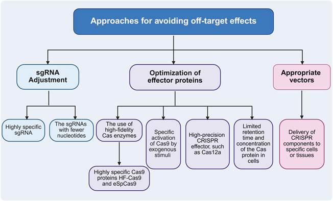
9.3. Delivery methods
Safe and efficient delivery of CRISPR/Cas components to target tissues in vivo represents another major challenge for clinical translation [368]. Specifically, programmable nucleases should be delivered in a transient manner at controlled doses to reduce off-target editing and immunogenicity while achieving sufficient therapeutic benefit [368]. CRISPR delivery methods have both advantages and limitations, which can be compared across three key dimensions. With respect to immunogenicity, AAV vectors carry a high risk of preexisting immunity and may elicit capsid-specific T cell responses [369,370]. LNPs exhibit low immunogenicity, with innate immune activation induced primarily by lipids that may be mitigated through formulation optimization [371,372]. Virus-like particles are considered safer owing to the absence of viral genetic material [370,373,374]. Regarding maximum tolerated dose, AAV administration at doses equal to or exceeding 5×10¹³ vg/kg has been associated with toxicity including thrombotic microangiopathies [375]. Hepatic accumulation represents the main factor limiting the maximum tolerated dose of LNPs, although NTLA-2001 was generally well-tolerated at 0.1–1.0 mg/kg [299,376]. Early preclinical data suggest that virus-like particles may offer increased maximum tolerated dose due to their biodegradability and reduced systemic inflammation [373,374]. Considering the feasibility of repeated administration, AAV vectors generally preclude repeated administration because of neutralizing antibodies [372]. Accordingly, EDIT-101 was designed as a single-administration therapy. In contrast, LNP-delivered CRISPR therapies have demonstrated the feasibility of repeated dosing, as their low immunogenicity permits effective booster administration [171,372]. Virus-like particles may still face constraints for repeated dosing owing to anti-vector immunity, although this is generally more modest than that associated with AAV [377].
Two pioneering in vivo CRISPR therapies illustrate how the delivery system fundamentally dictates clinical trial design [180,299]. NTLA-2001 employs LNPs to deliver Cas9 mRNA with liver tropism for the treatment of transthyretin amyloidosis, a design that maximizes therapeutic safety [299]. Conversely, EDIT-101 relies on an AAV5 vector to deliver the CRISPR components for the treatment of Leber congenital amaurosis 10, depending on sustained long-term expression to achieve durable therapeutic effects after a single dose [180]. This contrast demonstrates that the choice of delivery vector determines not only the pharmacokinetic profile of the editing components but also the entire therapeutic regimen, including dosing frequency, long-term safety monitoring, and patient follow-up strategies. These differences underscore the need to develop suitable delivery systems that combine specific targeting, robust transfection, superior cargo loading efficiency, and minimal immunogenicity. Furthermore, the long-term biosafety and minimal toxic effects of delivery vehicles must be rigorously evaluated before advancing CRISPR-based therapies into clinical practice [20].
9.4. Safety and ethical issues
While CRISPR faces numerous challenges related to its utilization, safety and ethical issues of genomic editing seem to present the greatest challenge. In particular, CRISPR/Cas may cause severe damage to tissues and result in systemic side effects due to host immunogenicity and genome integration. One major concern for safety that needs to be addressed is the immunogenicity of the Cas protein itself or its delivery components [35]. CRISPR therapies that require the sustained expression of Cas9 need to overcome preexisting immunity [378]. Therefore, removing antigen regions of the CRISPR system or designing Cas9 variants to avoid universally immunogenic epitopes may be effective strategies [378]. With the great progress in CRISPR technology, ethical issues have become a primary consideration for clinical applications [379]. Investigating the effects of genome editing on nontarget organisms is also essential, as these effects may persist and expand with each generation, leading to disastrous effects on the balance of ecosystems [380]. Another key concern is the possibility of modifying human germ cells. To date, genome editing experiments have been primarily conducted on somatic cells, and alterations to the germline genome in a zygote or embryo is widely considered unacceptable because genome editing could introduce mutations with unpredictable adverse reactions in future generations [381].
10. Conclusions and perspectives
The advancement of CRISPR technology is mainly driven by the continuous optimization of Cas nucleases. High-fidelity variants such as HF-Cas9 and eSpCas9 markedly improve specificity and minimize off-target effects. Compact Cas enzymes, including Cas12f variants, represent a significant breakthrough in overcoming AAV packaging limitations. Furthermore, chemical and physical control strategies have expanded the spatiotemporal precision of CRISPR systems. Precise editing strategies, particularly base editing and prime editing, have achieved unprecedented accuracy in genomic modifications. Apart from genome editing, CRISPR technology can also be utilized for transcriptional activation and inhibition, such as CRISPRa and CRISPRi systems. Collectively, with the constant progress of CRISPR technology, genome editing has become highly useful in fundamental biological research. Applications ranging from CRISPR screening to disease model construction have accelerated not only basic research but also innovation across the life sciences. Furthermore, CRISPR technology has demonstrated remarkable prospects for diverse diseases, including hematological, eye-related, metabolic, neurodegenerative, and autoimmune diseases, as well as other intractable disorders. Ongoing preclinical and clinical trials will help determine the full capabilities of CRISPR.
Despite the considerable capabilities of CRISPR technology, challenges persist in terms of its widespread application. Delivery remains a major bottleneck, with inherent trade-offs between viral and non-viral systems. Viral vectors offer high transfection efficiency but are limited by restricted cargo capacity, whereas non-viral systems exhibit improved safety profiles but display substantially lower efficacy. In terms of viral vectors, dual-AAV system has emerged as an effective strategy to circumvent packaging constraints. Parallel advancements in non-viral approaches include stimulus-responsive polymers enabling microenvironment-dependent payload release, LNP optimization by modifying the surface charge, and virus-like particles with superior targeting specificity. Rapid progress in delivery systems not only facilitates fundamental research but also promotes the clinical application of CRISPR technology.
Beyond delivery, off-target effects remain a critical concern even with high-fidelity Cas variants, particularly for systemic in vivo applications. Notably, rapidly developing artificial intelligence (AI) techniques, such as machine learning and deep learning, address these challenges by leveraging large-scale experimental datasets to improve gRNA design, predict off-target activity, and increase editing efficiency [382,383]. Trained on comprehensive datasets, machine learning algorithms are able to predict gRNA activity and off-target risks, optimizing gRNA design in terms of both efficiency and precision [382]. Moreover, AI has also facilitated the development of engineered editing proteins. Deep learning tools for predicting protein structure, including AlphaFold [384-386] and RoseTTAFold [387], provide widespread access to high-resolution structural information for Cas nucleases and other genome editing enzymes. The integration of AI and CRISPR technology provides novel approaches for personalized gene therapies.
Collectively, ongoing advancements in CRISPR technology can not only optimize therapeutic efficacy and precision but also surmount the current limitations. Multiple preclinical studies and clinical trials worldwide are significantly expanding the therapeutic prospects of CRISPR technology. These investigations have demonstrated the effectiveness and safety of CRISPR technology in disease treatment. With continuous refinement, CRISPR technology holds immense potential to achieve unprecedented breakthroughs in biomedical research and alleviate previously intractable diseases.
Acknowledgements
This work was financially supported by the National Natural Science Foundation of China (22205151 and 32201148), the Sichuan Natural Science Foundation (2024NSFSC1718) and Chengdu Science and Technology Program (2026-YF05-00807-SN).
Author contributions
Xue Wu: Writing – original draft, Validation, Conceptualization. Xiaorong Kou: Writing – original draft, Validation, Conceptualization. Liping Bai: Writing – review & editing. Chao Liu: Writing – review & editing. Qinjie Wu: Writing – review & editing. Ming Zhang: Supervision, Project administration. Ning Wang: Supervision, Project administration. Changyang Gong: Supervision, Project administration, Conceptualization.
Competing Interests
The authors have declared that no competing interest exists.
References
1. Katayama S, Watanabe M, Kato Y, Nomura W, Yamamoto T. Engineering of zinc finger nucleases through structural modeling improves genome editing efficiency in cells. Adv Sci (Weinh). 2024;11:e2310255
2. Li B, Niu Y, Ji W, Dong Y. Strategies for the CRISPR-based therapeutics. Trends Pharmacol Sci. 2020;41:55-65
3. Ma X, Miao L, Liu X. Teach plants to fish based on CRISPR-Cas system self-evolution. Plant Biotechnol J. 2025;23:2361-3
4. Jansen R, Embden JD, Gaastra W, Schouls LM. Identification of genes that are associated with DNA repeats in prokaryotes. Mol Microbiol. 2002;43:1565-75
5. Mojica FJ, Díez-Villaseñor C, García-Martínez J, Soria E. Intervening sequences of regularly spaced prokaryotic repeats derive from foreign genetic elements. J Mol Evol. 2005;60:174-82
6. Barrangou R, Fremaux C, Deveau H, Richards M, Boyaval P, Moineau S. et al. CRISPR provides acquired resistance against viruses in prokaryotes. Science. 2007;315:1709-12
7. Cong L, Ran FA, Cox D, Lin S, Barretto R, Habib N. et al. Multiplex genome engineering using CRISPR/Cas systems. Science. 2013;339:819-23
8. Gilbert LA, Larson MH, Morsut L, Liu Z, Brar GA, Torres SE. et al. CRISPR-mediated modular RNA-guided regulation of transcription in eukaryotes. Cell. 2013;154:442-51
9. Jinek M, Chylinski K, Fonfara I, Hauer M, Doudna JA, Charpentier E. A programmable dual-RNA-guided DNA endonuclease in adaptive bacterial immunity. Science. 2012;337:816-21
10. Perez-Pinera P, Kocak DD, Vockley CM, Adler AF, Kabadi AM, Polstein LR. et al. RNA-guided gene activation by CRISPR-Cas9-based transcription factors. Nat Methods. 2013;10:973-6
11. Lu Y, Xue J, Deng T, Zhou X, Yu K, Deng L. et al. Safety and feasibility of CRISPR-edited t cells in patients with refractory non-small-cell lung cancer. Nat Med. 2020;26:732-40
12. Komor AC, Kim YB, Packer MS, Zuris JA, Liu DR. Programmable editing of a target base in genomic DNA without double-stranded DNA cleavage. Nature. 2016;533:420-4
13. Anzalone AV, Randolph PB, Davis JR, Sousa AA, Koblan LW, Levy JM. et al. Search-and-replace genome editing without double-strand breaks or donor DNA. Nature. 2019;576:149-57
14. Villiger L, Joung J, Koblan L, Weissman J, Abudayyeh OO, Gootenberg JS. CRISPR technologies for genome, epigenome and transcriptome editing. Nat Rev Mol Cell Biol. 2024;25:464-87
15. Gori JL, Haddad E, Frangoul H, Kohn DB, Morris EC, Martin BN. et al. Prime editing for p47(phox)-deficient chronic granulomatous disease. N Engl J Med. 2026;394:1195-203
16. Scholz P, Thompson J, Crosby KT, Fauth T, Krah NM, Schlauderaff G. et al. RNA-triggered cell killing with CRISPR-Cas12a2. Nature. 2026;655:230-9
17. Yan M, Li J. The evolving CRISPR technology. Protein Cell. 2019;10:783-6
18. Wang M, Zhang R, Li J. CRISPR/cas systems redefine nucleic acid detection: Principles and methods. Biosens Bioelectron. 2020;165:112430
19. Gilbert LA, Horlbeck MA, Adamson B, Villalta JE, Chen Y, Whitehead EH. et al. Genome-scale CRISPR-mediated control of gene repression and activation. Cell. 2014;159:647-61
20. Lino CA, Harper JC, Carney JP, Timlin JA. Delivering CRISPR: A review of the challenges and approaches. Drug Deliv. 2018;25:1234-57
21. Behan FM, Iorio F, Picco G, Gonçalves E, Beaver CM, Migliardi G. et al. Prioritization of cancer therapeutic targets using CRISPR-Cas9 screens. Nature. 2019;568:511-6
22. Santiago-Fernández O, Osorio FG, Quesada V, Rodríguez F, Basso S, Maeso D. et al. Development of a CRISPR/Cas9-based therapy for hutchinson-gilford progeria syndrome. Nat Med. 2019;25:423-6
23. Beyret E, Liao HK, Yamamoto M, Hernandez-Benitez R, Fu Y, Erikson G. et al. Single-dose CRISPR-Cas9 therapy extends lifespan of mice with hutchinson-gilford progeria syndrome. Nat Med. 2019;25:419-22
24. Hendriks D, Clevers H, Artegiani B. CRISPR-Cas tools and their application in genetic engineering of human stem cells and organoids. Cell Stem Cell. 2020;27:705-31
25. Locatelli F, Lang P, Wall D, Meisel R, Corbacioglu S, Li AM. et al. Exagamglogene autotemcel for transfusion-dependent β-thalassemia. N Engl J Med. 2024;390:1663-76
26. Frangoul H, Altshuler D, Cappellini MD, Chen YS, Domm J, Eustace BK. et al. CRISPR-Cas9 gene editing for sickle cell disease and β-thalassemia. N Engl J Med. 2021;384:252-60
27. Frangoul H, Locatelli F, Sharma A, Bhatia M, Mapara M, Molinari L. et al. Exagamglogene autotemcel for severe sickle cell disease. N Engl J Med. 2024;390:1649-62
28. Zhang Y, Yu LC. Microinjection as a tool of mechanical delivery. Curr Opin Biotechnol. 2008;19:506-10
29. Wang L, Liu C, Wang X, Ma S, Liu F, Zhang Y. et al. Tumor-specific activated nano-domino-CRISPR to amplify intrinsic oxidative and activate endogenous apoptosis for spatiotemporally specific therapy. Biomaterials. 2023;295:122056
30. Ma S, Li Y, Liu F, Wang X, Qin Z, Wang L. et al. Hierarchical-unlocking virus-esque nanocrispr precisely disrupts autocrine and paracrine pathway of vegf for tumor inhibition and antiangiogenesis. J Control Release. 2024;366:505-18
31. Li X, Duan Z, Chen X, Pan D, Luo Q, Gu L. et al. Impairing tumor metabolic plasticity via a stable metal-phenolic-based polymeric nanomedicine to suppress colorectal cancer. Adv Mater. 2023;35:e2300548
32. Shen X, Cai H, Wang Y, Xie M, Li Y, Pan D. et al. Metabolic targeting of oxidative phosphorylation enhances chemosensitivity in triple-negative breast cancer via a synergistic nanomedicine. Theranostics. 2025;15:7607-26
33. Yang J, Bai L, Shen M, Gou X, Xiang Z, Ma S. et al. A multiple stimuli-responsive nanocrispr overcomes tumor redox heterogeneity to augment photodynamic therapy. ACS Nano. 2023;17:11414-26
34. Kosicki M, Tomberg K, Bradley A. Repair of double-strand breaks induced by CRISPR-Cas9 leads to large deletions and complex rearrangements. Nat Biotechnol. 2018;36:765-71
35. Chehelgerdi M, Chehelgerdi M, Khorramian-Ghahfarokhi M, Shafieizadeh M, Mahmoudi E, Eskandari F. et al. Comprehensive review of CRISPR-based gene editing: Mechanisms, challenges, and applications in cancer therapy. Mol Cancer. 2024;23:9
36. Makarova KS, Wolf YI, Alkhnbashi OS, Costa F, Shah SA, Saunders SJ. et al. An updated evolutionary classification of CRISPR-Cas systems. Nat Rev Microbiol. 2015;13:722-36
37. Shmakov S, Smargon A, Scott D, Cox D, Pyzocha N, Yan W. et al. Diversity and evolution of class 2 CRISPR-Cas systems. Nat Rev Microbiol. 2017;15:169-82
38. Zhang F, Huang Z. Mechanistic insights into the versatile class ii CRISPR toolbox. Trends Biochem Sci. 2022;47:433-50
39. van Dongen JE, Berendsen JTW, Steenbergen RDM, Wolthuis RMF, Eijkel JCT, Segerink LI. Point-of-care CRISPR/Cas nucleic acid detection: Recent advances, challenges and opportunities. Biosens Bioelectron. 2020;166:112445
40. Kim D, Kim J, Hur JK, Been KW, Yoon SH, Kim JS. Genome-wide analysis reveals specificities of cpf1 endonucleases in human cells. Nat Biotechnol. 2016;34:863-8
41. Kleinstiver BP, Tsai SQ, Prew MS, Nguyen NT, Welch MM, Lopez JM. et al. Genome-wide specificities of CRISPR-Cas cpf1 nucleases in human cells. Nat Biotechnol. 2016;34:869-74
42. Harrington LB, Burstein D, Chen JS, Paez-Espino D, Ma E, Witte IP. et al. Programmed DNA destruction by miniature CRISPR-Cas14 enzymes. Science. 2018;362:839-42
43. Makarova KS, Wolf YI, Iranzo J, Shmakov SA, Alkhnbashi OS, Brouns SJJ. et al. Evolutionary classification of CRISPR-Cas systems: A burst of class 2 and derived variants. Nat Rev Microbiol. 2020;18:67-83
44. Paul B, Montoya G. CRISPR-Cas12a: Functional overview and applications. Biomed J. 2020;43:8-17
45. Liu S, He Y, Fan T, Zhu M, Qi C, Ma Y. et al. PAM-relaxed and temperature-tolerant CRISPR-mb3cas12a single transcript unit systems for efficient singular and multiplexed genome editing in rice, maize, and tomato. Plant Biotechnol J. 2025;23:156-73
46. Pandey V, Sharma S, Pokharel YR. Exploring CRISPR-Cas: The transformative impact of gene editing in molecular biology. Mol Ther Nucleic Acids. 2025;36:102717
47. Abudayyeh OO, Gootenberg JS, Essletzbichler P, Han S, Joung J, Belanto JJ. et al. RNA targeting with CRISPR-cas13. Nature. 2017;550:280-4
48. Konermann S, Lotfy P, Brideau NJ, Oki J, Shokhirev MN, Hsu PD. Transcriptome engineering with RNA-targeting type vi-d CRISPR effectors. Cell. 2018;173:665-76.e14
49. Wang Q, Liu X, Zhou J, Yang C, Wang G, Tan Y. et al. The CRISPR-Cas13a gene-editing system induces collateral cleavage of RNA in glioma cells. Adv Sci (Weinh). 2019;6:1901299
50. Zhao X, Liu L, Lang J, Cheng K, Wang Y, Li X. et al. A CRISPR-Cas13a system for efficient and specific therapeutic targeting of mutant kras for pancreatic cancer treatment. Cancer Lett. 2018;431:171-81
51. Mali P, Yang L, Esvelt KM, Aach J, Guell M, DiCarlo JE. et al. RNA-guided human genome engineering via Cas9. Science. 2013;339:823-6
52. Liu JJ, Orlova N, Oakes BL, Ma E, Spinner HB, Baney KLM. et al. CasX enzymes comprise a distinct family of RNA-guided genome editors. Nature. 2019;566:218-23
53. Su M, Li F, Wang Y, Gao Y, Lan W, Shao Z. et al. Molecular basis and engineering of miniature Cas12f with c-rich PAM specificity. Nat Chem Biol. 2024;20:180-9
54. Xiao R, Li Z, Wang S, Han R, Chang L. Structural basis for substrate recognition and cleavage by the dimerization-dependent CRISPR-Cas12f nuclease. Nucleic Acids Res. 2021;49:4120-8
55. Xin C, Yin J, Yuan S, Ou L, Liu M, Zhang W. et al. Comprehensive assessment of miniature CRISPR-Cas12f nucleases for gene disruption. Nat Commun. 2022;13:5623
56. Zhang X, Li M, Chen K, Liu Y, Liu J, Wang J. et al. Engineered circular guide rnas enhance miniature CRISPR/Cas12f-based gene activation and adenine base editing. Nat Commun. 2025;16:3016
57. Gaj T, Gersbach CA, Barbas CF 3rd. ZFN, TALEN, and CRISPR/Cas-based methods for genome engineering. Trends Biotechnol. 2013;31:397-405
58. Symington LS, Gautier J. Double-strand break end resection and repair pathway choice. Annu Rev Genet. 2011;45:247-71
59. Pankowicz FP, Jarrett KE, Lagor WR, Bissig KD. CRISPR/Cas9: At the cutting edge of hepatology. Gut. 2017;66:1329-40
60. Young CS, Pyle AD, Spencer MJ. CRISPR for neuromuscular disorders: Gene editing and beyond. Physiology (Bethesda). 2019;34:341-53
61. Komor AC, Zhao KT, Packer MS, Gaudelli NM, Waterbury AL, Koblan LW. et al. Improved base excision repair inhibition and bacteriophage mu gam protein yields c:G-to-t:A base editors with higher efficiency and product purity. Sci Adv. 2017;3:eaao4774
62. Xu W, Zhang S, Qin H, Yao K. From bench to bedside: Cutting-edge applications of base editing and prime editing in precision medicine. J Transl Med. 2024;22:1133
63. Laurent M, Geoffroy M, Pavani G, Guiraud S. CRISPR-based gene therapies: From preclinical to clinical treatments. Cells. 2024;13:800
64. Gaudelli NM, Komor AC, Rees HA, Packer MS, Badran AH, Bryson DI. et al. Programmable base editing of a•t to g•c in genomic DNA without DNA cleavage. Nature. 2017;551:464-71
65. Maji B, Moore CL, Zetsche B, Volz SE, Zhang F, Shoulders MD. et al. Multidimensional chemical control of CRISPR-Cas9. Nat Chem Biol. 2017;13:9-11
66. Jain PK, Ramanan V, Schepers AG, Dalvie NS, Panda A, Fleming HE. et al. Development of light-activated CRISPR using guide rnas with photocleavable protectors. Angew Chem Int Ed Engl. 2016;55:12440-4
67. Wang P, Zhang L, Zheng W, Cong L, Guo Z, Xie Y. et al. Thermo-triggered release of CRISPR-Cas9 system by lipid-encapsulated gold nanoparticles for tumor therapy. Angew Chem Int Ed Engl. 2018;57:1491-6
68. Ryu JY, Won EJ, Lee HAR, Kim JH, Hui E, Kim HP. et al. Ultrasound-activated particles as CRISPR/Cas9 delivery system for androgenic alopecia therapy. Biomaterials. 2020;232:119736
69. Aubrey BJ, Kelly GL, Kueh AJ, Brennan MS, O'Connor L, Milla L. et al. An inducible lentiviral guide RNA platform enables the identification of tumor-essential genes and tumor-promoting mutations in vivo. Cell Rep. 2015;10:1422-32
70. Dow LE, Fisher J, O'Rourke KP, Muley A, Kastenhuber ER, Livshits G. et al. Inducible in vivo genome editing with CRISPR-Cas9. Nat Biotechnol. 2015;33:390-4
71. Nguyen DP, Miyaoka Y, Gilbert LA, Mayerl SJ, Lee BH, Weissman JS. et al. Ligand-binding domains of nuclear receptors facilitate tight control of split CRISPR activity. Nat Commun. 2016;7:12009
72. Huang HC, Wu LF, Liu K, Ma BG. Opto-CRISPR: New prospects for gene editing and regulation. Trends Biotechnol. 2026;44:22-39
73. Chen X, Chen Y, Xin H, Wan T, Ping Y. Near-infrared optogenetic engineering of photothermal nanocrispr for programmable genome editing. Proc Natl Acad Sci U S A. 2020;117:2395-405
74. Hoffmann MD, Mathony J, Upmeier Zu Belzen J, Harteveld Z, Aschenbrenner S, Stengl C. et al. Optogenetic control of neisseria meningitidis Cas9 genome editing using an engineered, light-switchable anti-CRISPR protein. Nucleic Acids Res. 2021;49:e29
75. Zhuo C, Zhang J, Lee JH, Jiao J, Cheng D, Liu L. et al. Spatiotemporal control of CRISPR/Cas9 gene editing. Signal Transduct Target Ther. 2021;6:238
76. Yuan L, Xiong Y, Zhang Y, Gu S, Lei Y. Epigenome editing based treatment: Progresses and challenges. Mol Ther. 2026;34:46-67
77. Bendixen L, Jensen TI, Bak RO. CRISPR-Cas-mediated transcriptional modulation: The therapeutic promises of CRISPRa and CRISPRi. Mol Ther. 2023;31:1920-37
78. Liu C, Wang N, Luo R, Li L, Yang W, Wang X. et al. A programmable hierarchical-responsive nanocrispr elicits robust activation of endogenous target to treat cancer. Theranostics. 2021;11:9833-46
79. Wu Q, Wu J, Karim K, Chen X, Wang T, Iwama S. et al. Massively parallel characterization of CRISPR activator efficacy in human induced pluripotent stem cells and neurons. Mol Cell. 2023;83:1125-39.e8
80. Savell KE, Bach SV, Zipperly ME, Revanna JS, Goska NA, Tuscher JJ. et al. A neuron-optimized CRISPR/dCas9 activation system for robust and specific gene regulation. eNeuro. 2019;6:e0495-18.2019
81. Clark T, Waller MA, Loo L, Moreno CL, Denes CE, Neely GG. CRISPR activation screens: Navigating technologies and applications. Trends Biotechnol. 2024;42:1017-34
82. Beerli RR, Dreier B, Barbas CF 3rd. Positive and negative regulation of endogenous genes by designed transcription factors. Proc Natl Acad Sci U S A. 2000;97:1495-500
83. Hilton IB, D'Ippolito AM, Vockley CM, Thakore PI, Crawford GE, Reddy TE. et al. Epigenome editing by a CRISPR-Cas9-based acetyltransferase activates genes from promoters and enhancers. Nat Biotechnol. 2015;33:510-7
84. Wang K, Escobar M, Li J, Mahata B, Goell J, Shah S. et al. Systematic comparison of CRISPR-based transcriptional activators uncovers gene-regulatory features of enhancer-promoter interactions. Nucleic Acids Res. 2022;50:7842-55
85. Duellman T, Doll A, Chen X, Wakamiya R, Yang J. dCas9-mediated transcriptional activation of tissue inhibitor of metalloproteinases. Metalloproteinases Med. 2017;4:63-73
86. Sajwan S, Mannervik M. Gene activation by dCas9-cbp and the SAM system differ in target preference. Sci Rep. 2019;9:18104
87. Zhou M, Tao X, Sui M, Cui M, Liu D, Wang B. et al. Reprogramming astrocytes to motor neurons by activation of endogenous ngn2 and isl1. Stem Cell Reports. 2021;16:1777-91
88. Zhang R, Liu J, Xue G, Yang J, Li D, Tian T. et al. Forced activation of dystrophin transcription by CRISPR/dCas9 reduced arrhythmia susceptibility via restoring membrane nav1.5 distribution. Gene Ther. 2023;30:142-9
89. Liao HK, Hatanaka F, Araoka T, Reddy P, Wu MZ, Sui Y. et al. In vivo target gene activation via CRISPR/Cas9-mediated trans-epigenetic modulation. Cell. 2017;171:1495-507.e15
90. Lee MH, Lin CC, Thomas JL, Li JA, Lin HY. Cellular reprogramming with multigene activation by the delivery of CRISPR/dCas9 ribonucleoproteins via magnetic peptide-imprinted chitosan nanoparticles. Mater Today Bio. 2021;9:100091
91. Groner AC, Meylan S, Ciuffi A, Zangger N, Ambrosini G, Dénervaud N. et al. Krab-zinc finger proteins and kap1 can mediate long-range transcriptional repression through heterochromatin spreading. PLoS Genet. 2010;6:e1000869
92. Drobna-Śledzińska M, Maćkowska-Maślak N, Jaksik R, Dąbek P, Witt M, Dawidowska M. CRISPRi for specific inhibition of mirna clusters and mirnas with high sequence homology. Sci Rep. 2022;12:6297
93. Moreno AM, Fu X, Zhu J, Katrekar D, Shih YV, Marlett J. et al. In situ gene therapy via AAV-CRISPR-Cas9-mediated targeted gene regulation. Mol Ther. 2018;26:1818-27
94. Liu Q, Zhao K, Wang C, Zhang Z, Zheng C, Zhao Y. et al. Multistage delivery nanoparticle facilitates efficient CRISPR/dCas9 activation and tumor growth suppression in vivo. Adv Sci (Weinh). 2019;6:1801423
95. Lu H, Zhang Q, He S, Liu S, Xie Z, Li X. et al. Reduction-sensitive fluorinated-pt(iv) universal transfection nanoplatform facilitating ct45-targeted CRISPR/dCas9 activation for synergistic and individualized treatment of ovarian cancer. Small. 2021;17:e2102494
96. Perrin A, Rousseau J, Tremblay JP. Increased expression of laminin subunit alpha 1 chain by dCas9-vp160. Mol Ther Nucleic Acids. 2017;6:68-79
97. Backstrom JR, Sheng J, Wang MC, Bernardo-Colón A, Rex TS. Optimization of s. aureus dCas9 and CRISPRi elements for a single adeno-associated virus that targets an endogenous gene. Mol Ther Methods Clin Dev. 2020;19:139-48
98. Hackett JA, Surani MA. DNA methylation dynamics during the mammalian life cycle. Philos Trans R Soc Lond B Biol Sci. 2013;368:20110328
99. Liu XS, Wu H, Ji X, Stelzer Y, Wu X, Czauderna S. et al. Editing DNA methylation in the mammalian genome. Cell. 2016;167:233-47.e17
100. Vojta A, Dobrinić P, Tadić V, Bočkor L, Korać P, Julg B. et al. Repurposing the CRISPR-Cas9 system for targeted DNA methylation. Nucleic Acids Res. 2016;44:5615-28
101. Lei Y, Zhang X, Su J, Jeong M, Gundry MC, Huang YH. et al. Targeted DNA methylation in vivo using an engineered dCas9-mq1 fusion protein. Nat Commun. 2017;8:16026
102. Xu X, Tao Y, Gao X, Zhang L, Li X, Zou W. et al. A CRISPR-based approach for targeted DNA demethylation. Cell Discov. 2016;2:16009
103. Zhao S, Allis CD, Wang GG. The language of chromatin modification in human cancers. Nat Rev Cancer. 2021;21:413-30
104. Allis CD, Jenuwein T. The molecular hallmarks of epigenetic control. Nat Rev Genet. 2016;17:487-500
105. Nuñez JK, Chen J, Pommier GC, Cogan JZ, Replogle JM, Adriaens C. et al. Genome-wide programmable transcriptional memory by CRISPR-based epigenome editing. Cell. 2021;184:2503-19.e17
106. O'Geen H, Tomkova M, Combs JA, Tilley EK, Segal DJ. Determinants of heritable gene silencing for krab-dcas9 + dnmt3 and ezh2-dcas9 + dnmt3 hit-and-run epigenome editing. Nucleic Acids Res. 2022;50:3239-53
107. Tremblay F, Xiong Q, Shah SS, Ko CW, Kelly K, Morrison MS. et al. A potent epigenetic editor targeting human pcsk9 for durable reduction of low-density lipoprotein cholesterol levels. Nat Med. 2025;31:1329-38
108. Doench JG, Fusi N, Sullender M, Hegde M, Vaimberg EW, Donovan KF. et al. Optimized sgRNA design to maximize activity and minimize off-target effects of CRISPR-Cas9. Nat Biotechnol. 2016;34:184-91
109. Evers B, Jastrzebski K, Heijmans JP, Grernrum W, Beijersbergen RL, Bernards R. CRISPR knockout screening outperforms shrna and CRISPRi in identifying essential genes. Nat Biotechnol. 2016;34:631-3
110. Braun CJ, Bruno PM, Horlbeck MA, Gilbert LA, Weissman JS, Hemann MT. Versatile in vivo regulation of tumor phenotypes by dCas9-mediated transcriptional perturbation. Proc Natl Acad Sci U S A. 2016;113:E3892-900
111. Katigbak A, Cencic R, Robert F, Sénécha P, Scuoppo C, Pelletier J. A CRISPR/Cas9 functional screen identifies rare tumor suppressors. Sci Rep. 2016;6:38968
112. Song CQ, Li Y, Mou H, Moore J, Park A, Pomyen Y. et al. Genome-wide CRISPR screen identifies regulators of mitogen-activated protein kinase as suppressors of liver tumors in mice. Gastroenterology. 2017;152:1161-73.e1
113. Kodama M, Kodama T, Newberg JY, Katayama H, Kobayashi M, Hanash SM. et al. In vivo loss-of-function screens identify kpnb1 as a new druggable oncogene in epithelial ovarian cancer. Proc Natl Acad Sci U S A. 2017;114:E7301-e10
114. Yau EH, Kummetha IR, Lichinchi G, Tang R, Zhang Y, Rana TM. Genome-wide CRISPR screen for essential cell growth mediators in mutant kras colorectal cancers. Cancer Res. 2017;77:6330-9
115. Patel SJ, Sanjana NE, Kishton RJ, Eidizadeh A, Vodnala SK, Cam M. et al. Identification of essential genes for cancer immunotherapy. Nature. 2017;548:537-42
116. Chow RD, Chen S. Cancer CRISPR screens in vivo. Trends Cancer. 2018;4:349-58
117. Swiech L, Heidenreich M, Banerjee A, Habib N, Li Y, Trombetta J. et al. In vivo interrogation of gene function in the mammalian brain using CRISPR-Cas9. Nat Biotechnol. 2015;33:102-6
118. Montalbano A, Canver MC, Sanjana NE. High-throughput approaches to pinpoint function within the noncoding genome. Mol Cell. 2017;68:44-59
119. Burr ML, Sparbier CE, Chan YC, Williamson JC, Woods K, Beavis PA. et al. Cmtm6 maintains the expression of pd-l1 and regulates anti-tumour immunity. Nature. 2017;549:101-5
120. Zou Y, Li H, Graham ET, Deik AA, Eaton JK, Wang W. et al. Cytochrome p450 oxidoreductase contributes to phospholipid peroxidation in ferroptosis. Nat Chem Biol. 2020;16:302-9
121. Chow JT, Desjardins A, Lee DKC, Grigore IA, Lee L, Fu NJ. et al. A microrna CRISPR screen reveals microrna-483-3p as an apoptotic regulator in prostate cancer cells. Cell Death Dis. 2025;16:752
122. Teng F, Li J, Cui T, Xu K, Guo L, Gao Q. et al. Enhanced mammalian genome editing by new Cas12a orthologs with optimized crRNA scaffolds. Genome Biol. 2019;20:15
123. Bikard D, Euler CW, Jiang W, Nussenzweig PM, Goldberg GW, Duportet X. et al. Exploiting CRISPR-Cas nucleases to produce sequence-specific antimicrobials. Nat Biotechnol. 2014;32:1146-50
124. Boos SL, Loevenich LP, Vosberg S, Engleitner T, Öllinger R, Kumbrink J. et al. Disease modeling on tumor organoids implicates aurka as a therapeutic target in liver metastatic colorectal cancer. Cell Mol Gastroenterol Hepatol. 2022;13:517-40
125. Zhang H, Qin C, An C, Zheng X, Wen S, Chen W. et al. Application of the CRISPR/Cas9-based gene editing technique in basic research, diagnosis, and therapy of cancer. Mol Cancer. 2021;20:126
126. Aleksakhina SN, Kashyap A, Imyanitov EN. Mechanisms of acquired tumor drug resistance. Biochim Biophys Acta Rev Cancer. 2019;1872:188310
127. Zou Y, Zheng S, Xie X, Ye F, Hu X, Tian Z. et al. N6-methyladenosine regulated fgfr4 attenuates ferroptotic cell death in recalcitrant her2-positive breast cancer. Nat Commun. 2022;13:2672
128. Adnan Awad S, Dufva O, Klievink J, Karjalainen E, Ianevski A, Pietarinen P. et al. Integrated drug profiling and CRISPR screening identify bcr::Abl1-independent vulnerabilities in chronic myeloid leukemia. Cell Rep Med. 2024;5:101521
129. Tyagi A, Kaushal K, Chandrasekaran AP, Sarodaya N, Das S, Park CH. et al. CRISPR/Cas9-based genome-wide screening for deubiquitinase subfamily identifies usp1 regulating mast1-driven cisplatin-resistance in cancer cells. Theranostics. 2022;12:5949-70
130. Chan YT, Lu Y, Wu J, Zhang C, Tan HY, Bian ZX. et al. CRISPR-Cas9 library screening approach for anti-cancer drug discovery: Overview and perspectives. Theranostics. 2022;12:3329-44
131. Deng Z, Mao X, Yang Y, Wu G, Meng H, Hou J. et al. Amplification-free CRISPR/Cas12a biosensor integrating aunps-mediated surface plasmon resonance for human papillomavirus detection and genotyping. Biosens Bioelectron. 2025;290:117960
132. Chertow DS. Next-generation diagnostics with CRISPR. Science. 2018;360:381-2
133. Scheler O, Glynn B, Kurg A. Nucleic acid detection technologies and marker molecules in bacterial diagnostics. Expert Rev Mol Diagn. 2014;14:489-500
134. Martzy R, Kolm C, Krska R, Mach RL, Farnleitner AH, Reischer GH. Challenges and perspectives in the application of isothermal DNA amplification methods for food and water analysis. Anal Bioanal Chem. 2019;411:1695-702
135. Zanoli LM, Spoto G. Isothermal amplification methods for the detection of nucleic acids in microfluidic devices. Biosensors (Basel). 2013;3:18-43
136. Li SY, Cheng QX, Wang JM, Li XY, Zhang ZL, Gao S. et al. CRISPR-Cas12a-assisted nucleic acid detection. Cell Discov. 2018;4:20
137. Chen JS, Ma E, Harrington LB, Da Costa M, Tian X, Palefsky JM. et al. CRISPR-Cas12a target binding unleashes indiscriminate single-stranded dnase activity. Science. 2018;360:436-9
138. Xiong Y, Zhang J, Yang Z, Mou Q, Ma Y, Xiong Y. et al. Functional DNA regulated CRISPR-Cas12a sensors for point-of-care diagnostics of non-nucleic-acid targets. J Am Chem Soc. 2020;142:207-13
139. Myhrvold C, Freije CA, Gootenberg JS, Abudayyeh OO, Metsky HC, Durbin AF. et al. Field-deployable viral diagnostics using CRISPR-cas13. Science. 2018;360:444-8
140. Liang M, Li Z, Wang W, Liu J, Liu L, Zhu G. et al. A CRISPR-Cas12a-derived biosensing platform for the highly sensitive detection of diverse small molecules. Nat Commun. 2019;10:3672
141. Yadav N, Narang J, Chhillar AK, Rana JS. CRISPR: A new paradigm of theranostics. Nanomedicine. 2021;33:102350
142. Bonafont J, Mencía A, Chacón-Solano E, Srifa W, Vaidyanathan S, Romano R. et al. Correction of recessive dystrophic epidermolysis bullosa by homology-directed repair-mediated genome editing. Mol Ther. 2021;29:2008-18
143. Smirnikhina SA, Kondrateva EV, Adilgereeva EP, Anuchina AA, Zaynitdinova MI, Slesarenko YS. et al. P.F508del editing in cells from cystic fibrosis patients. PLoS One. 2020;15:e0242094
144. Devine H, Patani R. The translational potential of human induced pluripotent stem cells for clinical neurology: The translational potential of hipscs in neurology. Cell Biol Toxicol. 2017;33:129-44
145. Kumar D, Anand T, Kues WA. Clinical potential of human-induced pluripotent stem cells: Perspectives of induced pluripotent stem cells. Cell Biol Toxicol. 2017;33:99-112
146. Mora C, Serzanti M, Consiglio A, Memo M, Dell'Era P. Clinical potentials of human pluripotent stem cells. Cell Biol Toxicol. 2017;33:351-60
147. Hockemeyer D, Jaenisch R. Induced pluripotent stem cells meet genome editing. Cell Stem Cell. 2016;18:573-86
148. Byrne SM, Mali P, Church GM. Genome editing in human stem cells. Methods Enzymol. 2014;546:119-38
149. Wilkinson AC, Dever DP, Baik R, Camarena J, Hsu I, Charlesworth CT. et al. Cas9-aav6 gene correction of beta-globin in autologous hscs improves sickle cell disease erythropoiesis in mice. Nat Commun. 2021;12:686
150. Clevers H. Modeling development and disease with organoids. Cell. 2016;165:1586-97
151. Driehuis E, Clevers H. CRISPR/Cas 9 genome editing and its applications in organoids. Am J Physiol Gastrointest Liver Physiol. 2017;312:G257-g65
152. Sachs N, de Ligt J, Kopper O, Gogola E, Bounova G, Weeber F. et al. A living biobank of breast cancer organoids captures disease heterogeneity. Cell. 2018;172:373-86.e10
153. Yan HHN, Siu HC, Law S, Ho SL, Yue SSK, Tsui WY. et al. A comprehensive human gastric cancer organoid biobank captures tumor subtype heterogeneity and enables therapeutic screening. Cell Stem Cell. 2018;23:882-97.e11
154. Lee SH, Hu W, Matulay JT, Silva MV, Owczarek TB, Kim K. et al. Tumor evolution and drug response in patient-derived organoid models of bladder cancer. Cell. 2018;173:515-28.e17
155. Fujii M, Clevers H, Sato T. Modeling human digestive diseases with CRISPR-Cas9-modified organoids. Gastroenterology. 2019;156:562-76
156. Pelletier S, Gingras S, Green DR. Mouse genome engineering via CRISPR-Cas9 for study of immune function. Immunity. 2015;42:18-27
157. Yang H, Wang H, Shivalila CS, Cheng AW, Shi L, Jaenisch R. One-step generation of mice carrying reporter and conditional alleles by CRISPR/Cas-mediated genome engineering. Cell. 2013;154:1370-9
158. Wang H, Yang H, Shivalila CS, Dawlaty MM, Cheng AW, Zhang F. et al. One-step generation of mice carrying mutations in multiple genes by CRISPR/Cas-mediated genome engineering. Cell. 2013;153:910-8
159. Modzelewski AJ, Chen S, Willis BJ, Lloyd KCK, Wood JA, He L. Efficient mouse genome engineering by CRISPR-ez technology. Nat Protoc. 2018;13:1253-74
160. Chen S, Lee B, Lee AY, Modzelewski AJ, He L. Highly efficient mouse genome editing by CRISPR ribonucleoprotein electroporation of zygotes. J Biol Chem. 2016;291:14457-67
161. Platt RJ, Chen S, Zhou Y, Yim MJ, Swiech L, Kempton HR. et al. CRISPR-Cas9 knockin mice for genome editing and cancer modeling. Cell. 2014;159:440-55
162. Chen J, Du Y, He X, Huang X, Shi YS. A convenient Cas9-based conditional knockout strategy for simultaneously targeting multiple genes in mouse. Sci Rep. 2017;7:517
163. Katigbak A, Robert F, Paquet M, Pelletier J. Inducible genome editing with conditional CRISPR/Cas9 mice. G3 (Bethesda). 2018;8:1627-35
164. Seki A, Rutz S. Optimized RNP transfection for highly efficient CRISPR/Cas9-mediated gene knockout in primary t cells. J Exp Med. 2018;215:985-97
165. Park H, Shin J, Kim Y, Saito T, Saido TC, Kim J. CRISPR/dCas9-dnmt3a-mediated targeted DNA methylation of app rescues brain pathology in a mouse model of alzheimer's disease. Transl Neurodegener. 2022;11:41
166. Park SH, Lee CM, Dever DP, Davis TH, Camarena J, Srifa W. et al. Highly efficient editing of the β-globin gene in patient-derived hematopoietic stem and progenitor cells to treat sickle cell disease. Nucleic Acids Res. 2019;47:7955-72
167. Zhao H, Li Y, He L, Pu W, Yu W, Li Y. et al. In vivo AAV-CRISPR/Cas9-mediated gene editing ameliorates atherosclerosis in familial hypercholesterolemia. Circulation. 2020;141:67-79
168. Harris VM, Koelsch KA, Kurien BT, Harley ITW, Wren JD, Harley JB. et al. Characterization of cxorf21 provides molecular insight into female-bias immune response in sle pathogenesis. Front Immunol. 2019;10:2160
169. Wu T, Hu Y, Tang LV. Gene therapy for polygenic or complex diseases. Biomark Res. 2024;12:99
170. Gao Z. Strategies for enhanced gene delivery to the central nervous system. Nanoscale Adv. 2024;6:3009-28
171. Cabré-Romans JJ, Cuella-Martin R. CRISPR-dependent base editing as a therapeutic strategy for rare monogenic disorders. Front Genome Ed. 2025;7:1553590
172. Raigani M, Eftekhari Z, Adeli A, Kazemi-Lomedasht F. Advancing gene editing therapeutics: Clinical trials and innovative delivery systems across diverse diseases. Mol Ther Nucleic Acids. 2025;36:102666
173. Frati G, Miccio A. Genome editing for β-hemoglobinopathies: Advances and challenges. J Clin Med. 2021;10:482
174. Piel FB, Steinberg MH, Rees DC. Sickle cell disease. N Engl J Med. 2017;376:1561-73
175. Chernyi N, Gavrilova D, Saruhanyan M, Oloruntimehin ES, Karabelsky A, Bezsonov E. et al. Recent advances in gene therapy for hemophilia: Projecting the perspectives. Biomolecules. 2024;14:854
176. Zhang H, McCarty N. CRISPR-Cas9 technology and its application in haematological disorders. Br J Haematol. 2016;175:208-25
177. Huai C, Jia C, Sun R, Xu P, Min T, Wang Q. et al. CRISPR/Cas9-mediated somatic and germline gene correction to restore hemostasis in hemophilia b mice. Hum Genet. 2017;136:875-83
178. Tang Q, Hu Z, Zhao J, Zhou T, Tang S, Wang P. et al. CRISPR-mediated in situ introduction or integration of f9-padua in human ipscs for gene therapy of hemophilia b. Int J Mol Sci. 2023;24:9013
179. Drivas TG, Bennett J. Cep290 and the primary cilium. Adv Exp Med Biol. 2014;801:519-25
180. Pierce EA, Aleman TS, Jayasundera KT, Ashimatey BS, Kim K, Rashid A. et al. Gene editing for cep290-associated retinal degeneration. N Engl J Med. 2024;390:1972-84
181. Maeder ML, Stefanidakis M, Wilson CJ, Baral R, Barrera LA, Bounoutas GS. et al. Development of a gene-editing approach to restore vision loss in leber congenital amaurosis type 10. Nat Med. 2019;25:229-33
182. Suzuki M, Nagai N, Izumi-Nagai K, Shinoda H, Koto T, Uchida A. et al. Predictive factors for non-response to intravitreal ranibizumab treatment in age-related macular degeneration. Br J Ophthalmol. 2014;98:1186-91
183. Huang X, Zhou G, Wu W, Duan Y, Ma G, Song J. et al. Genome editing abrogates angiogenesis in vivo. Nat Commun. 2017;8:112
184. Brandts J, Ray KK. Familial hypercholesterolemia: Jacc focus seminar 4/4. J Am Coll Cardiol. 2021;78:1831-43
185. Villiger L, Grisch-Chan HM, Lindsay H, Ringnalda F, Pogliano CB, Allegri G. et al. Treatment of a metabolic liver disease by in vivo genome base editing in adult mice. Nat Med. 2018;24:1519-25
186. Vockley J, Andersson HC, Antshel KM, Braverman NE, Burton BK, Frazier DM. et al. Phenylalanine hydroxylase deficiency: Diagnosis and management guideline. Genet Med. 2014;16:188-200
187. Bulcaen M, Carlon MS. Gene editing flows to the lungs. Science. 2024;384:1175-6
188. Sun Y, Chatterjee S, Lian X, Traylor Z, Sattiraju SR, Xiao Y. et al. In vivo editing of lung stem cells for durable gene correction in mice. Science. 2024;384:1196-202
189. Oppel F, Schürmann M, Goon P, Albers AE, Sudhoff H. Specific targeting of oncogenes using CRISPR technology. Cancer Res. 2018;78:5506-12
190. Roth TL, Puig-Saus C, Yu R, Shifrut E, Carnevale J, Li PJ. et al. Reprogramming human t cell function and specificity with non-viral genome targeting. Nature. 2018;559:405-9
191. De Ravin SS, Li L, Wu X, Choi U, Allen C, Koontz S. et al. CRISPR-Cas9 gene repair of hematopoietic stem cells from patients with x-linked chronic granulomatous disease. Sci Transl Med. 2017;9:eaah3480
192. Hong M, Clubb JD, Chen YY. Engineering CAR-T cells for next-generation cancer therapy. Cancer Cell. 2020;38:473-88
193. Hu KJ, Yin ETS, Hu YX, Huang H. Combination of CRISPR/Cas9 system and CAR-T cell therapy: A new era for refractory and relapsed hematological malignancies. Curr Med Sci. 2021;41:420-30
194. Stadtmauer EA, Fraietta JA, Davis MM, Cohen AD, Weber KL, Lancaster E. et al. CRISPR-engineered t cells in patients with refractory cancer. Science. 2020;367:eaba7365
195. Zhang Y, Fang Z, Pan D, Li Y, Zhou J, Chen H. et al. Dendritic polymer-based nanomedicines remodel the tumor stroma: Improve drug penetration and enhance antitumor immune response. Adv Mater. 2024;36:2401304
196. Li P, Zhang Y, Huang C, Wang G. Crosstalk between colorectal tumor metabolism and immune cells in the tumor microenvironment. Oncol Transl Med. 2025;11:260-70
197. Li Y, Wu Y, Fang Z, Zhang Y, Ding H, Ren L. et al. Dendritic nanomedicine with boronate bonds for augmented chemo-immunotherapy via synergistic modulation of tumor immune microenvironment. Adv Mater. 2024;36:2307263
198. Gong S, Liang X, Zhang M, Li L, He T, Yuan Y. et al. Tumor microenvironment-activated hydrogel platform with programmed release property evokes a cascade-amplified immune response against tumor growth, metastasis and recurrence. Small. 2022;18:e2107061
199. Yi K, Kong H, Lao YH, Li D, Mintz RL, Fang T. et al. Engineered nanomaterials to potentiate CRISPR/Cas9 gene editing for cancer therapy. Adv Mater. 2024;36:e2300665
200. Li Y, Shen X, Ding H, Zhang Y, Pan D, Su L. et al. Dendritic nanomedicine enhances chemo-immunotherapy by disturbing metabolism of cancer-associated fibroblasts for deep penetration and activating function of immune cells. Acta Pharm Sin B. 2024;14:3680-96
201. Li Z, Zhang Q, Li Z, Ren L, Pan D, Gong Q. et al. Branched glycopolymer prodrug-derived nanoassembly combined with a sting agonist activates an immuno-supportive status to boost anti-pd-l1 antibody therapy. Acta Pharm Sin B. 2024;14:2194-209
202. Tabebordbar M, Zhu K, Cheng JKW, Chew WL, Widrick JJ, Yan WX. et al. In vivo gene editing in dystrophic mouse muscle and muscle stem cells. Science. 2016;351:407-11
203. Nelson CE, Hakim CH, Ousterout DG, Thakore PI, Moreb EA, Castellanos Rivera RM. et al. In vivo genome editing improves muscle function in a mouse model of duchenne muscular dystrophy. Science. 2016;351:403-7
204. Long C, Amoasii L, Mireault AA, McAnally JR, Li H, Sanchez-Ortiz E. et al. Postnatal genome editing partially restores dystrophin expression in a mouse model of muscular dystrophy. Science. 2016;351:400-3
205. Li HL, Fujimoto N, Sasakawa N, Shirai S, Ohkame T, Sakuma T. et al. Precise correction of the dystrophin gene in duchenne muscular dystrophy patient induced pluripotent stem cells by TALEN and CRISPR-Cas9. Stem Cell Reports. 2015;4:143-54
206. Young CS, Hicks MR, Ermolova NV, Nakano H, Jan M, Younesi S. et al. A single CRISPR-Cas9 deletion strategy that targets the majority of dmd patients restores dystrophin function in hipsc-derived muscle cells. Cell Stem Cell. 2016;18:533-40
207. Morton CC, Nance WE. Newborn hearing screening-a silent revolution. N Engl J Med. 2006;354:2151-64
208. Kremer H. Hereditary hearing loss; about the known and the unknown. Hear Res. 2019;376:58-68
209. Zheng Z, Li G, Cui C, Wang F, Wang X, Xu Z. et al. Preventing autosomal-dominant hearing loss in bth mice with CRISPR/casrx-based RNA editing. Signal Transduct Target Ther. 2022;7:79
210. Jiang L, Wang D, He Y, Shu Y. Advances in gene therapy hold promise for treating hereditary hearing loss. Mol Ther. 2023;31:934-50
211. Bhardwaj S, Kesari KK, Rachamalla M, Mani S, Ashraf GM, Jha SK. et al. CRISPR/Cas9 gene editing: New hope for alzheimer's disease therapeutics. J Adv Res. 2022;40:207-21
212. Chacko L, Chaudhary A, Singh B, Dewanjee S, Kandimalla R. CRISPR-Cas9 in alzheimer's disease: Therapeutic trends, modalities, and challenges. Drug Discov Today. 2023;28:103652
213. György B, Lööv C, Zaborowski MP, Takeda S, Kleinstiver BP, Commins C. et al. CRISPR/Cas9 mediated disruption of the swedish app allele as a therapeutic approach for early-onset alzheimer's disease. Mol Ther Nucleic Acids. 2018;11:429-40
214. Pinjala P, Tryphena KP, Prasad R, Khatri DK, Sun W, Singh SB. et al. CRISPR/Cas9 assisted stem cell therapy in parkinson's disease. Biomater Res. 2023;27:46
215. Gordon R, Neal ML, Luo J, Langley MR, Harischandra DS, Panicker N. et al. Prokineticin-2 upregulation during neuronal injury mediates a compensatory protective response against dopaminergic neuronal degeneration. Nat Commun. 2016;7:12932
216. Chen Y, Dolt KS, Kriek M, Baker T, Downey P, Drummond NJ. et al. Engineering synucleinopathy-resistant human dopaminergic neurons by CRISPR-mediated deletion of the snca gene. Eur J Neurosci. 2019;49:510-24
217. Shin JW, Kim KH, Chao MJ, Atwal RS, Gillis T, MacDonald ME. et al. Permanent inactivation of huntington's disease mutation by personalized allele-specific CRISPR/Cas9. Hum Mol Genet. 2016;25:4566-76
218. Monteys AM, Ebanks SA, Keiser MS, Davidson BL. CRISPR/Cas9 editing of the mutant huntingtin allele in vitro and in vivo. Mol Ther. 2017;25:12-23
219. Heman-Ackah SM, Bassett AR, Wood MJ. Precision modulation of neurodegenerative disease-related gene expression in human iPSC-derived neurons. Sci Rep. 2016;6:28420
220. Kolli N, Lu M, Maiti P, Rossignol J, Dunbar GL. CRISPR-Cas9 mediated gene-silencing of the mutant huntingtin gene in an in vitro model of huntington's disease. Int J Mol Sci. 2017;18:754
221. Tsokos GC, Lo MS, Costa Reis P, Sullivan KE. New insights into the immunopathogenesis of systemic lupus erythematosus. Nat Rev Rheumatol. 2016;12:716-30
222. Wang YF, Zhang Y, Lin Z, Zhang H, Wang TY, Cao Y. et al. Identification of 38 novel loci for systemic lupus erythematosus and genetic heterogeneity between ancestral groups. Nat Commun. 2021;12:772
223. Modell AE, Lim D, Nguyen TM, Sreekanth V, Choudhary A. CRISPR-based therapeutics: Current challenges and future applications. Trends Pharmacol Sci. 2022;43:151-61
224. Hogrebe NJ, Augsornworawat P, Maxwell KG, Velazco-Cruz L, Millman JR. Targeting the cytoskeleton to direct pancreatic differentiation of human pluripotent stem cells. Nat Biotechnol. 2020;38:460-70
225. Ma S, Viola R, Sui L, Cherubini V, Barbetti F, Egli D. Β cell replacement after gene editing of a neonatal diabetes-causing mutation at the insulin locus. Stem Cell Reports. 2018;11:1407-15
226. Maxwell KG, Augsornworawat P, Velazco-Cruz L, Kim MH, Asada R, Hogrebe NJ. et al. Gene-edited human stem cell-derived β cells from a patient with monogenic diabetes reverse preexisting diabetes in mice. Sci Transl Med. 2020;12:eaax9106
227. Zhang X. Development of CRISPR-mediated nucleic acid detection technologies and their applications in the livestock industry. Genes (Basel). 2022;13:2007
228. Tyagi S, Kumar R, Kumar V, Won SY, Shukla P. Engineering disease resistant plants through CRISPR-Cas9 technology. GM Crops Food. 2021;12:125-44
229. Xu L, Wang J, Liu Y, Xie L, Su B, Mou D. et al. CRISPR-edited stem cells in a patient with HIV and acute lymphocytic leukemia. N Engl J Med. 2019;381:1240-7
230. Lin SR, Yang HC, Kuo YT, Liu CJ, Yang TY, Sung KC. et al. The CRISPR/Cas9 system facilitates clearance of the intrahepatic HBV templates in vivo. Mol Ther Nucleic Acids. 2014;3:e186
231. Seijas A, Cora D, Novo M, Al-Soufi W, Sánchez L, Arana Á J. CRISPR/Cas9 delivery systems to enhance gene editing efficiency. Int J Mol Sci. 2025;26:4420
232. Asmamaw Mengstie M. Viral vectors for the in vivo delivery of CRISPR components: Advances and challenges. Front Bioeng Biotechnol. 2022;10:895713
233. Wang D, Tai PWL, Gao G. Adeno-associated virus vector as a platform for gene therapy delivery. Nat Rev Drug Discov. 2019;18:358-78
234. Naso MF, Tomkowicz B, Perry WL 3rd, Strohl WR. Adeno-associated virus (AAV) as a vector for gene therapy. BioDrugs. 2017;31:317-34
235. Haley RM, Padilla MS, El-Mayta RD, Joseph RA, Weber JA, Figueroa-Espada CG. et al. Lipid nanoparticles for in vivo lung delivery of CRISPR-Cas9 ribonucleoproteins allow gene editing of clinical targets. ACS Nano. 2025;19:13790-804
236. Han J, Sul JH, Lee J, Kim E, Kim HK, Chae M. et al. Engineered exosomes with a photoinducible protein delivery system enable CRISPR-Cas-based epigenome editing in alzheimer's disease. Sci Transl Med. 2024;16:eadi4830
237. Kong J, Wang Y, Zhang J, Qi W, Su R, He Z. Rationally designed peptidyl virus-like particles enable targeted delivery of genetic cargo. Angew Chem Int Ed Engl. 2018;57:14032-6
238. Wang X, Li Y, Li Q, Neufeld CI, Pouli D, Sun S. et al. Hyaluronic acid modification of rnase a and its intracellular delivery using lipid-like nanoparticles. J Control Release. 2017;263:39-45
239. Chen Z, Zhu J, Chen Z, Du M, Yao R, Fu W. et al. High-throughput sequencing revealed low-efficacy genome editing using Cas9 rnps electroporation and single-celled microinjection provided an alternative to deliver CRISPR reagents into euglena gracilis. Plant Biotechnol J. 2022;20:2048-50
240. Foss DV, Muldoon JJ, Nguyen DN, Carr D, Sahu SU, Hunsinger JM. et al. Peptide-mediated delivery of CRISPR enzymes for the efficient editing of primary human lymphocytes. Nat Biomed Eng. 2023;7:647-60
241. Kamimura K, Suda T, Xu W, Zhang G, Liu D. Image-guided, lobe-specific hydrodynamic gene delivery to swine liver. Mol Ther. 2009;17:491-9
242. Suda T, Gao X, Stolz DB, Liu D. Structural impact of hydrodynamic injection on mouse liver. Gene Ther. 2007;14:129-37
243. Zhang G, Gao X, Song YK, Vollmer R, Stolz DB, Gasiorowski JZ. et al. Hydroporation as the mechanism of hydrodynamic delivery. Gene Ther. 2004;11:675-82
244. Kay MA. State-of-the-art gene-based therapies: The road ahead. Nat Rev Genet. 2011;12:316-28
245. Mingozzi F, High KA. Therapeutic in vivo gene transfer for genetic disease using AAV: Progress and challenges. Nat Rev Genet. 2011;12:341-55
246. Kotterman MA, Schaffer DV. Engineering adeno-associated viruses for clinical gene therapy. Nat Rev Genet. 2014;15:445-51
247. Zhang Y, Nishiyama T, Li H, Huang J, Atmanli A, Sanchez-Ortiz E. et al. A consolidated AAV system for single-cut CRISPR correction of a common duchenne muscular dystrophy mutation. Mol Ther Methods Clin Dev. 2021;22:122-32
248. Nelson CE, Wu Y, Gemberling MP, Oliver ML, Waller MA, Bohning JD. et al. Long-term evaluation of AAV-CRISPR genome editing for duchenne muscular dystrophy. Nat Med. 2019;25:427-32
249. Costa Verdera H, Kuranda K, Mingozzi F. AAV vector immunogenicity in humans: A long journey to successful gene transfer. Mol Ther. 2020;28:723-46
250. Sheridan C. The world's first CRISPR therapy is approved: Who will receive it? Nat Biotechnol. 2024;42:3-4
251. Tasfaout H, McMillen TS, Reyes TR, Halbert CL, Tian R, Regnier M. et al. Expression of full-length dystrophin reverses muscular dystrophy defects in young and old mdx4cv mice. J Clin Invest. 2025;135:e189075
252. Raguram A, Banskota S, Liu DR. Therapeutic in vivo delivery of gene editing agents. Cell. 2022;185:2806-27
253. Wang SW, Gao C, Zheng YM, Yi L, Lu JC, Huang XY. et al. Current applications and future perspective of CRISPR/Cas9 gene editing in cancer. Mol Cancer. 2022;21:57
254. Voets O, Tielen F, Elstak E, Benschop J, Grimbergen M, Stallen J. et al. Highly efficient gene inactivation by adenoviral CRISPR/Cas9 in human primary cells. PLoS One. 2017;12:e0182974
255. Maddalo D, Manchado E, Concepcion CP, Bonetti C, Vidigal JA, Han YC. et al. In vivo engineering of oncogenic chromosomal rearrangements with the CRISPR/Cas9 system. Nature. 2014;516:423-7
256. Wang D, Mou H, Li S, Li Y, Hough S, Tran K. et al. Adenovirus-mediated somatic genome editing of pten by CRISPR/Cas9 in mouse liver in spite of Cas9-specific immune responses. Hum Gene Ther. 2015;26:432-42
257. Ding Q, Strong A, Patel KM, Ng SL, Gosis BS, Regan SN. et al. Permanent alteration of pcsk9 with in vivo CRISPR-Cas9 genome editing. Circ Res. 2014;115:488-92
258. Maggio I, Stefanucci L, Janssen JM, Liu J, Chen X, Mouly V. et al. Selection-free gene repair after adenoviral vector transduction of designer nucleases: Rescue of dystrophin synthesis in dmd muscle cell populations. Nucleic Acids Res. 2016;44:1449-70
259. Li C, Guan X, Du T, Jin W, Wu B, Liu Y. et al. Inhibition of HIV-1 infection of primary cd4+ t-cells by gene editing of ccr5 using adenovirus-delivered CRISPR/Cas9. J Gen Virol. 2015;96:2381-93
260. Cheng R, Peng J, Yan Y, Cao P, Wang J, Qiu C. et al. Efficient gene editing in adult mouse livers via adenoviral delivery of CRISPR/Cas9. FEBS Lett. 2014;588:3954-8
261. Shalem O, Sanjana NE, Hartenian E, Shi X, Scott DA, Mikkelson T. et al. Genome-scale CRISPR-Cas9 knockout screening in human cells. Science. 2014;343:84-7
262. Kabadi AM, Ousterout DG, Hilton IB, Gersbach CA. Multiplex CRISPR/Cas9-based genome engineering from a single lentiviral vector. Nucleic Acids Res. 2014;42:e147
263. Heckl D, Kowalczyk MS, Yudovich D, Belizaire R, Puram RV, McConkey ME. et al. Generation of mouse models of myeloid malignancy with combinatorial genetic lesions using CRISPR-Cas9 genome editing. Nat Biotechnol. 2014;32:941-6
264. Roehm PC, Shekarabi M, Wollebo HS, Bellizzi A, He L, Salkind J. et al. Inhibition of HSV-1 replication by gene editing strategy. Sci Rep. 2016;6:23146
265. Koike-Yusa H, Li Y, Tan EP, Velasco-Herrera Mdel C, Yusa K. Genome-wide recessive genetic screening in mammalian cells with a lentiviral CRISPR-guide RNA library. Nat Biotechnol. 2014;32:267-73
266. Ma H, Dang Y, Wu Y, Jia G, Anaya E, Zhang J. et al. A CRISPR-based screen identifies genes essential for west-nile-virus-induced cell death. Cell Rep. 2015;12:673-83
267. Zhang D, Li Z, Li JF. Targeted gene manipulation in plants using the CRISPR/Cas technology. J Genet Genomics. 2016;43:251-62
268. Li L, He ZY, Wei XW, Gao GP, Wei YQ. Challenges in CRISPR/cas9 delivery: Potential roles of nonviral vectors. Hum Gene Ther. 2015;26:452-62
269. Glass Z, Lee M, Li Y, Xu Q. Engineering the delivery system for CRISPR-based genome editing. Trends Biotechnol. 2018;36:173-85
270. Wang HX, Li M, Lee CM, Chakraborty S, Kim HW, Bao G. et al. CRISPR/Cas9-based genome editing for disease modeling and therapy: Challenges and opportunities for nonviral delivery. Chem Rev. 2017;117:9874-906
271. Elahi N, Kamali M, Baghersad MH. Recent biomedical applications of gold nanoparticles: A review. Talanta. 2018;184:537-56
272. Kanu GA, Parambath JBM, Abu Odeh RO, Mohamed AA. Gold nanoparticle-mediated gene therapy. Cancers (Basel). 2022;14:5366
273. Panicker S, Ahmady IM, Almehdi AM, Workie B, Sahle-Demessie E, Han C. et al. Gold-aryl nanoparticles coated with polyelectrolytes for adsorption and protection of DNA against nuclease degradation. Appl Organomet Chem. 2019;33:e4803
274. Parambath JBM, Kanu GA, Abu Odeh RO, Kim S, Han C, Mohamed AA. Single-stranded DNA recognition over fluorescent gold-aryl nanoparticles. Colloids Interfaces. 2022;6:42
275. Mout R, Ray M, Yesilbag Tonga G, Lee YW, Tay T, Sasaki K. et al. Direct cytosolic delivery of CRISPR/Cas9-ribonucleoprotein for efficient gene editing. ACS Nano. 2017;11:2452-8
276. Mout R, Rotello VM. Cytosolic and nuclear delivery of CRISPR/Cas9-ribonucleoprotein for gene editing using arginine functionalized gold nanoparticles. Bio Protoc. 2017;7:e2586
277. Lee B, Lee K, Panda S, Gonzales-Rojas R, Chong A, Bugay V. et al. Nanoparticle delivery of CRISPR into the brain rescues a mouse model of fragile x syndrome from exaggerated repetitive behaviours. Nat Biomed Eng. 2018;2:497-507
278. Lee K, Conboy M, Park HM, Jiang F, Kim HJ, Dewitt MA. et al. Nanoparticle delivery of Cas9 ribonucleoprotein and donor DNA in vivo induces homology-directed DNA repair. Nat Biomed Eng. 2017;1:889-901
279. Shahbazi R, Sghia-Hughes G, Reid JL, Kubek S, Haworth KG, Humbert O. et al. Targeted homology-directed repair in blood stem and progenitor cells with CRISPR nanoformulations. Nat Mater. 2019;18:1124-32
280. Wirth J, Garwe F, Meyer R, Csáki A, Stranik O, Fritzsche W. Plasmonically enhanced electron escape from gold nanoparticles and their polarization-dependent excitation transfer along DNA nanowires. Nano Lett. 2014;14:3809-16
281. Zhang Z, Wang L, Wang J, Jiang X, Li X, Hu Z. et al. Mesoporous silica-coated gold nanorods as a light-mediated multifunctional theranostic platform for cancer treatment. Adv Mater. 2012;24:1418-23
282. Huang L, Zhou M, Abbas G, Li C, Cui M, Zhang XE. et al. A cancer cell membrane-derived biomimetic nanocarrier for synergistic photothermal/gene therapy by efficient delivery of CRISPR/Cas9 and gold nanorods. Adv Healthc Mater. 2022;11:e2201038
283. Lei W, Liu G, Zhang J, Liu M. Black phosphorus nanostructures: Recent advances in hybridization, doping and functionalization. Chem Soc Rev. 2017;46:3492-509
284. Zhou W, Cui H, Ying L, Yu XF. Enhanced cytosolic delivery and release of CRISPR/Cas9 by black phosphorus nanosheets for genome editing. Angew Chem Int Ed Engl. 2018;57:10268-72
285. Yue H, Huang R, Shan Y, Xing D. Delivery of Cas13a/crRNA by self-degradable black phosphorus nanosheets to specifically inhibit mcl-1 for breast cancer therapy. J Mater Chem B. 2020;8:11096-106
286. Li J, Røise JJ, He M, Das R, Murthy N. Non-viral strategies for delivering genome editing enzymes. Adv Drug Deliv Rev. 2021;168:99-117
287. Song N, Fan X, Guo X, Tang J, Li H, Tao R. et al. A DNA/upconversion nanoparticle complex enables controlled co-delivery of CRISPR-Cas9 and photodynamic agents for synergistic cancer therapy. Adv Mater. 2024;36:e2309534
288. Sohrabi M, Babaei Z, Haghpanah V, Larijani B, Abbasi A, Mahdavi M. Recent advances in gene therapy-based cancer monotherapy and synergistic bimodal therapy using upconversion nanoparticles: Structural and biological aspects. Biomed Pharmacother. 2022;156:113872
289. Madigan V, Zhang F, Dahlman JE. Drug delivery systems for CRISPR-based genome editors. Nat Rev Drug Discov. 2023;22:875-94
290. Gabrielson NP, Lu H, Yin L, Li D, Wang F, Cheng J. Reactive and bioactive cationic α-helical polypeptide template for nonviral gene delivery. Angew Chem Int Ed Engl. 2012;51:1143-7
291. Liu Q, Cai J, Zheng Y, Tan Y, Wang Y, Zhang Z. et al. Nanornp overcomes tumor heterogeneity in cancer treatment. Nano Lett. 2019;19:7662-72
292. Tang H, Zhao X, Jiang X. Synthetic multi-layer nanoparticles for CRISPR-Cas9 genome editing. Adv Drug Deliv Rev. 2021;168:55-78
293. Zhang Z, Wang Q, Liu Q, Zheng Y, Zheng C, Yi K. et al. Dual-locking nanoparticles disrupt the pd-1/pd-l1 pathway for efficient cancer immunotherapy. Adv Mater. 2019;31:e1905751
294. Liang S, Ma N, Li X, Yun K, Meng QF, Ma K. et al. A guanidinobenzol-rich polymer overcoming cascade delivery barriers for CRISPR-Cas9 genome editing. Nano Lett. 2024;24:6872-80
295. Wang N, Liu C, Li Y, Huang D, Wu X, Kou X. et al. A cooperative nano-CRISPR scaffold potentiates immunotherapy via activation of tumour-intrinsic pyroptosis. Nat Commun. 2023;14:779
296. Hołubowicz R, Du SW, Felgner J, Smidak R, Choi EH, Palczewska G. et al. Safer and efficient base editing and prime editing via ribonucleoproteins delivered through optimized lipid-nanoparticle formulations. Nat Biomed Eng. 2025;9:57-78
297. Gong N, Zhong W, Alameh MG, Han X, Xue L, El-Mayta R. et al. Tumour-derived small extracellular vesicles act as a barrier to therapeutic nanoparticle delivery. Nat Mater. 2024;23:1736-47
298. Zhang L, Feng Q, Wang J, Zhang S, Ding B, Wei Y. et al. Microfluidic synthesis of hybrid nanoparticles with controlled lipid layers: Understanding flexibility-regulated cell-nanoparticle interaction. ACS Nano. 2015;9:9912-21
299. Gillmore JD, Gane E, Taubel J, Kao J, Fontana M, Maitland ML. et al. CRISPR-Cas9 in vivo gene editing for transthyretin amyloidosis. N Engl J Med. 2021;385:493-502
300. Chen K, Han H, Zhao S, Xu B, Yin B, Lawanprasert A. et al. Lung and liver editing by lipid nanoparticle delivery of a stable CRISPR-Cas9 ribonucleoprotein. Nat Biotechnol. 2025;43:1445-57
301. Loughrey D, Dahlman JE. Non-liver mRNA delivery. Acc Chem Res. 2022;55:13-23
302. Cheng Q, Wei T, Farbiak L, Johnson LT, Dilliard SA, Siegwart DJ. Selective organ targeting (sort) nanoparticles for tissue-specific mRNA delivery and CRISPR-Cas gene editing. Nat Nanotechnol. 2020;15:313-20
303. Masarwy R, Breier D, Stotsky-Oterin L, Ad-El N, Qassem S, Naidu GS. et al. Targeted CRISPR/Cas9 lipid nanoparticles elicits therapeutic genome editing in head and neck cancer. Adv Sci (Weinh). 2025;12:e2411032
304. Huang Y, Li L, Do CW, Luo Q, Zheng Z, Xiong W. Lipid nanoparticle-mediated CRISPR/Cas9 delivery enables efficient trabecular meshwork gene editing in mice. J Control Release. 2026;389:114499
305. Streiber M, Liu N, Simon L, Adermann F, Bachmann V, Gath L. et al. Extrahepatic gene editing in vivo using organic solvent-free lipid nanoparticles. Small. 2026;22:e11489
306. Su Z, Liang Z, Wu Q, Xu S, Li C, Zheng H. et al. Metal-organic frameworks for CRISPR/Cas9 gene editing delivery: Innovations in therapeutic and diagnostic applications. Acta Biomater. 2026;210:516-34
307. Furukawa H, Cordova KE, O'Keeffe M, Yaghi OM. The chemistry and applications of metal-organic frameworks. Science. 2013;341:1230444
308. Yuan S, Feng L, Wang K, Pang J, Bosch M, Lollar C. et al. Stable metal-organic frameworks: Design, synthesis, and applications. Adv Mater. 2018;30:e1704303
309. Li Y, Liu L, Meng T, Wang L, Xie Z. Structural engineering of ionic MOF@cof heterointerface for exciton-boosting sunlight-driven photocatalytic filter. ACS Nano. 2023;17:2932-42
310. Safarkhani M, Dana N, Taghavimandi F, Najaflu M, Esmaeili Y, Zare EN. et al. Exploring metal-organic frameworks in gene delivery: From prostate to lung therapeutics. Appl Mater Today. 2024;41:102449
311. Wang Y, Yan J, Wen N, Xiong H, Cai S, He Q. et al. Metal-organic frameworks for stimuli-responsive drug delivery. Biomaterials. 2020;230:119619
312. Pu Y, Yin H, Dong C, Xiang H, Wu W, Zhou B. et al. Sono-controllable and ros-sensitive CRISPR-Cas9 genome editing for augmented/synergistic ultrasound tumor nanotherapy. Adv Mater. 2021;33:e2104641
313. Yu J, Wei Z, Li Q, Wan F, Chao Z, Zhang X. et al. Advanced cancer starvation therapy by simultaneous deprivation of lactate and glucose using a MOF nanoplatform. Adv Sci (Weinh). 2021;8:e2101467
314. Chen TT, Yi JT, Zhao YY, Chu X. Biomineralized metal-organic framework nanoparticles enable intracellular delivery and endo-lysosomal release of native active proteins. J Am Chem Soc. 2018;140:9912-20
315. Yu J, Li Q, Wei Z, Fan G, Wan F, Tian L. Ultra-stable MOF@MOF nanoplatform for photodynamic therapy sensitized by relieved hypoxia due to mitochondrial respiration inhibition. Acta Biomater. 2023;170:330-43
316. Liu C, Xu X, Koivisto O, Zhou W, Jacquemet G, Rosenholm JM. et al. Improving the knock-in efficiency of the MOF-encapsulated CRISPR/Cas9 system through controllable embedding structures. Nanoscale. 2021;13:16525-32
317. Alyami MZ, Alsaiari SK, Li Y, Qutub SS, Aleisa FA, Sougrat R. et al. Cell-type-specific CRISPR/Cas9 delivery by biomimetic metal organic frameworks. J Am Chem Soc. 2020;142:1715-20
318. Alsaiari SK, Patil S, Alyami M, Alamoudi KO, Aleisa FA, Merzaban JS. et al. Endosomal escape and delivery of CRISPR/Cas9 genome editing machinery enabled by nanoscale zeolitic imidazolate framework. J Am Chem Soc. 2018;140:143-6
319. Rothemund PW. Folding DNA to create nanoscale shapes and patterns. Nature. 2006;440:297-302
320. Shih WM, Lin C. Knitting complex weaves with DNA origami. Curr Opin Struct Biol. 2010;20:276-82
321. Tørring T, Voigt NV, Nangreave J, Yan H, Gothelf KV. DNA origami: A quantum leap for self-assembly of complex structures. Chem Soc Rev. 2011;40:5636-46
322. Hong F, Zhang F, Liu Y, Yan H. DNA origami: Scaffolds for creating higher order structures. Chem Rev. 2017;117:12584-640
323. Praetorius F, Kick B, Behler KL, Honemann MN, Weuster-Botz D, Dietz H. Biotechnological mass production of DNA origami. Nature. 2017;552:84-7
324. Ji J, Karna D, Mao H. DNA origami nano-mechanics. Chem Soc Rev. 2021;50:11966-78
325. Knappe GA, Wamhoff EC, Bathe M. Functionalizing DNA origami to investigate and interact with biological systems. Nat Rev Mater. 2023;8:123-38
326. Kearney CJ, Lucas CR, O'Brien FJ, Castro CE. DNA origami: Folded DNA-nanodevices that can direct and interpret cell behavior. Adv Mater. 2016;28:5509-24
327. Wang F, Hao Y, Li Q, Li J, Zhang H, Zhang X. et al. Programming PAM antennae for efficient CRISPR-Cas9 DNA editing. Sci Adv. 2020;6:eaay9948
328. Xiong Q, Xie C, Zhang Z, Liu L, Powell JT, Shen Q. et al. DNA origami post-processing by CRISPR-Cas12a. Angew Chem Int Ed Engl. 2020;59:3956-60
329. Tang W, Tong T, Wang H, Lu X, Yang C, Wu Y. et al. A DNA origami-based gene editing system for efficient gene therapy in vivo. Angew Chem Int Ed Engl. 2023;62:e202315093
330. Abe K, Sugiyama H, Endo M. Construction of an optically controllable CRISPR-Cas9 system using a DNA origami nanostructure. Chem Commun (Camb). 2021;57:5594-6
331. Herrmann IK, Wood MJA, Fuhrmann G. Extracellular vesicles as a next-generation drug delivery platform. Nat Nanotechnol. 2021;16:748-59
332. Luan X, Sansanaphongpricha K, Myers I, Chen H, Yuan H, Sun D. Engineering exosomes as refined biological nanoplatforms for drug delivery. Acta Pharmacol Sin. 2017;38:754-63
333. Kučuk N, Primoič M, Knez, Leitgeb M. Exosomes engineering and their roles as therapy delivery tools, therapeutic targets, and biomarkers. Int J Mol Sci. 2021;22:9543
334. van der Meel R, Fens MH, Vader P, van Solinge WW, Eniola-Adefeso O, Schiffelers RM. Extracellular vesicles as drug delivery systems: Lessons from the liposome field. J Control Release. 2014;195:72-85
335. Tan A, Rajadas J, Seifalian AM. Exosomes as nano-theranostic delivery platforms for gene therapy. Adv Drug Deliv Rev. 2013;65:357-67
336. Kumar S, Michael IJ, Park J, Granick S, Cho YK. Cloaked exosomes: Biocompatible, durable, and degradable encapsulation. Small. 2018;14:e1802052
337. Bunggulawa EJ, Wang W, Yin T, Wang N, Durkan C, Wang Y. et al. Recent advancements in the use of exosomes as drug delivery systems. J Nanobiotechnology. 2018;16:81
338. Jafari D, Shajari S, Jafari R, Mardi N, Gomari H, Ganji F. et al. Designer exosomes: A new platform for biotechnology therapeutics. BioDrugs. 2020;34:567-86
339. Ha D, Yang N, Nadithe V. Exosomes as therapeutic drug carriers and delivery vehicles across biological membranes: Current perspectives and future challenges. Acta Pharm Sin B. 2016;6:287-96
340. Ma Y, Wei W, Yang Z, Zhou Y, Dong T, Wang T. et al. Exosomes as nonviral carrier for targeted delivery of CRISPR-Cas12a for therapeutic HIV-1 proviral DNA editing. Mol Ther. 2026;34:1152-71
341. Lou C, Wang J, Dai C, Wang J, Yang J, Fang Y. et al. Engineered Cas9 exosome vesicles as a novel gene editing tool for targeted aspn editing in osteoarthritis. J Nanobiotechnology. 2026;24:165
342. Chung YH, Cai H, Steinmetz NF. Viral nanoparticles for drug delivery, imaging, immunotherapy, and theranostic applications. Adv Drug Deliv Rev. 2020;156:214-35
343. Taha EA, Lee J, Hotta A. Delivery of CRISPR-Cas tools for in vivo genome editing therapy: Trends and challenges. J Control Release. 2022;342:345-61
344. Lee SH, Park YH, Jin YB, Kim SU, Hur JK. CRISPR diagnosis and therapeutics with single base pair precision. Trends Mol Med. 2020;26:337-50
345. Nishimasu H, Shi X, Ishiguro S, Gao L, Hirano S, Okazaki S. et al. Engineered CRISPR-Cas9 nuclease with expanded targeting space. Science. 2018;361:1259-62
346. Hu JH, Miller SM, Geurts MH, Tang W, Chen L, Sun N. et al. Evolved Cas9 variants with broad PAM compatibility and high DNA specificity. Nature. 2018;556:57-63
347. Ma D, Liu F. Genome editing and its applications in model organisms. Genomics Proteomics Bioinformatics. 2015;13:336-44
348. Shamshad A, Kang C, Jenny LA, Persad-Paisley EM, Tsang SH. Translatability barriers between preclinical and clinical trials of AAV gene therapy in inherited retinal diseases. Vision Res. 2023;210:108258
349. Kimmelman J, Bodilly Kane P, Bicer S, Carlisle BG. Preclinical assessment for translation to humans: The path approach for assessing supporting evidence for early-phase trials and innovative care. Med. 2024;5:1227-36
350. Yin J, Wen H, Zeng J, Sun Y, Chen X, Jin L. et al. CRISPR-based genome editing in human embryos: A review of efficiency, safety, and ethical implications. Biol Reprod. 2026;114:ioag056
351. Xu X. Species barriers in AAV tropism: Mechanisms, models, and emerging solutions for clinical translation. Acta Biochim Pol. 2026;73:15939
352. Perlman RL. Mouse models of human disease: An evolutionary perspective. Evol Med Public Health. 2016;2016:170-6
353. Feng G, Jensen FE, Greely HT, Okano H, Treue S, Roberts AC. et al. Opportunities and limitations of genetically modified nonhuman primate models for neuroscience research. Proc Natl Acad Sci U S A. 2020;117:24022-31
354. Prescott MJ. Ethical and welfare implications of genetically altered non-human primates for biomedical research. J Appl Anim Ethics Res. 2020;2:151-76
355. Fan X, Hou K, Liu G, Shi R, Wang W, Liang G. Strategies to overcome the limitations of current organoid technology - engineered organoids. J Tissue Eng. 2025;16:20417314251319475
356. Li T, Yang Y, Qi H, Cui W, Zhang L, Fu X. et al. CRISPR/Cas9 therapeutics: Progress and prospects. Signal Transduct Target Ther. 2023;8:36
357. Chen J, Liao S, Xiao Z, Pan Q, Wang X, Shen K. et al. The development and improvement of immunodeficient mice and humanized immune system mouse models. Front Immunol. 2022;13:1007579
358. Hosur V, Low BE, Avery C, Shultz LD, Wiles MV. Development of humanized mice in the age of genome editing. J Cell Biochem. 2017;118:3043-8
359. Hsu PD, Scott DA, Weinstein JA, Ran FA, Konermann S, Agarwala V. et al. DNA targeting specificity of RNA-guided Cas9 nucleases. Nat Biotechnol. 2013;31:827-32
360. Fu Y, Sander JD, Reyon D, Cascio VM, Joung JK. Improving CRISPR-Cas nuclease specificity using truncated guide rnas. Nat Biotechnol. 2014;32:279-84
361. Kleinstiver BP, Pattanayak V, Prew MS, Tsai SQ, Nguyen NT, Zheng Z. et al. High-fidelity CRISPR-Cas9 nucleases with no detectable genome-wide off-target effects. Nature. 2016;529:490-5
362. Slaymaker IM, Gao L, Zetsche B, Scott DA, Yan WX, Zhang F. Rationally engineered Cas9 nucleases with improved specificity. Science. 2016;351:84-8
363. Cho SW, Kim S, Kim Y, Kweon J, Kim HS, Bae S. et al. Analysis of off-target effects of CRISPR/Cas-derived RNA-guided endonucleases and nickases. Genome Res. 2014;24:132-41
364. Lin S, Staahl BT, Alla RK, Doudna JA. Enhanced homology-directed human genome engineering by controlled timing of CRISPR/Cas9 delivery. Elife. 2014;3:e04766
365. Kim S, Kim D, Cho SW, Kim J, Kim JS. Highly efficient RNA-guided genome editing in human cells via delivery of purified Cas9 ribonucleoproteins. Genome Res. 2014;24:1012-9
366. Davis KM, Pattanayak V, Thompson DB, Zuris JA, Liu DR. Small molecule-triggered Cas9 protein with improved genome-editing specificity. Nat Chem Biol. 2015;11:316-8
367. Hemphill J, Borchardt EK, Brown K, Asokan A, Deiters A. Optical control of CRISPR/Cas9 gene editing. J Am Chem Soc. 2015;137:5642-5
368. Chen M, Mao A, Xu M, Weng Q, Mao J, Ji J. CRISPR-Cas9 for cancer therapy: Opportunities and challenges. Cancer Lett. 2019;447:48-55
369. Li A, Tanner MR, Lee CM, Hurley AE, De Giorgi M, Jarrett KE. et al. AAV-CRISPR gene editing is negated by pre-existing immunity to Cas9. Mol Ther. 2020;28:1432-41
370. Mandalawatta HP, Rajendra KC, Fairfax K, Hewitt AW. Emerging trends in virus and virus-like particle gene therapy delivery to the brain. Mol Ther Nucleic Acids. 2024;35:102280
371. Lee Y, Jeong M, Park J, Jung H, Lee H. Immunogenicity of lipid nanoparticles and its impact on the efficacy of mRNA vaccines and therapeutics. Exp Mol Med. 2023;55:2085-96
372. Kenjo E, Hozumi H, Makita Y, Iwabuchi KA, Fujimoto N, Matsumoto S. et al. Low immunogenicity of LNP allows repeated administrations of CRISPR-Cas9 mRNA into skeletal muscle in mice. Nat Commun. 2021;12:7101
373. Li DF, Yang MF, Yue NN, Huang LB, Kong C, Zhang Y. et al. Virus-like nanoparticles for vaccine development and drug delivery. Rev Med Virol. 2026;36:e70139
374. Ling S, Zhang X, Dai Y, Jiang Z, Zhou X, Lu S. et al. Customizable virus-like particles deliver CRISPR-Cas9 ribonucleoprotein for effective ocular neovascular and huntington's disease gene therapy. Nat Nanotechnol. 2025;20:543-53
375. Ertl HCJ. Immunogenicity and toxicity of AAV gene therapy. Front Immunol. 2022;13:975803
376. Abdelhady AM, Phillips JA, Xu Y, Stroh M. Clinical pharmacology and translational considerations in the development of CRISPR-based therapies. Clin Pharmacol Ther. 2023;114:591-603
377. Lummerstorfer M, Lächelt U. Non-viral CRISPR carriers: Transient delivery with lasting effects. Drug Deliv. 2026;33:2614125
378. Simhadri VL, McGill J, McMahon S, Wang J, Jiang H, Sauna ZE. Prevalence of pre-existing antibodies to CRISPR-associated nuclease Cas9 in the USA population. Mol Ther Methods Clin Dev. 2018;10:105-12
379. Chandrasekaran AP, Karapurkar JK, Chung HY, Ramakrishna S. The role of the CRISPR-Cas system in cancer drug development: Mechanisms of action and therapy. Biotechnol J. 2022;17:e2100468
380. Oye KA, Esvelt K, Appleton E, Catteruccia F, Church G, Kuiken T. et al. Biotechnology. Regulating gene drives. Science. 2014;345:626-8
381. El Ouar I, Djekoun A. Therapeutic and diagnostic relevance of crispr technology. Biomed Pharmacother. 2021;138:111487
382. Thomson T, Li G, Strilchuk A, Cui H, Wang B, Li B. Harnessing artificial intelligence to advance CRISPR-based genome editing technologies. Nat Rev Genet. 2026;27:212-30
383. Kim MG, Go MJ, Kang SH, Jeong SH, Lim K. Revolutionizing CRISPR technology with artificial intelligence. Exp Mol Med. 2025;57:1419-31
384. Senior AW, Evans R, Jumper J, Kirkpatrick J, Sifre L, Green T. et al. Improved protein structure prediction using potentials from deep learning. Nature. 2020;577:706-10
385. Jumper J, Evans R, Pritzel A, Green T, Figurnov M, Ronneberger O. et al. Highly accurate protein structure prediction with AlphaFold. Nature. 2021;596:583-9
386. Abramson J, Adler J, Dunger J, Evans R, Green T, Pritzel A. et al. Accurate structure prediction of biomolecular interactions with AlphaFold 3. Nature. 2024;630:493-500
387. Baek M, DiMaio F, Anishchenko I, Dauparas J, Ovchinnikov S, Lee GR. et al. Accurate prediction of protein structures and interactions using a three-track neural network. Science. 2021;373:871-6
Author contact
Corresponding authors: zhangmingscu0905com (Ming Zhang), ningwang_labcom (Ning Wang), chygong14com (Changyang Gong).