Impact Factor




Theranostics 2027; 17(1):488-523. doi:10.7150/thno.138850 This issue Cite
Review
Targeting macropinocytosis for cancer drug delivery from biology to translational challenges
Hongfei Tan1, Xia Yao1, Yueyan Wu1, Qiuyu Cheng1, Mengmeng Shi1, Shi Li1, Hao Lyu1, Dong Guo1, Qi Zhang1, Chaojun Yan1, Haichuan Zhu1, Rachael Mira McLean2, Zhenhuan Zhao1, Jingfeng Tang1,  , Rui Zhang1, , Cefan Zhou1,
1. National "111" Center for Cellular Regulation and Molecular Pharmaceutics, Key Laboratory of Fermentation Engineering (Ministry of Education), Cooperative Innovation Center of Industrial Fermentation (Ministry of Education & Hubei Province), Hubei Key Laboratory of Industrial Microbiology, Hubei University of Technology, No. 28 Nanli Road, Hongshan District, Wuhan, Hubei 430068, China.
2. Department of Public Health, University of Otago, PO Box 56, Dunedin 9054, New Zealand.
Received 2026-6-4; Accepted 2026-10-9; Published 2027-1-1
Abstract
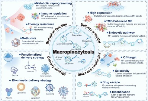
Precise drug delivery to tumors and efficient accumulation within tumor cells remain major challenges in cancer therapy. Macropinocytosis is a high-capacity endocytic pathway that is frequently upregulated in cancer cells and has therefore emerged as a promising route for intracellular drug delivery. However, the biological complexity of this pathway and the difficulty of achieving precise targeting hinder the clinical translation of delivery systems that exploit it. This review systematically summarizes the biological functions and regulatory mechanisms of macropinocytosis in cancer, along with related drug delivery strategies. It also critically examines the associated risks and challenges from biological and mechanistic perspectives. Through a comprehensive assessment of the field, this review aims to provide a theoretical framework and practical research directions for advancing the clinical translation of macropinocytosis-mediated delivery systems.
Keywords: macropinocytosis, cancer, drug delivery, tumor microenvironment, nanomedicine
1. Introduction
Achieving precise and efficient drug delivery remains a major challenge for improving the clinical efficacy of anticancer therapies [1]. Most anticancer drugs and some delivery systems exhibit nonspecific biodistribution, resulting in accumulation in healthy tissues and off-target toxicity [2]. At the cellular level, conventional targeting strategies are further constrained by receptor heterogeneity, inefficient membrane translocation, and size-related barriers that limit the entry of macromolecules and nanoparticles [3]. Furthermore, even when drugs successfully enter cells, overexpressed efflux pumps, such as P-glycoprotein (P-gp), can mediate the efflux of these drugs, thereby reducing intracellular exposure and retention [4]. Consequently, these barriers at both the tissue and cellular levels pose major obstacles to improving therapeutic efficacy against solid tumors.
Macropinocytosis (MP) is a non-selective, fluid-phase endocytic pathway driven by actin-dependent membrane ruffling and lamellipodial protrusions, which is frequently activated in a variety of solid tumors [5]. Its activity is regulated by key signaling pathways, such as RAS and phosphoinositide 3-kinase (PI3K) [6], and is further enhanced under stress conditions, including nutrient deprivation, hypoxia, and acidic tumor microenvironments. Unlike classical receptor-mediated endocytosis, macropinocytosis generates macropinosomes that typically exceed 0.2 μm in diameter. This feature enables large-volume, non-selective uptake of extracellular fluid and associated cargo [7]. By scavenging extracellular nutrients, macropinocytosis provides metabolic support for tumor cells, thereby sustaining proliferation, promoting immune suppression, and fostering therapeutic resistance [8, 9]. These properties also make macropinocytosis an attractive pathway for intracellular drug delivery. Its receptor-independent nature may also reduce the limitations imposed by target heterogeneity. Entry into cells via macropinosomes may also reduce early drug loss through plasma membrane efflux pumps.
Interest in macropinocytosis-based delivery has grown, but the connections between its biology, carrier design, and intracellular trafficking remain poorly understood. This review considers how pathway regulation can inform delivery design and clinical translation. We examine the effects of carrier physicochemical properties and the tumor microenvironment on uptake, followed by the consequences of cellular distribution and intracellular trafficking for delivery efficiency. Connecting these mechanisms with current delivery strategies may help guide carrier optimization, selection of suitable tumors, and combination approaches for chemotherapy.
2. Biological foundations and molecular regulation of macropinocytosis
2.1 Definition and physiological significance
First described by Warren Lewis in macrophages and tumor cells, macropinocytosis is a non-selective endocytic pathway with a high uptake capacity [10]. It does not depend on the specific cargo-receptor recognition involved in clathrin- or caveolin-mediated endocytosis [11]. Actin drives lamellipodial protrusions and membrane ruffles, which fold back and fuse to enclose extracellular material in macropinosomes. This allows cells to internalize extracellular materials, including soluble nutrients, cellular debris, bacteria, and viruses [12, 13]. Three-dimensional structured illumination microscopy (3D-SIM) and lattice light-sheet microscopy (LLSM) have revealed the spatial and temporal dynamics of macropinocytosis [14, 15]. Phosphatidylinositol-3,4,5-trisphosphate (PIP3) accumulates at the ruffle rim and activates RAC1 and CDC42, promoting membrane closure and vesicle formation. Newly formed macropinosomes undergo maturation and intracellular sorting before fusing with lysosomes, where their contents are degraded for cellular use. However, undegraded material is returned to the cell surface and subsequently released via recycling pathways [9, 16].
Macropinocytosis is thought to have evolved from a conserved mechanism for nutrient acquisition [17, 18]. Different cells use it for nutrient uptake, antigen presentation [19, 20], synaptic regulation [21], plasma protein recycling [22], and cell migration [23]. Various pathogens, including viruses and bacteria, have also evolved mechanisms to manipulate host endocytic signaling and cytoskeletal remodeling, thereby exploiting macropinocytosis for cellular entry [24, 25]. In cancer, the significance of macropinocytosis is particularly evident in oncogene-driven tumors. KRAS-mutant cells often show increased activity, taking up extracellular macromolecules for lysosomal degradation to support proliferation and metabolic adaptation [9]. Accumulating evidence indicates that macropinocytosis also contributes to cancer invasion, therapeutic resistance, and other malignant phenotypes [26-28].
2.2 Biological characteristics and regulatory mechanisms
In different cell types, the regulatory mechanism and physiological function of macropinocytosis are different. However, its core molecular machinery is highly conserved [29]. Macropinocytosis can be described as a dynamic process comprising three stages: initiation and macropinosome formation, tubulation and maturation, and recycling and degradation. This section reviews the molecular networks involved in this process and discusses their biological significance in cancer (Figure 1 and Table S1).
Molecular regulatory mechanisms of macropinocytosis. (A) Macropinosome formation: Receptor tyrosine kinase or oncogenic KRAS signaling activates RAC1, which drives branched actin polymerization. This promotes membrane ruffling and macropinocytic cup formation. (B) Macropinosome maturation: Na+ and Cl- efflux through TPC1/2 and ASOR drives osmotic shrinkage. Sequential conversion of PIP3 to PI(3,4)P2 and then PI(3)P, together with replacement of Rab5 by Rab7, supports maturation. (C) Macropinosome degradation: PIKfyve converts PI(3)P to PI(3,5)P2 and promotes TRPML1 activation, helping maintain lysosomal calcium homeostasis and acidity to support fusion with macropinosomes. A second wave of WASH activity recycles V-ATPase and prepares the compartment for exocytosis of non-degradable remnants. (D) Macropinosome recycling: WASH-dependent actin polymerization drives rapid membrane recycling. SNX-Retromer selectively retrieves cargo, while Rab4 and Rab11 regulate fast and slow recycling, respectively. Abbreviations: HOPS: homotypic fusion and protein sorting complex; INPP4: inositol polyphosphate 4-phosphatase; ERC: endocytic recycling compartment; TPC1/2: two-pore channels 1 and 2; ASOR: acid-sensitive outwardly rectifying anion channel; TMEM206: transmembrane protein 206; V-ATPase: vacuolar H⁺-ATPase.
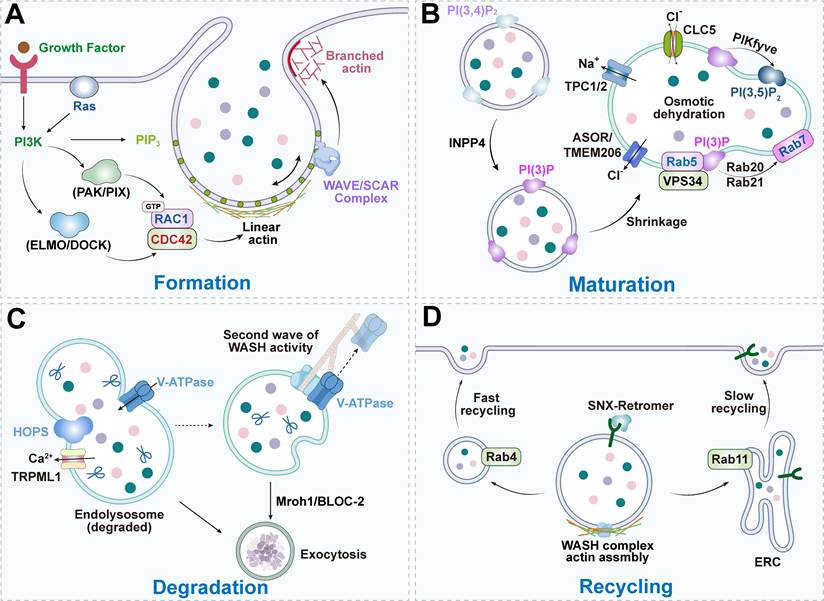
The formation of macropinosomes depends on actin-driven membrane remodeling [30]. PI3K promotes PIP3 accumulation in specific membrane regions and local exclusion of phosphatase and tensin homolog (PTEN), thus establishing stable lipid-signaling microdomains [29]. At the same time, RhoG and CDC42 guide RAC1 localization and activation through guanine nucleotide exchange factor (GEF) complexes (including ELMO/DOCK and PAK/PIX) [31]. Activated RAC1 plays a dual role: it triggers the SCAR/WAVE-Arp2/3 pathway to drive branched actin nucleation and works with Ena/VASP to promote membrane protrusion. SCAR/WAVE localization is tightly regulated by the spatial offset of RAS/PIP3 signaling and its preference for saddle-shaped negative curvature at the cell edge [32]. RAS/RAC signaling also promotes formin-mediated assembly of linear actin filaments at the cup base, which helps stabilize the open portion of the cup. Protein kinases A, G, and C (AGC) family kinases further fine-tune these cytoskeletal dynamics [33]. During closure, Rho family members are activated sequentially [11, 31]. RhoC is transiently activated before closure, and then RhoA is activated to generate contractile force and cortical tension through the ROCK-MLC pathway, sealing the vesicle. Morphologically, one mode of closure involves inward retraction of the cup margin to internalize the entire PIP3 domain. In the other, the cup continues to deepen and subsequently undergoes fission. Residual PIP3 remains at the cell surface and may prime subsequent cycles. Leep2A/Leep2B, RasGAPs, and PTEN limit the spatial extent and duration of these signaling microdomains, helping control when and where cups form and close [34, 35].
After closure, macropinosomes undergo membrane tubulation and progressive shrinkage and rapidly mature. Their volume and surface area decrease substantially. Their volume decreases through ion-driven osmotic water loss, whereas membrane is retrieved through tubulation and recycling. Na+ and Cl- flow out through two-pore channels 1 and 2 (TPC1/2) and acid-sensitive outwardly rectifying anion channel (ASOR) ion channels, causing osmotic water loss, which is a key factor in promoting shrinkage [36]. This shrinkage facilitates luminal acidification and hydrolase concentration, while also supporting surface protein retrieval. Maturation is accompanied by sequential changes in phosphoinositides and Rab GTPases: after closure, PIP3 is converted to PI(3,4)P2 and PI(3)P, and the latter is converted to PI(3,5)P2 by enzymes such as PIKfyve [37]. Rab5 recruits PIK3C3/VPS34 to generate PI(3)P [38], and PI(3)P recruits effector proteins containing FYVE or PX domains [39]. After that, Rab20 and Rab21 associate with the vesicle membrane to regulate the transition phase [40, 41]. Eventually, Rab5 is replaced by Rab7, marking the transition to a late endosomal compartment. At this stage, the maturing macropinosome acquires the machinery required for lysosomal fusion [42, 43]. It is this coordinated network that enables the macropinosomes to complete the functional transformation and become specialized compartments for sorting or degradation. This process may also provide a window for intracellular escape by drug delivery systems.
There are two main fates for mature macropinosomes: recycling or degradation. During recycling, the WASH complex sorts membrane components and returns them to the cell surface [16], and fast and slow recycling are regulated by Rab4 and Rab11, respectively [44]. The SNX-Retromer complex mediates selective cargo retrieval, which helps maintain membrane homeostasis [45]. The degradative pathway depends on the PIKfyve–PI(3,5)P2 axis to regulate lysosomal fusion [46]. The TRPML1 channel maintains lysosomal ion homeostasis and acidity, supporting efficient proteolysis [47]. In addition, recent studies have reported several post-degradative mechanisms. A second wave of WASH activity drives vacuolar H+-ATPase (V-ATPase) recycling, which helps neutralize the compartment and promotes exocytosis of indigestible remnants [48]. This process is jointly regulated by Mroh1 and the BLOC-2 complex and supports waste clearance and resource recovery [49, 50]. Through these mechanisms, cells can balance nutrient acquisition with the removal of harmful substances and maintain homeostasis.
3. Multiple roles of macropinocytosis in cancer
3.1 Nutritional support and metabolic reprogramming
In the nutrient-deprived tumor microenvironment, cancer cells face substantial metabolic stress because the proliferation process is accompanied by high metabolic demand [51]. In order to survive under these conditions, tumor cells reprogram their metabolism through oncogenic signaling pathways such as RAS [52], MYC [53], and PI3K [54]. At the same time, they also obtain nutrients through autophagy [55] and macropinocytosis [9]. Autophagy mainly recycles damaged intracellular organelles and macromolecules to replenish amino acids, fatty acids, and nucleosides [56]. In contrast, macropinocytosis complements this process by taking up large amounts of extracellular material, including nutrient-rich substrates. This broadens the range of available metabolic substrates and helps tumor cells survive and proliferate under nutrient stress [57].
In KRAS-mutant pancreatic cancer, macropinocytosis mediates the uptake of serum albumin and a variety of extracellular matrix (ECM) components [58, 59]. These substrates are degraded by lysosomes to release free amino acids (especially glutamine), which provide fuel for the tricarboxylic acid cycle and anabolic pathways. The above findings establish the role of macropinocytosis as a key route for tumor cells to acquire extracellular macromolecules, so that they can maintain metabolic fitness under nutrient limitation.
Severe starvation or treatment can extend the range of cargo taken up through macropinocytosis. In ovarian cancer, ataxia-telangiectasia mutated (ATM) inhibition and mechanistic target of rapamycin (mTOR) inactivation promote uptake of branched-chain amino acids and support tumor growth [60]. Non-small cell lung cancer (NSCLC) cells internalize extracellular ATP (eATP), raising intracellular ATP levels and resistance to chemotherapy and targeted therapy [26, 27]. Activated epidermal growth factor receptor (EGFR) and KRAS can cooperate to promote uptake of extracellular vesicles (EVs) [61]. Tumor cells also scavenge necrotic cell debris, which may support growth during severe nutrient depletion [28]. In pancreatic ductal adenocarcinoma (PDAC), macropinocytic acquisition of exogenous fatty acids helps cells resist alkalosis-induced death [62]. These findings suggest that macropinocytosis is a highly adaptive pathway that enables tumor cells to broaden their nutrient sources under metabolic stress.
3.2 Remodeling of the tumor immune microenvironment
Macropinocytosis affects tumor immunity through nutrient use and signaling between cells. Depending on the cells involved, it can suppress or activate immune responses.
Tumor cells degrade macropinocytosed proteins into amino acids that support glycolysis and one-carbon metabolism. Increased metabolic activity leads to lactate accumulation and efflux [63]. Acidity and nutrient depletion impair cytotoxic T-cell and natural killer (NK) cell function, suppress inflammatory responses, and favor immune escape [64]. Tumor-derived extracellular vesicles provide another route for communication with immune cells [65]. Dendritic cells (DCs) and tumor-associated macrophages (TAMs) can internalize these EVs through macropinocytosis, while the microRNA and protein cargo carried by EVs promote the formation of immunosuppressive phenotypes by upregulating the expression of programmed death-ligand 1 (PD-L1) [66].
In antigen-presenting cells, macropinocytosis supplies antigens for immune activation. Immature DCs take up soluble antigens through macropinocytosis and concentrate them in compartments associated with major histocompatibility complex (MHC) class II antigen presentation. This uptake declines as DCs mature [20]. Exogenous antigens can also be cross-presented on MHC class I molecules to activate CD8+ T cells [67].
Therefore, the therapeutic strategy targeting macropinocytosis needs to fully consider its dual impact on tumor metabolism and immune regulation: tumor cells and immunosuppressive stromal cells can support immune escape by internalizing substances through macropinocytosis, and antigen-presenting cells can help achieve immune priming through antigen uptake.
3.3 Therapy resistance in cancer
3.3.1 Chemotherapy resistance
Drug resistance remains a major cause of treatment failure and recurrence in cancer therapy [68]. During chemotherapy, cancer cells often enhance drug efflux by upregulating ATP-binding cassette (ABC) transporters [69]. This reduces intracellular drug exposure and weakens cytotoxic effects [70]. Extracellular ATP (eATP) in the tumor microenvironment acts as both a nutrient source and a signaling molecule. Tumor cells can internalize eATP through macropinocytosis. Elevated intracellular ATP enhances drug efflux and supports glutathione (GSH) synthesis and epithelial-mesenchymal transition (EMT), thereby contributing to chemoresistance [27, 71].
In addition, many commonly used chemotherapeutic agents, including gemcitabine, 5-fluorouracil (5-FU), doxorubicin (DOX), and platinum compounds, exert antitumor effects by disrupting nucleotide synthesis or directly damaging DNA [72]. However, tumor cells can internalize necrotic cell debris through macropinocytosis to support DNA and RNA synthesis. This reduces reliance on endogenous nucleotide synthesis pathways and contributes to chemoresistance [28]. A similar compensatory adaptation has also been reported in osteosarcoma. Benzethonium chloride (BZN), a Food and Drug Administration (FDA) approved preservative, suppresses tumor cell proliferation, migration, and invasion in vitro and in vivo. However, osteosarcoma cells can activate macropinocytosis as a compensatory survival pathway, thereby reducing the antitumor effects of BZN [73]. This finding suggests that combining BZN with a macropinocytosis inhibitor may enhance its therapeutic potential.
3.3.2 Resistance to targeted therapy
Adaptive drug resistance is the main problem that limits the efficacy of therapies targeting oncogenic drivers. Its development often involves a variety of mechanisms such as bypass signaling activation and metabolic reprogramming [74]. In many types of cancer, macropinocytosis can serve as a compensatory route for nutrient acquisition to support tumor cells under the pressure of targeted therapy.
This phenomenon is particularly evident in PDAC. Autophagy inhibitors, such as chloroquine (CQ), can enhance therapeutic efficacy in the short term. CQ treatment also activates the nuclear factor erythroid 2-related factor 2 (NRF2) pathway and upregulates key regulators of macropinocytosis, including RAC1 and p21-activated kinase 1 (PAK1) [75]. This process enables tumor cells to obtain alternative nutrients by internalizing necrotic cell debris. This compensatory mechanism explains why autophagy inhibition produces only transient therapeutic effects. Similarly, resistance to mTOR inhibitors (such as rapamycin) arises when tumor cells scavenge albumin via macropinocytosis, thereby activating the LAMTOR1-SLC38A9-dependent mTORC1 bypass signaling [76, 77]. Activation of macropinocytosis can also reduce the efficacy of JTC801, a small-molecule drug that induces alkaliptosis. Inhibition of this process or the knockout of RAC1 can restore cellular sensitivity to treatment [62]. The efficacy of MRTX1133, a selective KRASG12D inhibitor, is also limited by advanced glycation end-product receptor (AGER)-mediated macropinocytosis. Blocking the AGER-DIAPH1 axis, or combining MRTX1133 with macropinocytosis inhibitors, can restore antitumor efficacy. In addition, the deubiquitinating enzyme ubiquitin-specific peptidase 21 (USP21) maintains amino acid supply and anabolic metabolism through microtubule affinity-regulating kinase 3 (MARK3)-mediated macropinocytosis, thereby supporting KRAS-independent tumor growth and contributing to resistance to KRAS inhibition [78].
In hepatocellular carcinoma (HCC), sorafenib induces mitochondrial dysfunction, thereby activating the PI3K-RAC1-PAK1 axis and enhancing macropinocytosis. The macropinocytosis inhibitor 5-(N-ethyl-N-isopropyl)amiloride (EIPA) can re-induce ferroptosis and enhance the antitumor efficacy of sorafenib [79]. A recent study further revealed that the hyperglycemic tumor microenvironment remodeled by transarterial chemoembolization (TACE) elevates lysophosphatidic acid (LPA) levels. Elevated LPA activates ADP-ribosylation factor 6 (Arf6), thereby significantly enhancing macropinocytosis in hepatocellular carcinoma cells. Tumor cells exploit this heightened macropinocytic activity to internalize CD147⁺ EVs. The fucosylated CD147 carried by these EVs constitutively activates the AKT/mTOR/4EBP1 signaling axis, antagonizing the cytotoxic effects of sorafenib and promoting drug resistance [80].
Similar adaptive mechanisms have also been reported in other tumor types. Osimertinib is a third-generation irreversible epidermal growth factor receptor tyrosine kinase inhibitor (EGFR-TKI) used in EGFR-mutant non-small cell lung cancer, but acquired resistance is almost inevitable [81, 82]. In NSCLC, tumor cells can internalize type I collagen and other extracellular matrix components through macropinocytosis. This activates mTOR bypass signaling, thereby promoting adaptive resistance to EGFR-TKIs [59]. In triple-negative breast cancer (TNBC), loss of the glutamine transporter alanine-serine-cysteine transporter 2 (ASCT2) leads to sustained glutamine supply through increased macropinocytosis, thereby conferring tolerance to single-agent glutamine uptake inhibition [83]. In colorectal cancer (CRC), KRAS inhibition can induce resistance through Yes-associated protein 1 (YAP1) activation. YAP1 restores syndecan-1 (SDC1) surface localization and thereby enhances macropinocytosis [84].
Taken together, these findings indicate that macropinocytosis is a recurrent adaptive response to targeted therapeutic pressure across multiple tumor types. This adaptive upregulation may contribute to acquired resistance, but it may also create opportunities for therapeutic intervention and delivery strategies that exploit macropinocytosis.
3.4 Methuosis
3.4.1 Mechanisms and characteristics of methuosis
In tumor cells, excessive activation of macropinocytosis can cause macropinosome formation to exceed lysosomal processing capacity. This can cause progressive vacuole accumulation and a distinct form of non-apoptotic cell death known as methuosis [85, 86]. Overmeyer and colleagues first described this process in glioblastoma cells driven by oncogenic RASG12V [87]. Unlike other forms of non-apoptotic cell death, methuosis has distinct morphological characteristics. Its main morphological hallmark is that the cell is filled with numerous optically clear, single-membrane vacuoles. These vacuoles are usually positive for late endosomal or lysosomal markers (such as Rab7 and LAMP1) [88]. As vacuolization progresses, the cytoplasm is compressed and the cells continue to swell, which leads to rupture of the plasma membrane, but this process does not exhibit apoptotic features such as nuclear fragmentation or apoptotic body formation.
At present, methuosis has been observed in various cancer models, and some cancer cells appear more susceptible to this form of death than normal cells [89, 90]. Small-molecule compounds have been developed as methuosis inducers, many of which have shown significant anticancer activity [91]. There are many triggers of methuosis, but a proposed common mechanism involves upstream RAS-RAC1 signaling that enhances membrane ruffling and macropinosome formation. Meanwhile, impaired Arf6-mediated trafficking leads to an imbalance between uptake and recycling, promoting progressive macropinosome enlargement and ultimately resulting in vacuolization [92].
3.4.2 Biological complexity of methuosis
Most studies on methuosis use cancer models, but similar vacuolization phenotypes also occur in cardiomyocytes and non-mammalian systems [93-95]. Due to the lack of specific molecular markers, it is difficult to distinguish methuosis from other forms of cell death [96, 97]. In addition, DNA fragmentation and elevated autophagy markers can occur concurrently, further complicating classification [88, 98]. Methuosis can also interact with other death pathways. For example, SGI-1027 induces macropinocytic vacuolization and methuosis in renal cancer cells [99]. When combined with everolimus, enhanced vacuolization and lysosomal dysfunction further induce lysosomal membrane permeabilization, followed by activation of caspase-3-mediated apoptosis and gasdermin E (GSDME)-dependent pyroptosis. These findings suggest that lysosomal membrane permeabilization may link methuosis to apoptosis and GSDME-dependent pyroptosis. In contrast, direct mechanistic links between methuosis and ferroptosis remain insufficiently established. In KRAS-mutant cells, ionizing radiation can induce methuosis-like vacuolization accompanied by ferroptosis-like features characterized by lipid peroxidation [100]. However, this process is not effectively prevented by Ferrostatin-1 or vitamin E, suggesting that it differs from classical ferroptosis. In addition, recent studies suggest that excessive vacuolization may contribute to cytoskeletal disruption and late-stage plasma membrane damage, together with damage-associated molecular patterns (DAMPs), such as ATP release and calreticulin exposure. However, this membrane damage does not depend on GSDMD or p-MLKL, suggesting that methuosis can be accompanied by immunogenic damage signals [101]. Recent in vivo studies showed that immunogenic methuosis in KRAS-mutant tumors can accompany pro-inflammatory tumor-associated macrophage (TAM) reprogramming and enhanced antitumor immunity [102]. This indicates that methuosis may not only be a vacuolization-associated cell death process, but may also promote antitumor immune responses by inducing immunogenic damage.
3.4.3 Challenges in identifying methuosis
The lack of specific biomarkers makes methuosis difficult to define. Its identification therefore requires multiple lines of evidence beyond vacuole morphology or individual molecular markers.
First, the origin of accumulated vacuoles should be confirmed using fluid-phase tracers and live-cell imaging. This helps distinguish methuosis from other processes, such as endoplasmic reticulum expansion, mitochondrial swelling, or autophagosome accumulation.
Second, the subsequent trafficking and processing of macropinocytosis-derived vacuoles should be further evaluated. Methuosis-associated vacuoles may acquire late endosomal or lysosomal markers such as Rab7 and LAMP1/2, yet fail to undergo normal recycling or degradation. Therefore, these markers alone are insufficient to define methuosis. Pulse-chase experiments with fluid-phase tracers can be used to monitor macropinosome fate and evaluate whether these vesicles undergo normal recycling and degradation [103]. For persistently enlarged vesicles, LysoTracker, pH-sensitive probes, and proteolytic substrates can be used to assess lysosomal acidification and degradative capacity. The functional analysis of macropinocytosis regulators such as RAC1 and Arf6 helps to determine whether vesicle accumulation is related to defects in macropinosome recycling [92].
Transmission electron microscopy (TEM) can help distinguish methuosis from other forms of cell death by identifying single-membrane vacuoles and excluding double-membrane autophagosomes, expanded endoplasmic reticulum, or swollen mitochondria. Apoptosis and autophagy markers should be assessed alongside functional experiments to determine whether these pathways cause death or change secondarily during methuosis.
Excessive macropinocytosis may be a therapeutic vulnerability, but defining methuosis still requires better criteria, and the selectivity and safety of inducing it remain to be established [104].
4. The potential of macropinocytosis as a drug delivery target in cancer
4.1 Mechanisms regulating macropinocytosis in cancer
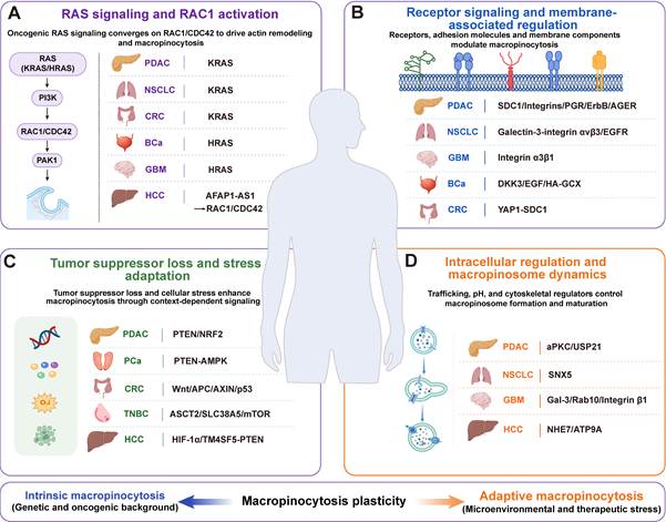
The molecular machinery described in Section 2.2 is relatively conserved, but cancers differ in the upstream signals and cellular conditions that increase macropinocytosis (Figure 2). This section examines those differences. Higher activity in tumors than in normal tissues may offer an opportunity to improve anticancer drug delivery, provided that delivery systems can exploit this difference.
Major mechanisms regulating enhanced macropinocytosis across cancer types. (A) RAS signaling and RAC1 activation: Oncogenic RAS signaling and related regulators activate RAC1/CDC42 to promote actin remodeling and macropinocytosis. (B) Receptor signaling and membrane-associated regulation: Cell-surface receptors, adhesion molecules, and membrane components regulate macropinocytosis through pathways that vary among cancer types. (C) Tumor suppressor loss and stress adaptation: Loss of tumor suppressor function and responses to nutrient limitation, hypoxia, or other cellular stresses enhance macropinocytosis through context-dependent signaling. (D) Intracellular regulation and macropinosome dynamics: Regulators of membrane trafficking, intracellular pH, and cytoskeletal organization control macropinosome formation and maturation.
4.1.1 RAS signaling and RAC1 activation
RAS-driven macropinocytosis has been studied extensively in PDAC. KRAS mutations occur in approximately 90–95% of cases and are closely associated with increased macropinocytosis [105]. KRAS-mutant MIA PaCa-2 cells show greater activity than KRAS wild-type BxPC-3 cells [8]. In the KRASG12D-driven KPC mouse model (KrasLSL-G12D/+; Trp53LSL-R172H/+; Pdx1-Cre mouse model), advanced pancreatic intraepithelial neoplasia (PanIN) lesions also show marked macropinocytosis, a phenotype absent from wild-type pancreatic tissues [106].
RAS also promotes macropinocytosis outside PDAC. Under glucose deprivation, KRAS-mutant NSCLC cells increase macropinocytosis and albumin uptake to obtain nutrients [107]. In CRC, KRAS mutations promote both macropinocytosis and tumor growth; blocking uptake increases the antitumor effect of disrupting asparagine biosynthesis [108]. Oncogenic HRAS enhances the pathway in bladder cancer [109]. Abnormal RAS signaling can also stimulate macropinocytosis in glioblastoma (GBM), despite RAS pathway mutations being less frequent than EGFR alterations [87]. Excessive activation can produce macropinosome accumulation and methuosis, as described in Section 3.4.
RAC1 and CDC42 act as common downstream effectors in the regulation of macropinocytosis. In HCC, actin filament associated protein 1 antisense RNA 1 (AFAP1-AS1) is associated with tumor recurrence after incomplete radiofrequency ablation. Sublethal heat stress induces AFAP1-AS1 expression and enhances macropinocytosis through RAC1/CDC42 signaling [110]. Thus, RAC1 and CDC42 represent a shared regulatory node, whereas the upstream signals that activate this axis vary among cancer types.
4.1.2 Receptor signaling and membrane-associated regulation
In addition to RAS signaling, cell surface receptors, adhesion molecules, and membrane-associated regulators can also modulate macropinocytosis in cancer. Syndecan-1, integrins, progesterone receptor (PGR), and ErbB family receptors have been reported to enhance macropinocytosis [111-115]. Upregulation of AGER also promotes macropinocytosis and extracellular albumin uptake in PDAC cells [116].
In NSCLC, Galectin-3 promotes macropinocytosis through interaction with integrin αvβ3 and contributes to nutrient uptake and redox homeostasis [117]. Gefitinib can reduce macropinocytosis in EGFR-mutant HCC827 cells, which supports the role of EGFR in regulating this pathway [118]. The researchers also examined EGFR expression in different breast cancer subtypes and its association with prognosis [119], although the existing data have not established an EGFR-dependent macropinocytic mechanism in triple-negative breast cancer (TNBC). Integrin α3β1 promotes macropinocytosis in glioblastoma (GBM)-associated endothelial cells through Ca²⁺ influx. It can also promote macropinosome maturation and lysosomal exocytosis, thus contributing to vessel formation [113]. In bladder cancer, DKK3 overexpression and epidermal growth factor (EGF) stimulation enhance macropinocytosis, while disruption of the hyaluronic acid-associated glycocalyx inhibits this process [120-122]. In colorectal cancer (CRC), following KRAS inhibition, YAP1 activation can restore SDC1 localization at the cell surface and enhance macropinocytosis, thus promoting acquired drug resistance [84].
4.1.3 Tumor suppressor loss and stress adaptation
The loss of tumor suppressor genes can enhance macropinocytosis. PTEN loss activates the PI3K signaling pathway and promotes macropinocytosis in pancreatic ductal adenocarcinoma (PDAC) [77]. PTEN loss is also common in metastatic castration-resistant prostate cancer, in which enhanced PI3K/AKT signaling and metabolic stress jointly activate AMP-activated protein kinase (AMPK) and promote RAC1-dependent macropinocytosis [123, 124]. PTEN-deficient prostate cancer cells can use this pathway to internalize necrotic cell debris under nutrient stress [125]. In hepatocellular carcinoma (HCC), transmembrane 4 L six family member 5 (TM4SF5) promotes PIP3 accumulation and macropinocytic albumin uptake by stabilizing nuclear receptor coactivator 3 (NCOA3) and suppressing PTEN [126]. In colorectal cancer (CRC), activation of Wnt/β-catenin signaling and loss of adenomatous polyposis coli (APC) or Axin enhance macropinocytosis and extracellular protein uptake and degradation [127, 128]. Loss of p53 also increases macropinocytosis and extracellular vesicle uptake in CRC [129]. Although mutations in the TP53 gene are common in HCC, direct evidence that TP53 loss enhances macropinocytosis in this cancer remains limited [130].
Nutrient availability and cellular stress also regulate this pathway. In triple-negative breast cancer (TNBC), ASCT2 loss limits glutamine transport and induces compensatory macropinocytosis [83]. Solute carrier family 38 member 5 (SLC38A5) promotes macropinocytosis by regulating amino acid transport, ion homeostasis, and pH near the plasma membrane [131]. Inhibiting mTORC1/2 can increase macropinocytosis and verteporfin uptake at the same time [132]. Stress responses vary among cancers: in PDAC, autophagy inhibition acts through NRF2-related signaling, while in HCC, hypoxia promotes macropinocytosis through HIF-1α/EHD2 signaling [75, 133].
4.1.4 Intracellular regulation and macropinosome dynamics
Intracellular trafficking and cytoskeletal organization regulate macropinosome formation and maturation. In pancreatic ductal adenocarcinoma (PDAC), atypical protein kinase C (aPKC) regulates microtubule dynamics through the Par3-dynein axis, thus promoting vesicle formation [134]. The deubiquitinase USP21 can stabilize MARK3, thereby enhancing cytoskeletal remodeling and macropinosome formation [78].
In NSCLC, sorting nexin 5 (SNX5) participates in membrane remodeling during macropinocytosis and promotes extracellular ATP (eATP) uptake through this pathway [26, 27]. Macropinocytic uptake of eATP increases intracellular ATP levels and supports tumor cell proliferation and treatment resistance. In GBM, the FOXG1-FOXO6-PAK1 axis promotes cell-cycle re-entry and enhances macropinocytosis in neural stem cell and malignant transformation models [135]. In patient-derived GBM stem cells, Galectin-3, Rab10, and integrin β1 form a complex that coordinates macropinosome formation and maturation together with integrin recycling. This complex also contributes to energy homeostasis and treatment resistance [136].
In HCC, sodium/hydrogen exchanger 7 (NHE7) regulates intracellular and endosomal pH to support macropinosome formation and maturation, thereby enhancing small extracellular vesicle (sEV) uptake [137]. ATPase phospholipid transporting 9A (ATP9A) promotes the transport of ATPase H+ transporting V1 subunit A (ATP6V1A) to the plasma membrane, resulting in cholesterol accumulation at the membrane and enhanced macropinocytosis through RAC1 activation [138].
Different cancers enhance macropinocytosis through distinct upstream mechanisms that converge on a relatively conserved downstream program. In addition to oncogene activation or loss of tumor suppressor function, which can provide a genetic basis for sustained macropinocytosis, nutrient limitation, hypoxia, mTOR inhibition, and treatment pressure can further modulate macropinocytosis. Thus, macropinocytosis reflects the combined effects of genetic background and cellular state rather than being determined by any single oncogenic alteration. This also means that KRAS, PTEN, or EGFR alterations alone may not accurately predict macropinocytic activity in a given tumor. Instead, the actual level of macropinocytic activity and its responsiveness to microenvironmental or therapeutic stress may better reflect the potential of this pathway for drug delivery.
4.2 Potential of the tumor microenvironment in enhancing macropinocytosis-mediated drug delivery
4.2.1 Nutrient deprivation as a driver of macropinocytosis
Nutrient deprivation in the tumor microenvironment (TME) arises mainly from the high metabolic demands of rapidly proliferating tumor cells and abnormal vasculature that restricts nutrient supply [139]. Tumor cells adapt to these conditions through pathways such as autophagy and macropinocytosis. Autophagy recycles intracellular components to supply metabolic substrates [55], while macropinocytosis enables the direct uptake of extracellular nutrients and complements intracellular recycling under microenvironmental stress [9].
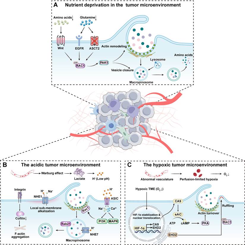
In addition to intrinsic drivers such as KRAS mutations, nutrient deprivation can further enhance macropinocytic activity, thus helping tumor cells alleviate metabolic stress [115, 140]. The signaling pathways involved vary according to the tumor type and the limiting nutrient. KRAS-mutant pancreatic cancer cells, for example, increase macropinocytosis to scavenge extracellular proteins when glucose or amino acids are scarce [141]. EGFR-PAK signaling can further amplify macropinocytosis under specific nutrient-deprivation conditions [115]. In NSCLC, glucose scarcity activates the PI3K-RAC1-PAK axis to promote protein uptake through macropinocytosis [107]. TNBC compensates for the loss of glutamine transport by increasing macropinocytosis [83]. In colorectal cancer, Wnt-PAK signaling stimulates macropinocytosis to cope with amino acid deficiency [128].
Although evidence supports nutrient deprivation as an important extrinsic inducer of macropinocytosis, most current models often mimic tumor nutrient conditions by withdrawing a single nutrient in vitro. These simplified conditions differ markedly from the complexity of the TME, and how this limitation affects our understanding of macropinocytosis regulation remains unclear [142]. Nevertheless, current evidence suggests that nutrient stress can enhance macropinocytic activity in biologically meaningful tumor contexts. This has important implications for drug delivery, as nutrient-deprived tumor regions may provide a favorable therapeutic window for the preferential uptake of extracellular carriers.
4.2.2 Acidic microenvironment as a driver of macropinocytosis
Acidity in the TME arises from the combined effects of hypoxia and the Warburg effect. Hypoxia increases glycolytic flux, while the Warburg effect sustains glycolysis even when oxygen is available. Both contribute to excess lactate production [143, 144].
Acidic conditions can promote macropinocytosis by altering cytoskeletal organization and activating signaling pathways. Acidic stress activates the integrin β1/cofilin/F-actin axis and downregulates cofilin, which in turn triggers F-actin redistribution and accumulation [145, 146]. Acidic conditions can also directly enhance macropinocytosis by activating signaling pathways. When mouse dendritic cells are exposed to pH 6.5, the PI3K/AKT and MAPK pathways are activated, which in turn stimulates macropinocytosis and enhances cellular uptake of exogenous tracers (such as FITC-OVA, FITC-dextran, and HRP) [147]. Therefore, acidic conditions can stimulate macropinocytic uptake by activating signaling pathways in dendritic cells, although the relevance of these findings to cancer cells requires further study.
Sodium/hydrogen exchanger 1 (NHE1) is a regulator of cellular pH homeostasis. NHE1-mediated H+ efflux helps maintain a relatively alkaline microenvironment beneath the plasma membrane [148], thus supporting cortical actin assembly and macropinosome closure. The widely used macropinocytosis inhibitor EIPA acts by blocking NHE1-mediated H+ efflux. The resulting cytosolic acidification disrupts actin remodeling and inhibits macropinosome formation [149]. Elevated expression of NHE7, a homolog of NHE1, also increases intracellular pH and decreases endosomal pH, thereby promoting macropinosome maturation [137]. The colocalization of NHE7 with endosomal markers such as Rab21 further supports its role in maturation.
Other pH-sensitive membrane proteins may contribute to acid-induced macropinocytosis. Acid-sensing ion channels (ASICs) respond to extracellular protons by allowing cation influx, including Na⁺ entry. In macrophages, acidity substantially increases FITC-dextran uptake, an effect reduced by ASIC1 or ASIC3 inhibition [150]. ASICs also participate in dendritic-cell responses to acidification, which enhances macropinocytosis and antigen uptake [147, 151]. In pancreatic cancer cells, ASIC1 and ASIC3 translate acidification into increased intracellular Ca²⁺ and RhoA activation [152]. These signals regulate cytoskeletal changes involved in migration and invasion, suggesting a possible route by which ASICs could regulate macropinocytosis. Proton-sensing GPCRs such as GPR4, GPR65, and GPR68 respond to acidification as well [153-155]. Their effects on the cytoskeleton and membrane dynamics suggest a role in macropinocytosis, although direct evidence is lacking. These sensors could offer targets for acidic tumors. Compared with EIPA-mediated NHE1 inhibition, their dependence on extracellular acidity and lack of direct disruption of cellular pH homeostasis may provide greater microenvironmental selectivity. Targeting individual ASIC or GPCR subtypes might also limit effects on other pathways. These proposed advantages require direct comparison in macropinocytosis models.
4.2.3 Hypoxic microenvironment as a driver of macropinocytosis
In solid tumors, rapid proliferation and abnormal vascular structure can also lead to local hypoxia [156]. This hypoxia will reduce the activity of glutamate oxaloacetate transaminase 2 (GOT2) and limit aspartate synthesis, thus affecting the growth of cancer cells [157]. However, cancer cells show elevated macropinocytic activity in hypoxic regions, thus compensating for the nutritional limitations caused by hypoxia. In KRAS-mutant cells, hypoxia can stabilize HIF-1α and upregulate the expression of its canonical target gene, carbonic anhydrase 9 (CA9). Subsequently, CA9 activates the soluble adenylyl cyclase (sAC)/protein kinase A (PKA) signaling pathway that is dependent on bicarbonate, thus promoting macropinocytosis [133, 158]. In addition, under hypoxic conditions, HIF-1α directly upregulates EHD2 transcription. And EHD2 enhances the cell's capacity for macropinocytosis by regulating cytoskeletal remodeling and promoting membrane ruffling. This mechanism has been confirmed in a variety of mouse models and does not depend on specific oncogenic drivers [133].
Although macropinocytosis is enhanced in a variety of cancers under hypoxic conditions, these hypoxic regions often also show increased acidity. Therefore, it is necessary to distinguish between the effects of hypoxia itself and accompanying acidification in future studies. For delivery, increased macropinocytic activity may favor carrier uptake after the carriers reach hypoxic cells. However, these regions are often located deep within tumors, are poorly perfused, and are difficult to access. The future delivery system needs to overcome these transport barriers first to realize the cellular uptake advantage offered by hypoxia-associated macropinocytosis (Figure 3).
Molecular mechanisms that promote macropinocytosis in the TME. This figure summarizes how nutrient deprivation, acidosis, and hypoxia in the tumor microenvironment promote macropinocytosis. (A) Nutrient-deprived microenvironment: Under conditions of amino acid, glutamine, or other nutrient limitation, tumor cells activate pathways such as Wnt and EGFR, or respond to transporter loss such as ASCT2 deficiency. These signals converge on RAC1-PAK1, drive actin remodeling, and enhance macropinocytosis. (B) Acidic microenvironment: Lactate accumulation acidifies the tumor microenvironment. Acidic stress promotes F-actin reorganization through integrin-related signaling and also activates PI3K and MAPK pathways. In parallel, NHE1-mediated H+ efflux supports submembrane alkalinization and macropinosome closure, whereas NHE7 contributes to macropinosome maturation. (C) Hypoxic microenvironment: Hypoxia stabilizes HIF-1α, which upregulates CA9 and induces EHD2. The resulting actin remodeling and membrane ruffling support macropinosome formation and increase macropinocytosis.
4.2.4 Spatial constraints on macropinocytosis-based drug delivery in the tumor microenvironment
Nutrient limitation, acidity, and hypoxia can enhance macropinocytosis at the cellular level. However, these conditions often occur in poorly perfused tumor regions with substantial tissue barriers. Abnormal vasculature, elevated interstitial fluid pressure, and dense extracellular matrix can limit vascular extravasation and tissue penetration of nanocarriers, preventing them from reaching tumor cells with enhanced macropinocytosis [159]. Therefore, the enhanced macropinocytosis induced by the tumor microenvironment can only be effectively exploited when delivery systems can access and distribute within these regions.
Carrier design must take into account penetration through heterogeneous tumor tissue and uptake by tumor cells. Stimuli-responsive carriers can change their physicochemical properties in response to the tumor microenvironment to overcome these barriers [160]. For example, larger carriers can remain in circulation for longer and accumulate at tumor sites, then transform into smaller structures in response to local signals and diffuse through the dense extracellular matrix [161]. In contrast, charge-transformable carriers remain electrically neutral or slightly negatively charged during circulation to reduce nonspecific interactions and systemic clearance [162]. In the acidic tumor microenvironment, their surfaces can become positively charged, thus strengthening interactions with the cell membrane and promoting macropinocytic uptake. These changes in properties may improve local carrier availability and facilitate subsequent cellular internalization.
Tumor matrix remodeling or degradation is another potential strategy to improve carrier access to regions with active macropinocytosis. Cross-linking of extracellular matrix components such as collagen and hyaluronic acid can increase stromal density, thereby limiting carrier penetration into deeper tumor regions. Controlled degradation of the extracellular matrix using collagenase or hyaluronidase may improve intratumoral carrier penetration and spatial distribution [163]. Beyond directly reducing matrix barriers, active modulation of the local tumor microenvironment may also improve conditions for subsequent carrier delivery. For example, disruption of local tumor vasculature can increase coagulation and hypoxia [164]. Since hypoxia can promote macropinocytosis in some tumors, such microenvironmental pretreatment could further enhance local macropinocytic activity.
Meanwhile, the evaluation of macropinocytosis-based delivery systems should also be extended from the cellular level to the tissue level. Many studies still evaluate carrier uptake using two-dimensional cell cultures. Two-dimensional cultures lack the nutrient, oxygen, and pH gradients present in tumors and cannot adequately reflect the effects of cell-matrix interactions on carrier behavior and macropinocytosis regulation. In contrast, three-dimensional tumor spheroids, matrix-embedded models, patient-derived organoids, and tumor-on-a-chip models can better reproduce tumor architecture and microenvironmental features [165]. In addition, physical properties of the extracellular matrix can influence carrier diffusion and membrane interactions, thereby affecting macropinocytic uptake.
4.3 Advantages of macropinocytosis in delivery
Drugs enter cells by passive diffusion, carrier-mediated transport, or endocytosis. Endocytosis accommodates cargo ranging from small molecules to nanoparticles, proteins, and gene-delivery vectors [166]. Selecting an uptake route therefore requires considering which pathway can deliver a given cargo most effectively in the tumor setting.
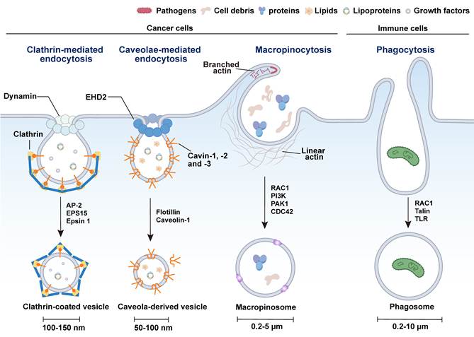
The four routes compared in Figure 4 are clathrin-mediated endocytosis (CME), caveolae-mediated endocytosis, phagocytosis, and macropinocytosis [167]. CME achieves selective uptake through ligand-receptor recognition at the cell surface. Adaptor proteins such as AP-2, Eps15, and Epsin recruit receptors and cargo to endocytic sites and promote membrane bending. Dynamin then constricts and severs the membrane neck, releasing the vesicle. However, its uptake capacity is limited by receptor abundance, receptor saturation, and tumor heterogeneity, making it particularly suitable for receptor-targeted or ligand-modified delivery systems [168]. Phagocytosis is mainly carried out by professional phagocytes (such as macrophages), which engulf large particles such as pathogens and cellular debris. Receptor-mediated particle recognition triggers RAC1-dependent F-actin remodeling, promoting pseudopod extension and phagosome formation. Because this process is largely restricted to specialized immune cells, its application in direct drug delivery to tumor cells is limited [169]. Caveolae-mediated endocytosis depends on specialized membrane invaginations called caveolae, whose main structural components are caveolin-1 and cavin family members; together with cholesterol- and sphingolipid-rich membrane domains, they maintain the flask-shaped morphology of caveolae. EHD2 is mainly localized at the caveolar neck, where it contributes to caveolar stability and membrane dynamics. Because caveolae-derived vesicles are typically only 50–100 nm in diameter, their small size imposes greater constraints on cargo dimensions than macropinocytosis [170]. Macropinocytosis generates large vesicles, typically 0.2–5 μm in diameter, through actin-dependent membrane ruffling [5]. PI3K-RAC1/CDC42-PAK1 signaling coordinates their formation and actin remodeling.
Comparison of the major endocytic pathways in cells. This figure summarizes the four major endocytic pathways: clathrin-mediated endocytosis, caveolae-mediated endocytosis, phagocytosis, and macropinocytosis. Among them, macropinocytosis generates large vesicles, typically 0.2–5 μm in diameter, through actin-dependent membrane ruffling. These vesicles are substantially larger than those formed by clathrin-mediated or caveolae-mediated endocytosis, which are typically 50–150 nm in diameter. This size difference supports high-capacity, non-selective fluid uptake. Phagocytosis also forms large vesicles, typically 0.2–10 μm in diameter, but is largely restricted to professional immune cells and primarily functions in the clearance of pathogens and debris. By contrast, macropinocytosis is active in multiple cancer types and may provide a promising route for tumor-selective drug delivery.
The size of macropinosomes permits bulk uptake of extracellular fluid and solutes, including nanoparticles and gene-delivery systems. Because capture does not require recognition of a specific receptor, low or heterogeneous receptor expression imposes fewer constraints. This may be useful in solid tumors containing cell populations with different receptor profiles. Macropinosomes also change substantially during maturation: shrinkage, membrane tubulation, and active sorting may provide a transient opportunity for cargo escape. As these compartments enter the endolysosomal network, established escape strategies may be adapted to promote cytosolic release [171, 172]. Efficient escape through this route nevertheless remains difficult, as discussed below.
Overall, the high capacity, receptor-independent uptake, and distinctive intracellular trafficking features of macropinocytosis make it a promising strategy for improving drug delivery into solid tumors.
5. Macropinocytosis-based delivery strategies
Macropinocytosis enables cells to internalize extracellular fluid and diverse cargo, providing a promising route for drug delivery. Current delivery strategies utilize macropinocytosis in different ways. Some platforms exploit intrinsic macropinocytic activity in target cells, whereas others actively enhance this process. For some carriers, however, macropinocytosis represents only one of multiple uptake routes involved in internalization. The contribution of macropinocytosis to carrier uptake depends on carrier properties and cellular context, and other endocytic pathways may also be involved. Some earlier preclinical studies inferred uptake mechanisms primarily from chemical inhibitor assays; these interpretations require corroboration through genetic evidence or cargo colocalization with fluid-phase tracers, as discussed in Section 6.4. The following subsections describe representative delivery systems, their design principles, and their advantages and limitations.
5.1 Biomimetic delivery strategies
Biomimetic delivery strategies use natural biomolecules or mimic biological structures to promote drug transport and cellular entry. Albumin and lipoproteins can serve as carriers for therapeutic cargo, while exosomes, liposomes, and viral platforms provide vesicular or capsid-based structures for cargo packaging and delivery. The relationship between these platforms and macropinocytosis varies with platform type and cellular context. In cells with active macropinocytosis, this pathway may contribute to carrier uptake to varying degrees; in addition, surface engineering can further enhance cell recognition or promote macropinocytic uptake in specific systems. Although the contribution of this pathway depends on the carrier and recipient cell, these features provide a versatile basis for macropinocytosis-based delivery strategies.
5.1.1 Albumin-based delivery strategies
Albumin is a natural protein that is widely used as a drug delivery carrier because of its biocompatibility and low immunogenicity. Its chemical versatility also supports a variety of drug-loading methods [173]. The preferential accumulation of albumin in tumors is attributed to the enhanced permeability and retention (EPR) effect and immune evasion [174]. As mentioned earlier, nutrient deprivation or KRAS mutations can enhance macropinocytic activity in cancer cells and promote extracellular albumin uptake [9]. This provides an active uptake route for albumin-based carriers. The abundant functional groups on the surface of albumin enable it to bind a variety of drugs through covalent or non-covalent interactions. This binding can improve drug solubility and stability, as well as pharmacokinetic behavior in vivo [175].
Nab-paclitaxel (Abraxane®) is a clinically established albumin-based formulation approved for metastatic breast cancer, non-small cell lung cancer, and metastatic pancreatic adenocarcinoma. Preclinical and clinical studies have demonstrated its therapeutic value [176]. It was developed for albumin-mediated delivery, without specifically targeting macropinocytosis. Early explanations of its delivery mechanism emphasized gp60-mediated endothelial transcytosis and a proposed contribution of SPARC-albumin binding to tumor accumulation [177, 178]. Later studies showed that KRAS-mutant pancreatic cancer cells take up and degrade albumin through macropinocytosis and that RAS-associated macropinocytosis contributes to nab-paclitaxel uptake [179]. However, the contribution of this pathway to its clinical efficacy remains unclear. Subsequent studies further clarified the mechanistic connection between albumin-based delivery and tumor-associated macropinocytosis. In KRAS-mutant pancreatic ductal adenocarcinoma, reduced neonatal Fc receptor (FcRn)-mediated recycling and increased macropinocytosis enhance the uptake and retention of albumin-associated cargo, broadening the therapeutic window of albumin-based systems [106]. This principle has been applied to albumin nanoparticles carrying the NAD(P)H quinone oxidoreductase 1 (NQO1)-targeted prodrug β-lapachone (nab-pro-β-lap) [180]. In BRAFV600E thyroid cancer, constitutively elevated macropinocytosis also promotes cellular uptake and intratumoral accumulation of an albumin-valine-citrulline-MMAE conjugate (Alb-vc-MMAE) [181]. Recombinant albumin fusion strategies address the structural heterogeneity caused by stochastic conjugation to native albumin. DF-HSA and DF₂-HSA retain macropinocytic uptake in KRAS-mutant tumors and improve intratumoral deposition, cargo retention, and antitumor activity [182, 183]. These systems are designed to use existing macropinocytic activity and do not necessarily stimulate the pathway. Other designs incorporate motifs that induce macropinocytosis, potentially enabling delivery to cells with lower baseline activity. One non-covalent approach uses a palmitoylated cyclic D-arginine peptide [Pal-cyclic-(D-Arg)12], which binds to the fatty-acid-binding pockets of human serum albumin (HSA), thereby forming a stable HSA-CPP complex. The complex enhances transmembrane transport and engages heparan sulfate proteoglycans (HSPGs) and C-X-C chemokine receptor type 4 (CXCR4) to activate PKC-PI3K-JNK-mTOR signaling. This triggers macropinocytosis and promotes the intracellular delivery of diverse cargo [184]. This approach may therefore extend albumin-based delivery to tumors with low baseline macropinocytosis.
Albumin carriers can incorporate targeting modules. Fv-LDP-D3 combines human serum albumin domain III (D3) with an anti-EGFR single-chain antibody (Fv), coupling receptor recognition to high-capacity macropinocytic uptake to increase tumor drug accumulation [185]. F-HSA-LDP-AE is composed of folate, HSA, LDP, and the enediyne toxin AE. Folate-receptor targeting and albumin-associated macropinocytosis can enhance its accumulation in cancer cells [186]. Uptake after arrival in a tumor is not restricted to malignant cells. Activated pancreatic stellate cells, a major stromal component, also internalize albumin-conjugated flumethasone through macropinocytosis. This uptake alters the local immune environment and improves antitumor efficacy [187]. Albumin carriers can therefore deliver to stromal targets, but improving cancer-cell selectivity within the tumor remains a separate design problem.
Albumin-based carriers combine compatibility with biological systems and flexible chemical modification with access to tumor nutrient-scavenging pathways. Their use requires accounting for uptake by stromal and other non-malignant cells, which can also actively scavenge albumin.
5.1.2 Lipoprotein-based delivery strategies
Lipoproteins (LPs) transport lipids and regulate metabolism. Lipoproteins are classified by density into chylomicrons (CM), very-low-density lipoproteins (VLDL), intermediate-density lipoproteins (IDL), low-density lipoproteins (LDL), and high-density lipoproteins (HDL) [188]. The advantages of these carrier materials are first of all reflected in their biodegradability and relatively low immunogenicity, so they have good biocompatibility. On the other hand, in order to maintain membrane synthesis, the demand for LDL in many tumor cells has increased significantly [189]. LDL and HDL themselves have nanoscale dimensions and can be recognized by cells through apolipoprotein-mediated recognition, which can promote the accumulation of lipoprotein-like carriers in tumor cells [190, 191].
Limited availability, variable structure, and imprecise targeting complicate the use of native lipoproteins. Reconstituted HDL- and LDL-like nanoparticles address these constraints [192]. Their stability, surface chemistry, and cargo release can be engineered, including modifications intended to exploit tumor-associated macropinocytosis.
ApoE3-reconstituted HDL enclosing an ATF5 siRNA-loaded calcium phosphate core was designed to exploit extracellular protein uptake by Ras-activated cancer cells. The carrier uses Ras-associated macropinocytosis and may also stimulate the pathway. It silenced ATF5 and inhibited growth in preclinical GBM models [193]. Self-disassembling porphyrin lipoprotein-coated calcium peroxide nanoparticles (PLCNPs) combine EPR-mediated accumulation with active targeting [194]. These nanoparticles are efficiently internalized via macropinocytosis and exhibit robust anti-GBM and relapse-prevention effects in murine models. The basic logic of these strategies is to use the strong macropinocytic capacity of tumor cells to design a delivery system that can be efficiently internalized by tumor cells, especially to let the carrier mimic extracellular proteins or lipoprotein-like cargo. On this basis, surface engineering can further introduce receptor specificity and a macropinocytosis-activating function into lipoprotein nanocarriers. For example, after modifying the surface of the particles with an SDF-1 mimetic peptide (FH38), the carrier can target CXCR4 and induce macropinocytosis, which significantly improves the uptake efficiency of particles by glioma-initiating cells (GICs) [195]. When loaded with miR-34a, this system downregulates stemness-associated factors such as SOX2 and Notch1, and demonstrates significant antitumor efficacy both in vitro and in vivo. In PDAC, 4F-modified reconstituted HDL (pGpC-rHDL) exploits pancreatic cancer cell macropinocytosis for selective tumor targeting [196].
Lipoprotein-based delivery strategies may also extend beyond tumor cells. DCs can also internalize the lipoprotein nanovaccine R837-αOVA-ApoE3-HNP through macropinocytosis, and thus promote DC maturation and antigen presentation [197]. The nanovaccine shows strong immunostimulatory activity in both prophylactic and combination treatment, and the antitumor effect can be further enhanced when combined with anti-PD-1 therapy. However, if the purpose of treatment is to kill cancer cells, uptake by immune cells may become a source of interference. After the carrier enters the tumor microenvironment, immune cells will compete with cancer cells for uptake opportunities. This phenomenon of being "intercepted" by non-malignant cells will cause the cargo to deviate from the intended target cells, ultimately weakening the selectivity of delivery.
5.1.3 Exosome-based delivery strategies
An exosome is an extracellular vesicle derived from the endosomal system. Its size ranges from 30 to 150 nm, and it is a nanoscale lipid-bilayer vesicle [198]. During its formation, some intracellular proteins and nucleic acids are selectively loaded into the exosome through ESCRT-dependent pathways, and then participate in intercellular communication [199]. An exosome has a natural membrane structure, so it has good biocompatibility, relatively low immunogenicity, and can cross certain biological barriers. In addition, exosomes can also carry therapeutic small molecules and macromolecular cargo, including nucleic acids and proteins [200], supporting their use as drug delivery vehicles.
Exosome uptake involves several endocytic pathways, including clathrin- and caveolae-mediated endocytosis, and usually depends on cell-surface receptor-ligand interactions. The dominant route varies with the type and state of the recipient cell [201]. Because vesicle size can affect the uptake route, macropinocytosis has been investigated as a means of internalizing larger exosomal vesicles in selected tumor settings [202, 203]. Macropinocytosis-associated genes can also regulate exosome uptake efficiency in some cancers [61, 204]. Fluorescent labeling studies provide evidence of exosome internalization through macropinocytosis in cancer cells [205]. In addition, exosomes from normal human fibroblasts have been loaded with KRAS-targeting siRNA. Surface CD47 reduced monocyte-mediated phagocytic clearance and prolonged circulation, while cancer-cell macropinocytosis supported siRNA delivery. These iExosomes showed antitumor activity in orthotopic and genetically engineered PDAC models. Uptake by α-SMA-positive cancer-associated fibroblasts (CAFs) showed that improved tumor delivery was accompanied by entry into non-malignant cells [206]. This iExoKRASG12D has entered Phase I/II clinical evaluation for pancreatic cancer (NCT03608631), with Phase I results now published [207].
For tumors with low baseline activity, exosomes can be modified to induce macropinocytosis. Co-loading EGF with therapeutic cargo activates EGFR-driven uptake [61]. Displaying the artificial leucine zipper peptide K4 on the surface promotes binding to E3-tagged EGFR, receptor clustering, and macropinocytosis [208, 209]. Cell-penetrating peptides can also stimulate the pathway and increase tumor-cell uptake [210-213]. Hybrid vesicles offer another design: M-LIP-CLT combines celastrol-loaded PEGylated liposomes with DC2.4 cell membranes. It mimics exosome-like targeting and accumulates through macropinocytosis in KRAS-mutant pancreatic cancer cells [214].
Even in cancer cells with high macropinocytic activity, the coexistence of multiple uptake pathways still makes it difficult to control the uptake process precisely. Future engineering strategies could favor macropinocytic uptake, with the aim of improving delivery selectivity and reducing nonspecific internalization. Whether this shift in uptake route also improves target-cell selectivity will need to be assessed directly.
5.1.4 Liposome-based delivery strategies
Liposomes are phospholipid-bilayer vesicles whose biocompatibility and cargo capacity have established them as a clinically used nanocarrier platform [215]. The ordinary liposomal delivery system mainly relies on passive tumor accumulation and lacks cell selectivity. Their surface chemistry and membrane composition can be adjusted to exploit existing macropinocytic activity, improving cargo uptake in selected cellular settings.
Surface ligands, such as glycyrrhetinic acid (GA) or arginine-glycine-aspartate (RGD) peptides, can improve tumor recognition and liposome association with cells [216-218]. By increasing liposome association with cancer cells, these modifications may make more carriers available for uptake through existing macropinocytic activity. Increased carrier uptake, however, does not necessarily indicate enhanced macropinocytic activity. In addition, surface modifications can exploit macropinocytic uptake of extracellular material to facilitate carrier entry into cancer cells. Liposomes modified with the penetratin-derived peptide 89WP and loaded with R8/siRNA complexes were developed for intranasal delivery of c-Myc siRNA. The 89WP modification improved nasal mucosal penetration, and the modified lipoplexes could also associate with necrotic cell debris. As glioma cells internalized this debris through macropinocytosis, the associated carriers were taken up with it. Subsequent cytoplasmic siRNA release enabled effective c-Myc silencing and prolonged survival in glioma-bearing mice [219]. In addition to chemical functionalization, exogenous physical stimulation may be another strategy to enhance macropinocytosis-mediated delivery. Modulated electro-hyperthermia (mEHT) significantly increases the uptake of liposomal doxorubicin (Lipodox®) by cancer cells. Although 70 kDa FITC-dextran is commonly used to monitor macropinocytosis, dextran uptake by macrophages can also occur through MRC1/CD206-mediated internalization [220]. Combined evidence from 70 kDa FITC-dextran uptake assays and wortmannin inhibition suggests that macropinocytosis is an important route contributing to this enhancement, although wortmannin is a broad PI3K inhibitor that can also affect clathrin-mediated endocytosis and autophagy [221]. This effect is likely related to membrane perturbation and may also involve EGFR activation and membrane ruffling. These findings suggest that physical stimulation may serve as a complementary strategy to modulate macropinocytosis and enhance local tumor uptake. Immune cells also exhibit pronounced macropinocytosis. A cationic core-shell lipid delivery platform incorporating mRNA condensed with poly(β-amino ester) (PBAE) is efficiently taken up by dendritic cells through macropinocytosis, thus improving vaccine delivery [222].
Liposome-based systems offer a highly engineerable platform for macropinocytosis-related delivery. Their main strengths lie in surface programmability and formulation flexibility, which can facilitate macropinocytic uptake in selected formulations and cellular contexts.
5.1.5 Virus-based delivery strategies
Macropinocytosis has a dual role in host-pathogen interactions. Host cells use macropinocytosis to sample extracellular material and clear pathogens. Viruses and bacteria can also exploit the same pathway by activating receptor signaling, actin remodeling, and membrane ruffling [24, 223]. In cancer cells, elevated macropinocytosis may therefore support nutrient acquisition while making these cells more susceptible to pathogen entry, a property that viral delivery systems could exploit.
Viral carriers designed specifically for macropinocytic tumor delivery remain at an early stage. Polymer-complexed oncolytic adenoviruses with pH-responsive and bioreducible properties have been developed for activation in acidic tumors [224]. After activation, macropinocytosis contributes to their internalization. An adenovirus-PEI-bile acid complex likewise improved transduction in CAR-low tumor cells through several endocytic routes, including macropinocytosis [225]. Studies of other viruses help explain how this entry mechanism might be used. Measles virus enters certain breast and colon cancer cell lines through PVRL4-mediated macropinocytosis [226]. The oncolytic vaccine strain SG33 enters pancreatic cancer cells through this pathway; phosphatidylserine (PS) on its envelope mimics apoptotic cells and further promotes uptake [227]. Viruses that induce receptor signaling and membrane remodeling may depend less on pre-existing macropinocytic activity.
Virus-based carriers still face several limitations in tumor delivery. First, macrophages and dendritic cells can clear viral carriers through macropinocytosis after systemic administration, reducing tumor exposure [228]. Second, many viruses need specific cell-surface receptors for recognition or entry signaling before macropinocytic internalization. Differences in receptor expression among tumors can therefore alter delivery. Viral carriers may also trigger innate immune responses and inflammation in off-target tissues. Replication-competent oncolytic viruses require control of infection and replication in normal tissue. Capsid or fiber modification, receptor retargeting, and multiligand designs may reduce dependence on native receptors and improve tumor selectivity. Tumor-specific promoters or miRNA-responsive regulation may help restrict replication and limit damage to normal cells [229].
Virus-based delivery through macropinocytosis remains under development. The ability of some viruses to induce this uptake pathway may offer advantages for nucleic acid delivery and oncolytic therapy. Translating these properties into effective delivery will require better tumor selectivity, less immune clearance and receptor dependence, and tighter control of off-target toxicity.
5.2 Functionalized delivery strategies
Functionalized carriers incorporate moieties that alter their interactions with cells to improve uptake, intracellular delivery, and treatment effects. These approaches emphasize engineered carrier-cell interactions more than the biological mimicry described above. Peptide modification, nutrient camouflage, and inorganic nanomaterials are commonly used to promote macropinocytic tumor-cell uptake and improve selectivity.
5.2.1 Peptide-based delivery strategies
Peptides and peptidomimetics, typically 5–50 amino acids long, provide biocompatible sequences that can be modified for particular functions [230]. Conjugation to drugs or nanoparticle surfaces allows these sequences to serve as modular components of delivery systems [231]. Their roles in macropinocytosis include receptor engagement, clustering of cell-surface components, and changes in membrane organization. The approaches discussed here comprise albumin-binding peptides, receptor-triggered macropinocytosis-inducing peptides, cell-penetrating peptides (CPPs), and peptides that induce bystander uptake.
First, compared with direct albumin-drug conjugates, albumin-binding peptide-drug conjugates (ABP-drug) bind endogenous albumin in vivo [232]. This method helps preserve FcRn-mediated long circulation and tumor accumulation via macropinocytosis, while avoiding stochastic conjugation sites and heterogeneous drug-to-albumin ratios. The efficacy of this strategy has been demonstrated in models of KRAS-mutant pancreatic ductal adenocarcinoma and PTEN-deficient metastatic triple-negative breast cancer [233-235]. In addition, co-delivery of the DNA-dependent protein kinase (DNA-PK) inhibitor AZD7648 with the ABP-drug can further potentiate macropinocytic activity through AMPK-PI3K activation, thereby enhancing tumor uptake and therapeutic efficacy [236]. In this setting, the peptide does not directly trigger macropinocytosis, but rather acts as a molecular hitchhiking module that couples endogenous albumin trafficking to tumor-associated macropinocytic dependency.
The second design logic is to use short peptides to induce macropinocytosis through receptor engagement directly. For example, K4-modified exosomes promote EGFR clustering and macropinocytic uptake [208]. KGF-ELP enhances macropinocytosis in cells expressing high levels of the keratinocyte growth factor receptor (KGFR) [237]. Similarly, CXCR4, a class A GPCR that serves as a receptor for CXCL12/SDF-1, is overexpressed in many cancers [238]. CXCL12/SDF-1 stimulation has been shown to induce macropinocytosis in macrophages [239]. The researchers coupled the peptide (SN21) composed of 21 residues at the N-terminus of SDF-1 with the membrane-lytic peptide LK15, which can trigger macropinocytosis and promote endosomal escape at the same time [240]. Another study further found that the two cysteines in the SN21 peptide play a key role in the induction of macropinocytosis. On this basis, the shortened octapeptide P4A (YRCACRFF) can form an intramolecular disulfide bond to yield oxP4A in the culture medium containing cystine, and show stronger macropinocytic activity [241]. It is worth noting that the response is not sensitive to CXCR4 inhibition, indicating that the conformational activation driven by disulfide bonds may be more important than receptor binding. In addition, N-terminal acylation can further reduce serum-mediated attenuation of activity [242]. However, in another study, it was found that FH38, an SDF-1 mimetic whose native Cys-Pro-Cys sequence was replaced by Ala-Pro-Ala, could still enhance macropinocytosis stimulated by CXCR4 and promote the uptake of lipoprotein-like nanostructures by glioma-initiating cells. This shows that cysteine residues are not the only determinant of macropinocytosis induction, and the relevant mechanism still needs to be further explored [195].
CPPs represent another major class of peptide-based macropinocytic delivery tools. Beyond direct membrane translocation via multivalent guanidinium-anion interactions, CPPs can also promote cellular uptake by inducing macropinocytosis [243, 244]. Carrier-bound arginine-rich CPPs, including Tat, bind HSPGs multivalently and cluster receptors, activating RAC1/PAK1 signaling to promote macropinocytic entry [184, 219]. In addition, these strategies have been extended to dimeric (sC18)2 peptides and stearylated R8 peptides presented on extracellular vesicles [211, 245].
A further extension of this concept is the bystander-effect strategy. Instead of covalently attaching peptides to every cargo, certain peptides can induce a local macropinocytic state that co-internalizes nearby unmodified molecules or nanocarriers. For instance, CendR-motif peptides bind and cluster neuropilin-1 (NRP1), recruit adaptor proteins such as GAIP-interacting protein C terminus 1 (GIPC1)/synectin, and trigger a fluid-phase uptake process that resembles macropinocytosis [246]. This process results in the internalization of co-delivered molecules or nanoparticles in a bystander fashion. Although the pathway has not yet been fully delineated, subsequent studies have strengthened the evidence for a central role of macropinocytosis in bystander uptake. Wei et al. reported that Tat-functionalized nanoparticles significantly enhance the uptake of various unmodified nanoparticles (AgNPs, AuNPs, IONPs, and QDs) via receptor-dependent macropinocytosis, which is regulated by the extracellular cysteine/cystine axis [247]. Notably, Tat-NPs did not enhance the uptake of fluid-phase markers such as dextran. One possible explanation is that high-density Tat induces strong multivalent clustering of HSPGs. This results in locally compact membrane microdomains and macropinocytic cups (>200 nm) that preferentially capture nearby nanoparticles while exerting minimal effect on freely diffusing solutes. Furthermore, an amphipathic "transportan" (TP) peptide enhances the bystander uptake of co-administered liposomes via macropinocytosis [248]. This peptide-assisted liposomal uptake does not generalize to cationic CPPs, consistent with the mechanistic framework outlined above. Free cationic CPPs bind HSPGs with relatively weaker and looser affinities, predominantly amplifying bulk fluid-phase uptake. Consequently, they can facilitate the entry of some inorganic nanoparticles but are less effective for larger, softer, PEGylated liposomes. In contrast, amphipathic CPPs reside stably at the plasma membrane and cluster receptors more efficiently, thereby eliciting stronger macropinocytic responses that enable high-efficiency co-internalization of larger carriers, including liposomes. These explanations need experimental testing before they can guide the design or clinical use of macropinocytosis-based bystander delivery.
Peptide-based strategies occupy a distinctive position in macropinocytic delivery. Their versatility allows peptides to facilitate delivery through albumin binding, stimulation of macropinocytosis, or enhanced co-internalization of nearby cargo. In addition, integrating macropinocytosis-inducing peptides with other functional modules, such as intracellular or endosomal escape peptides, may further improve overall delivery efficiency. Nevertheless, many aspects of short peptide-based delivery systems, including their mechanisms of action, structure-function relationships, and in vivo behavior, remain insufficiently understood. This poses a major challenge for future optimization of peptide design and delivery performance (Figure 5).
Peptide-based macropinocytic delivery strategies. (A) Albumin-binding peptide-drug conjugate strategy: Albumin-binding peptide-drug conjugates (ABP-drug) reversibly bind to endogenous albumin and are taken up via tumor cell macropinocytosis. (B) Macropinocytosis-inducing peptide strategy: Macropinocytosis-inducing peptides activate tumor cell macropinocytosis to enhance drug delivery. (C) Cell-penetrating peptide strategy: CPPs multivalently bind to HSPG, drive receptor clustering, and activate the RAC1/PAK1 signaling pathway, thereby activating macropinocytosis to enhance uptake. (D) Bystander effect strategy: Peptides without covalent conjugation to drugs or carriers induce local macropinocytosis, leading to co-internalization of nearby unmodified molecules or nanocarriers.
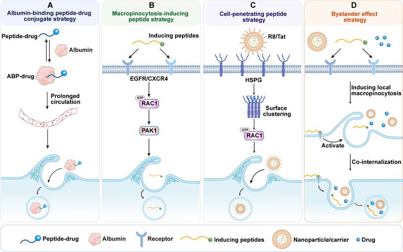
5.2.2 Nutrient-camouflaged delivery strategies
Nutrient-camouflaged delivery strategies exploit the metabolic dependencies of cancer cells by disguising carriers as nutrient-mimicking substrates. Dextran is a neutral, branched polysaccharide that is biosynthesized by certain lactic acid bacteria from sucrose. Its backbone consists of α-1,6-linked glucose residues with diverse side chains [249]. Dextran and its derivatives exhibit favorable biocompatibility and tunable molecular weight, making them valuable carrier materials in nanomedicine. Their value in tumor-targeted delivery has long been recognized. Compared with free doxorubicin, a doxorubicin-oxidized dextran conjugate achieved higher tumor accumulation with reduced cardiac exposure and subsequently advanced to phase I clinical evaluation [250, 251].
Based on the high uptake of 70 kDa dextran observed in macropinocytic PDAC cells, the dextran-triptolide conjugate (DEX-TP) was developed [252, 253]. In subcutaneous models, DEX-TP showed greater tumor accumulation and antitumor activity than free triptolide in KRAS-mutant MIA PaCa-2 tumors and BxPC-3 tumors expressing KRASG12V, whereas this advantage was absent in wild-type BxPC-3 tumors. In an orthotopic MIA PaCa-2 model, DEX-TP also suppressed tumor progression, prolonged survival, and showed better tolerability than free triptolide. Similar benefits were observed in an immunocompetent orthotopic KPC model, where DEX-TP prolonged survival and reduced the loss of T cells and effector cytokines induced by free triptolide at the same dose. These results indicate that the delivery advantage of DEX-TP through macropinocytosis is not limited to subcutaneous proof-of-concept models, but is also retained in orthotopic PDAC models with more complex stromal and immune microenvironments.
Glutamine has also been used to construct a related nutrient-camouflaged system. Iron nanoparticles were modified with glutamine to generate a glutamine-based iron delivery system (Gln-IDS), thereby exploiting cancer cells' metabolic dependence on glutamine for initial cellular entry [254]. The inhibitor V9302 carried by this system blocks exogenous glutamine uptake, thereby inducing metabolic reprogramming and significantly activating macropinocytosis. This feedback mechanism suggests that co-delivery systems combining therapeutic agents with nutrient-deprivation cues may induce macropinocytosis, thereby promoting carrier reuptake and subsequent accumulation. However, release timing and sequence would need careful control. Coupling delivery to this adaptive metabolic response may expand the therapeutic window of the formulation.
Nutrient-camouflaged systems represent a distinctive class of macropinocytic delivery strategies because they link tumor metabolic addiction with adaptive macropinocytic responses. Their major strength lies in integrating metabolic recognition and feedback-driven accumulation within a single design framework. This strategy offers a promising route to expand the therapeutic window of nanomedicines.
5.2.3 Inorganic nanomaterial-based strategies
Inorganic nanomaterials have a stable structure and unique physical properties such as magnetism and photothermal activity, so they have potential applications in drug delivery and theranostics. The macropinocytic uptake of such materials varies depending on the material properties and cell type [255]. For example, silica-coated superparamagnetic iron oxide nanoparticles (SPIONs) show faster internalization and more pronounced lysosomal trafficking in M-HeLa cells than in primary motor neurons [256]. The two cell types involve different endocytic pathways, but both include macropinocytosis. In another design, the researchers loaded hydroxychloroquine (HCQ) and doxorubicin (DOX) into mesoporous silica nanorods, combining particle geometry with the inhibitory effect of HCQ on clathrin-mediated endocytosis. On the one hand, the design limits uptake by healthy cells, and on the other hand, it uses the existing macropinocytic pathway in cancer cells to deliver DOX [257]. Surface modification can also further help recognize target cells before macropinocytic internalization. Folate-polyethylene glycol-modified carboxylated graphene quantum dots can achieve targeting through folate receptor recognition, and uptake experiments suggest that macropinocytosis is also involved in their internalization in HeLa cells [258]. In general, the structure and surface of inorganic nanomaterials are adjustable, which provides an opportunity for drug delivery through cellular macropinocytosis. If how particle size, shape, and surface chemistry affect macropinocytic uptake in different cell types can be clarified, it will help guide carrier design, thus improving delivery efficiency and selectivity.
Representative systems and the translational status of the different macropinocytosis-based delivery platforms discussed above are summarized in Table S2. Their main advantages, limitations, and representative cancer types are further compared in Table S3.
6. Risks and challenges
Macropinocytosis-based drug delivery is being explored for cancer treatment, and some systems have entered early clinical evaluation. Although this pathway enables high-capacity uptake and can improve intracellular delivery, several barriers still limit its clinical translation, including off-target effects, inefficient endosomal/lysosomal escape, difficulties in monitoring macropinocytosis, and the influence of carrier physicochemical properties on uptake efficiency. This section discusses these challenges and potential strategies to address them (Figure 6).
Risks and challenges of macropinocytosis-based delivery systems. (A) Off-target risk: Uptake by non-malignant stromal and immune cells can divert carriers from their intended targets and cause toxicity. (B) Non-selectivity paradox: Particle shape, size, molecular weight, charge, surface topology, and stiffness influence macropinocytic uptake. (C) Drug escape challenges: Endolysosomal acidification and enzymatic degradation limit cytoplasmic delivery of internalized cargo. (D) Identifying challenges: Insufficient specificity of available markers and inhibitors complicates pathway identification and quantification.
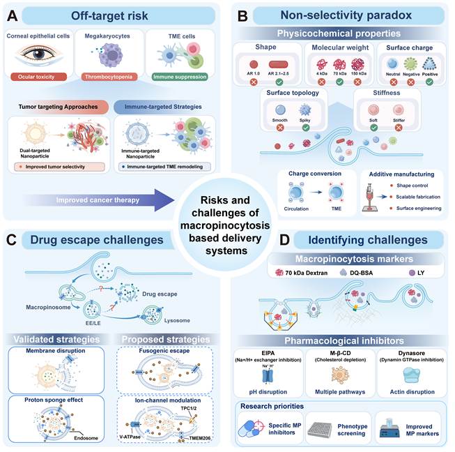
6.1 Off-target risk
Enhanced macropinocytosis in tumor cells provides a route for drug delivery, but its lack of tumor specificity limits its clinical application. Normal cells, including immune cells and other cell types such as endothelial cells and neurons, also exhibit macropinocytic activity [7]. Clinical observations also raise concerns about potential off-target effects. The antibody-drug conjugate (ADC) AGS-16C3F was evaluated in a randomized Phase II trial for metastatic renal cell carcinoma (ClinicalTrials.gov: NCT02639182). Although it showed antitumor activity, ocular toxicity was reported. This toxicity may be related to the internalization of antibody-drug conjugate (ADC) by human corneal epithelial cells (HCECs) through macropinocytosis [259]. The process is driven by the drug molecule's positive charge or hydrophobic residues, ultimately leading to inhibition of cell proliferation. Similarly, trastuzumab emtansine (T-DM1), an approved human epidermal growth factor receptor 2 (HER2)-targeted ADC evaluated in the Phase III EMILIA trial (NCT00829166), may enter megakaryocytes through macropinocytosis and contribute to thrombocytopenia [260].
Tumor-targeting modules have been incorporated into macropinocytosis-based delivery systems to increase tumor accumulation and reduce nonspecific distribution in normal tissues. However, accumulation of a carrier within tumor tissue does not necessarily translate into selective delivery to cancer cells. Tumor-associated macrophages and cancer-associated fibroblasts are abundant in many solid tumors. Their high accessibility to incoming carriers and strong endocytic capacity can create a “cellular sponge effect,” leading to competitive sequestration of nanocarriers within the tumor [261]. These non-malignant cells themselves exhibit varying levels of macropinocytosis. For instance, in pancreatic cancer, CAFs activate macropinocytosis under conditions of glutamine depletion via the ARHGEF2 RhoGEF and the CaMKK2-AMPK axis [262]. Under glutamine limitation, inhibition of macropinocytosis promotes the conversion of myofibroblastic cancer-associated fibroblasts (myCAFs) into inflammatory cancer-associated fibroblasts (iCAFs) through a MEK/ERK-dependent intermediate state [263]. Various immune cells also exhibit significant macropinocytosis. For example, activated T cells can acquire extracellular nutrients via macropinocytosis [264]. Meanwhile, DCs and macrophages can utilize this pathway to internalize exosomes and other extracellular substances, whereas immature DCs participate in antigen uptake and presentation through macropinocytosis [265, 266]. Several orthotopic models further demonstrate cell-level off-target uptake within the tumor microenvironment. For example, although AF647-labeled iExosomes were mainly distributed in pancreatic cancer cells, uptake signals were also detected in alpha-smooth muscle actin (α-SMA)-positive CAFs, as well as in normal acinar cells, ductal cells, and endocrine islets [206]. Similarly, in an orthotopic anaplastic thyroid cancer model, fluorescent albumin accumulation per cell was higher in TAMs than in cancer cells. Nevertheless, cancer cells accounted for most tumor-associated albumin because they were more abundant. Macrophage coculture also attenuated the increase in cancer-cell albumin uptake induced by inhibition of insulin-like growth factor 1 receptor (IGF1R). These findings suggest that non-malignant cells can influence both payload distribution and cancer-cell uptake responses [181]. In an orthotopic PDAC model, activated pancreatic stellate cells also showed strong albumin uptake, which was further exploited by Albumin-Flu for stromal targeting and remodeling [187].
Macropinocytosis-based delivery therefore faces two selectivity challenges: off-target uptake in normal tissues and competitive allocation among cells within tumors. Future delivery systems could address these challenges through sequential control of tissue and cellular selectivity. Tumor acidity, matrix metalloproteinases, or other microenvironmental signals could first trigger surface remodeling or removal of shielding layers after tumor accumulation [267]. Cancer-cell recognition modules could then promote selective binding, followed by exposure or activation of a macropinocytosis-inducing module to enhance internalization. This design could combine tumor-site activation with cancer-cell recognition. However, its ability to reduce off-target uptake in normal tissues and sequestration by non-malignant cells within tumors requires direct validation. Some stromal and immune cells can take up carriers through macropinocytosis and act as cellular sponges, making it necessary to limit their uptake after carriers enter the tumor. Differences in cell-surface receptor expression could help direct carriers preferentially toward cancer cells. For example, αvβ6 is enriched in PDAC tumor epithelium but shows comparatively limited expression in the surrounding stroma [268], making it a candidate recognition target. Coupling cancer-cell recognition to the subsequent exposure or activation of a macropinocytosis-inducing module could preferentially promote carrier internalization by cancer cells. This design aims to reduce sequestration by CAFs and TAMs, leaving more of the cargo delivered to the tumor available for cancer-cell uptake. Adjusting carrier dose or circulation half-life alone does not ensure preferential uptake by cancer cells over CAFs or TAMs. Comparing intracellular cargo amounts across these cell populations would help determine whether the proposed strategy improves cancer-cell delivery while reducing stromal sequestration.
In addition, macropinocytosis in non-malignant cells can also be therapeutically exploited. Targeting stromal or immune cells to remodel the tumor microenvironment provides an alternative therapeutic strategy [269]. However, because cancer cells and other nontarget cells also exhibit macropinocytic activity, relying only on this uptake pathway cannot ensure that carriers enter the intended stromal or immune cells. Therefore, when targeting these cells, their specific surface molecules can first be used to promote carrier accumulation, after which macropinocytosis can further enhance carrier internalization. For example, nanoparticles modified with FAP-α antibodies can preferentially deliver CXCL12 siRNA to cancer-associated fibroblasts [270]. Nanoparticles targeting CLEC9A and DEC205 can also enhance binding to and uptake by cDC1 cells, enabling the targeted delivery of STING agonists [271]. Although these studies have not yet directly demonstrated that the carriers are internalized via macropinocytosis, they indicate that the use of different cell surface molecules can enhance the cell-type specificity of the carriers. In the future, macropinocytosis-enhancing modules could be further incorporated based on these findings to promote carrier internalization while maintaining cell specificity, thereby improving overall delivery efficiency.
Therefore, although macropinocytosis can enhance carrier internalization, its limited cell-type selectivity creates two major risks: off-target uptake in normal tissues and competitive sequestration or redistribution among different cell populations within tumors. Future delivery systems could first use tumor microenvironment-responsive mechanisms for tumor-site activation, followed by cell-surface recognition to improve target-cell selectivity. Macropinocytosis could then be exploited or further induced to enhance carrier internalization. This framework can be applied to both cancer cells and stromal or immune cells selected as therapeutic targets. Evaluation should extend beyond overall tumor accumulation to cellular distribution, internalization, and cargo release in models that preserve relevant stromal and immune components.
6.2 Non-selectivity paradox
Macropinocytosis has long been regarded as a non-selective endocytic pathway. In theory, any extracellular material or carrier near the plasma membrane can be randomly internalized, and the uptake efficiency should not depend on its physicochemical properties. However, increasing evidence suggests that the physicochemical properties of delivery systems or cargo can substantially influence macropinocytic entry and the kinetics of uptake.
Particle geometry appears to be an important determinant of macropinocytic uptake. The amphiphilic peptide I2Q4 self-assembles into rod-like micelles via hydrogen bonding. The paclitaxel-loaded rod-like PTX@I2Q4 nanostructure exhibits stronger macropinocytosis-mediated uptake than spherical PTX-albumin nanoparticles [272]. The study of the aspect ratio (AR) of mesoporous silica nanoparticles (MSNPs) and its effect on macropinocytic uptake further supports this conclusion. Rod-shaped MSNPs with an aspect ratio of 2.1–2.5 show the highest level of macropinocytic uptake [273]. The authors propose that nanoparticles with an aspect ratio of 2.1–2.5 are more readily internalized through macropinocytosis because their geometry better matches the biomechanical requirements of membrane wrapping. Upon initial contact with the plasma membrane, these particles generate a more favorable local curvature (typically at an incident angle less than 45°), thereby facilitating membrane engulfment and the formation of a cup-like structure. At the same time, their increased contact line length provides more nucleation sites for actin. This amplifies RAC1/CDC42 activation, accelerates membrane invagination and pseudopod closure, and ultimately facilitates macropinocytosis. Compared with spheres or excessively elongated rods, particles with ARs of 2.1–2.5 overcome the dual challenges of initiation difficulty and closure failure, thereby enabling higher-throughput macropinocytic endocytosis. Based on this phenomenon, Cong et al. co-loaded hydroxychloroquine and doxorubicin into mesoporous silica nanorods measuring approximately 180 nm × 68 nm. Owing to their specific morphology and size window, these nanorods minimized uptake by healthy cells while enabling selective internalization by cancer cells via macropinocytosis [257].
In addition, physical curvature to drive macropinocytosis has also been reported in another study. Instead of exploring the curvature changes of nanoparticles to promote uptake, the authors seeded cells onto a nanotopographical surface with 400 nm blind pores [274]. They found that high-curvature membrane deformation mimicked the ruffling associated with macropinocytosis initiation, thereby triggering actin cytoskeleton rearrangement, inducing macropinocytosis, and enhancing the nonspecific uptake of various exogenous substances. This study further confirms, from a different perspective, the key role of membrane curvature in the initiation of macropinocytosis.
Particle shape alone, however, does not explain all differences in uptake. For example, the endocytic uptake of 70 kDa dextran was significantly higher than that of 4 kDa and 150 kDa dextran [253, 275]. The 4, 70, and 150 kDa dextrans adopted random-coil conformations, with hydrodynamic diameters of approximately 2.8 nm, 12 nm, and 17 nm, respectively, and no discernible difference in AR. This suggests that geometric anisotropy is not the sole determining factor. One possible explanation is that the process from macropinocytic cup formation to closure is extremely rapid, with a typical duration of only tens of seconds [30, 276, 277]. Whether cargo can be successfully internalized may depend on whether it can be effectively trapped in the cup during the brief existence of the macropinocytic cup. Taking dextran as an example, the molecular weight of 70 kDa dextran is higher than that of 4 kDa dextran, and its diffusion coefficient is lower, so it is less likely to diffuse out before the macropinocytic cup closes, and it is more likely to be retained in the cup. In contrast, the diffusion rate of 150 kDa dextran may be too slow for it to fully enter the cup before the macropinocytic cup closes, thus limiting its encapsulation. However, this diffusion-based explanation is still just a hypothesis and needs to be verified experimentally.
The influence of carrier surface topology on macropinocytosis mainly occurs through changes in carrier-membrane contact and retention at the membrane. Take virus-like spiky particles as an example. Compared with silica particles with smooth surfaces, they can form multiple contact points with the plasma membrane, thus enhancing adhesion and improving cellular uptake [278]. This uptake process involves both macropinocytosis and clathrin-mediated endocytosis. Similarly, asymmetric nanorobots have a sea urchin-like spiky head and a hollow tail, and their cellular uptake is also enhanced. At the same time, it was found that the greater the number of spikes and the longer their length, the more pronounced the increase in uptake [279]. After adding cytochalasin D, uptake decreased, suggesting that the process depends on actin. However, it should be noted that cytochalasin D is not a specific inhibitor of macropinocytosis, and the study did not use fluid-phase tracers or directly observe macropinocytic cup formation, so the contribution of macropinocytosis cannot be directly established. Another study on kaolinite provides new evidence from the perspective of membrane retention. Kinetic analysis found that multiple sites on the kaolinite surface continually form, break, and re-form hydrogen bonds with the membrane interface. On the one hand, this dynamic interaction maintains contact between the particles and the membrane, and on the other hand, it also allows interfacial rearrangement. Because the particles are less likely to detach and remain near membrane ruffles for longer, the chance of being captured during macropinocytic cup formation and closure also increases accordingly [280].
Carrier stiffness may also influence the route of cellular entry. Banquy et al. found that soft hydrogel nanoparticles with a Young’s modulus of approximately 18 kPa entered RAW264.7 macrophages mainly through macropinocytosis. Increasing stiffness to approximately 211 kPa shifted the predominant uptake route toward clathrin-mediated endocytosis [281]. One possible explanation involves the mechanics of membrane wrapping: rigid particles require greater membrane deformation, while softer particles can deform as they are engulfed, altering particle-membrane contact. However, few studies have directly examined how stiffness affects macropinocytic cup expansion and closure, so the mechanistic evidence remains limited.
Particle surface charge is another key factor in regulating the efficiency of macropinocytosis. The positive surface charge of the carrier can significantly enhance macropinocytic uptake. In HeLa cells, arginine-rich cationic cell-penetrating peptides such as R8/Tat show uptake that is highly sensitive to EIPA and actin inhibitors, indicating that they mainly enter cells through macropinocytosis [282, 283]. Extracellular vesicles modified with arginine-rich cationic cell-penetrating peptides also show a similar uptake process [213]. In HeLa cells, positively charged nanoparticles are internalized through macropinocytosis more efficiently than negatively charged particles, accompanied by typical pseudopod formation and F-actin rearrangement. The process is generally mediated by RAC1/CDC42, is sensitive to EIPA, and partly depends on microtubules and COX activity [284]. In addition, when neutral or negatively charged nanoparticles are coated with the cationic polymer polyethyleneimine (PEI), their primary internalization route shifts toward macropinocytosis [285]. One possible mechanism is the electrostatic interaction between positively charged particles and negatively charged tumor cell surfaces and stromal components in the acidic tumor microenvironment. These interactions can increase the adsorption and retention of positively charged particles, allowing greater cell contact and possibly promoting macropinocytosis. Cationic functional groups (such as guanidino groups) can form multivalent interactions with HSPGs on the cell surface [286]. The finding that tau aggregates are internalized through HSPG-dependent macropinocytosis further shows the broader relevance of this mechanism [287]. Secondary condensation of Tat/pDNA complexes with Ca²⁺ or cetyltrimethylammonium bromide (C16TAB) can further enhance macropinocytic uptake [288, 289]. Although the treated particles become smaller and more compact, this alone is not enough to explain why uptake increases. After Ca²⁺ or C16TAB treatment, the zeta potential of the particles shifts markedly toward more positive values; compared with the original Tat/pDNA complexes, this shows that these treatments not only promote secondary condensation but may also reorganize the complexes into smaller, more highly cationic nanoparticles by strengthening electrostatic and hydrophobic interactions. Similarly, high cationic density has shown similar effects in other studies. Liposomes with low R8 density are mainly internalized through clathrin-mediated endocytosis, while high R8 density is more conducive to macropinocytosis [290]. This shows that local cationic density helps determine the uptake route. Densely displayed cationic ligands are more likely to form stable multivalent interactions with anionic cell-surface components (especially HSPGs), thus prolonging membrane residence time and promoting RAC1/CDC42-dependent actin remodeling and membrane ruffling. Therefore, the improvement in uptake through secondary condensation may be related to both changes in particle compactness and stronger interactions between the carrier and the cell surface.
When designing carriers to improve macropinocytic uptake, in addition to considering the optimization of the above-mentioned physicochemical properties, attention should also be paid to the effects of these properties in vivo. Positive surface charge can promote uptake, but at the same time increase nonspecific tissue adsorption and systemic toxicity [291]. After cationic nanocarriers enter the blood, they will quickly adsorb plasma proteins and form protein coronas, thus changing their apparent surface properties. At the same time, the electrostatic interaction between them and red blood cells may also pose a risk of hemolysis. Protein adsorption will also enhance the recognition and clearance of carriers by the mononuclear phagocyte system (MPS), shortening their circulation time and reducing tumor exposure [292]. Therefore, the design of cationic carriers must achieve a balance between cellular uptake and stability during circulation. The charge conversion strategy discussed in Section 4.2.4 may help solve this problem. When optimizing particle size and shape, additive manufacturing methods such as nano-3D printing can be considered. Carriers prepared by this method can have their size and shape controlled more accurately, and this method is also conducive to systematically studying the effects of these properties on macropinocytosis-based delivery [293].
The mechanobiological basis by which carrier properties regulate macropinocytosis remains incompletely understood. Therefore, parameters such as stiffness, surface topology, and surface chemistry should not be examined in isolation, but should be systematically evaluated. If we can clarify how these properties affect membrane retention, macropinocytic cup formation, and cargo capture, we can provide a more rational basis for the design of macropinocytosis-based delivery systems.
6.3 Drug escape challenges
After internalization through macropinocytosis, therapeutic cargo may initially avoid direct exposure to plasma membrane efflux pumps such as P-gp. However, the resulting macropinosomes typically mature along the early-to-late endosomal pathway and ultimately enter lysosomal compartments with a pH of approximately 4.5-5.5. Although this process promotes lysosomal enrichment of lysosome-targeting drugs, the acidic, hydrolase-rich lysosomal environment readily leads to ion trapping or cargo degradation. Furthermore, macropinosomal recycling and cargo re-export further reduce the drug's effective cytoplasmic exposure. Consequently, intracellular escape remains a central bottleneck that limits therapeutic efficacy and represents a major challenge in the design of macropinocytosis-based delivery systems.
Among current strategies for endosomal or lysosomal escape, lysosomal membrane permeabilization (LMP) triggered by inorganic ions is mainly exploited at the lysosomal level. For instance, nanocarriers containing inorganic ions are internalized into lysosomes via cancer-associated macropinocytosis [294]. Upon lysosomal entry, these ions are released, inducing LMP to facilitate the escape of cargo from lysosomes into the cytoplasm. Experimentally studied approaches to endosomal escape include protonatable carriers and membrane-active peptides or lipids. One relies on the "proton sponge effect" to induce osmotic swelling and membrane rupture. For instance, after Janus base nanoparticles are internalized via macropinocytosis, the NPs escape from endosomes through a process analogous to the "proton sponge" effect of cationic polymers [295]. The alternative mechanism involves fusion peptides or cationic lipids, which are triggered by the low pH in endosomes, leading to their insertion into the membrane. By lowering the energy barrier to curvature, transient nanopores form, enabling the leakage of contents across the membrane into the cytoplasm [240, 296].
Compared with classical endocytic vesicles, macropinosomes are micrometer-scale single-membrane compartments. During maturation, macropinosomes undergo rapid osmotic shrinkage through Na⁺/Cl⁻ efflux mediated by ion channels such as TPC1/2 and transmembrane protein 206 (TMEM206) [36]. These giant vacuoles undergo substantial changes in membrane tension and curvature during intracellular transport. These features may make macropinosomes more amenable to mechanically assisted leakage or rupture than smaller coated endosomes [297]. Consequently, this characteristic offers a potential escape window for drugs.
Based on this mechanism, two possible design directions may be considered. First, the hypertonic and high-curvature window created by osmotic shrinkage could be exploited by modifying carrier surfaces with pH-responsive membrane fusion or pore-forming peptides, such as HA2, INF7, or amphipathic helical peptides with masking/unmasking functions. These peptides are activated in acidic environments and insert into the membrane, thereby temporarily increasing membrane permeability or promoting membrane fusion during a critical window. This process ultimately results in the release of the payload into the cytoplasm [298, 299]. Furthermore, liposome composition can be optimized by increasing conical lipids, such as DOPE, and reducing cholesterol, and by introducing reversible lysine/alkylation groups, thereby reducing the membrane bending modulus and enhancing insertion under low-pH conditions [300]. Another, as yet unvalidated, approach is to interfere with ion channels involved in osmotic shrinkage during macropinosome maturation to facilitate cargo escape. During this process, TPC1/2 mediate Na⁺ efflux, whereas ASOR/TMEM206 mediates Cl⁻ efflux, together driving osmotic water loss, macropinosome shrinkage, and subsequent membrane tubulation [36, 301]. Interfering with these ion transport pathways could reduce ion and water efflux and prolong the large-volume state of macropinosomes. When combined with protonatable materials, ion-channel inhibition may further increase luminal osmotic stress through the proposed proton sponge effect, potentially enhancing membrane destabilization.
Mechanistically, ion channel inhibition differs from the proton sponge effect and pH-responsive membrane disruption. Ion channel inhibition mainly delays macropinosome shrinkage by reducing ion and water efflux. By contrast, the proton sponge effect increases the luminal osmotic load, whereas fusion peptides or pore-forming materials directly disrupt the membrane. Therefore, ion channel inhibition alone may not be sufficient to cause membrane rupture and may be more suitable as an auxiliary escape mechanism. Ion-channel inhibition could therefore be combined with protonatable materials or pH-responsive fusogenic and pore-forming systems to further facilitate membrane destabilization and cargo release. However, this strategy is currently based mainly on the known mechanisms of macropinosome maturation, and direct evidence that ion channel inhibition can consistently improve cytosolic escape is still lacking. In addition, the roles of these channels are not limited to macropinocytosis. Therefore, sustained or systemic inhibition may disrupt endolysosomal trafficking and substrate clearance in normal cells, increasing the risk of storage-related dysfunction. For example, TPC2 deficiency impairs LDL-cholesterol trafficking and increases susceptibility to hepatic cholesterol overload and liver injury in mice fed a cholesterol-rich diet [302]. ASOR/TMEM206 deficiency causes endosomal hyperacidification and alters transferrin receptor-mediated endocytosis [303]. Although these genetic studies cannot directly predict the toxicity of pharmacological inhibition, they suggest that prolonged disruption of these channels could lead to substrate accumulation and cellular stress, compromising normal cell function and survival in healthy tissues. When combined with membrane-disruptive materials, excessive membrane permeabilization may also cause nonspecific cellular injury, which should be distinguished from effective cargo escape. Future studies should therefore prioritize local, transient, and reversible interventions, assessing endolysosomal pH, membrane integrity, and viability in normal cells alongside cytosolic cargo release to establish a safety window.
6.4 Identifying challenges
At present, pharmacological inhibitors are commonly used to distinguish endocytic pathways, with the entry route inferred from their effects on carrier uptake. In the study of macropinocytosis, EIPA is one of the most widely used tools. It mainly inhibits the Na+/H+ exchanger NHE1, and inhibits macropinosome formation by blocking proton efflux to disrupt pH homeostasis in the submembrane region, and then interferes with actin remodeling mediated by RAC1/CDC42. However, EIPA does not seem to only affect macropinocytosis. In epithelial cells originating from renal proximal tubules, EIPA will inhibit NHE3-dependent receptor-mediated albumin internalization [304]. At the same time, Na+/H+ exchange itself is also involved in the regulation of intracellular pH. In the spheroid model of triple-negative breast cancer, long-term EIPA treatment will lead to drug accumulation and organelle damage [305]. These broader cellular effects will interfere with the assessment of carrier uptake and treatment effects. Similar disputes over specificity also exist in the verification of other endocytic pathways. For example, methyl-β-cyclodextrin is a cholesterol scavenger commonly used to test clathrin-dependent and lipid-raft-dependent endocytosis, but cholesterol can also activate RAC1 by accumulating at the plasma membrane, thus activating macropinocytosis [306, 307]. The small-molecule inhibitor Dynasore, which is often used to inhibit clathrin-dependent endocytosis, has also been found to interfere with F-actin dynamics, thus affecting a variety of endocytic pathways that depend on cytoskeletal reorganization [41, 308]. These cross-interferences show that bias can arise from relying only on pharmacological inhibitors to clarify the uptake mechanism. At the same time, the effect of macropinocytosis inhibitors is also closely related to the cellular context. In A431 cells, rottlerin, a non-selective kinase inhibitor with mitochondrial uncoupling activity, inhibits the uptake of 70 kDa dextran even more strongly than EIPA [309]. However, in other cellular contexts, this stronger inhibitory effect is not consistently observed. Rottlerin can also reduce mitochondrial membrane potential and affect ATP availability in some cells [310]. These effects may also impair cytoskeletal remodeling and energy-dependent uptake. Therefore, when interpreting pharmacological evidence, the effects on overall cell physiology and other endocytic pathways must also be considered. The reduction in uptake after treatment with a single inhibitor alone is not enough to prove that uptake depends on macropinocytosis, and the percentage inhibition cannot be directly equated with the proportion of macropinocytosis in total uptake.
Therefore, highly selective macropinocytosis inhibitors should not be developed solely by targeting broad upstream regulators. Future studies can focus more on targets involved in membrane ruffling, cup closure, and macropinosome maturation. For example, CARMIL1 is an actin cytoskeletal scaffold protein that is recruited to the cell front through its membrane-binding region. It further regulates capping protein (CP) activity via its CP-interacting domains, thereby maintaining the dynamic balance of barbed ends at the cell edge [311]. A CP-binding-defective mutant, CARMIL1-AA, has been shown to selectively block macropinocytosis-dependent tumor growth. Compared with EIPA, it exhibits greater selectivity in suppressing macropinocytosis, while having limited effects on RAC activation, autophagy, and other basic cellular processes [312]. However, the inhibitory effect of CARMIL1-AA currently requires genetic expression. Pharmacological approaches that mimic its effect may therefore provide a more selective alternative to EIPA. In addition, key regulators of membrane remodeling may represent another class of candidate targets. SNX5 is recruited to phosphoinositide-enriched plasma membrane regions following receptor activation and promotes Arp2/3-dependent dorsal ruffle formation, thereby facilitating macropinosome biogenesis. In bone-marrow-derived macrophages, SNX5 deficiency reduced macropinocytic uptake of dextran or ovalbumin by approximately 60–70%, while phagocytosis was unaffected [313]. However, this effect is dependent on cell type and macropinocytosis subtype, as SNX5 deficiency does not impair macropinocytosis in DCs, and SNX5 also participates in other endosomal sorting processes. Its translational potential will therefore depend on identifying the macropinocytosis-relevant functional interface of SNX5 and developing pharmacological tools that interfere with this activity without broadly disrupting endosomal trafficking. Together, these findings suggest that targeting macropinocytosis-specific membrane and cytoskeletal machinery may provide greater selectivity than conventional inhibitors, although further translation will require pharmacologically tractable targets, broader validation across cell types, and rigorous assessment of their effects on other endocytic pathways. In addition to developing inhibitors against known targets, direct phenotype-based screening may represent a more effective strategy for identifying highly selective macropinocytosis inhibitors. For example, 70 kDa dextran uptake can serve as a readout in high-content screening for bioactive small molecules, enabling the identification of candidate compounds with stronger inhibitory effects than EIPA [314]. In parallel, some studies have developed screening models for virus entry via macropinocytosis and identified novel inhibitory candidates using this approach. These findings suggest that screening strategies based on pathogen entry phenotypes may also provide a promising avenue for the discovery of macropinocytosis inhibitors [315]. Such phenotype-based approaches should be followed by mechanistic and pathway-selectivity validation to distinguish genuine macropinocytosis inhibitors from compounds that broadly suppress endocytosis or cell viability.
In addition to the use of inhibitors, colocalization analysis of delivery systems with 70 kDa dextran remains the mainstream approach for qualitative and quantitative assessment of macropinocytosis. However, recent studies have revealed that this tracer can undergo MRC1 receptor-mediated endocytosis in specific cell types, such as macrophages [220]. This may blur the distinction between macropinocytosis and receptor-mediated endocytosis. In addition, other commonly used macropinocytosis indicators, such as DQ-BSA and Lucifer Yellow, exhibit varying degrees of nonspecificity across different cellular or tissue contexts [316, 317]. Therefore, future macropinocytosis markers should be developed through rational design based on the physicochemical preferences of macropinocytic uptake. Based on the principles discussed above, probes may be rationally engineered by optimizing parameters such as size, shape, surface charge, and molecular weight. Such designed macropinocytosis markers can be further combined with fluorescent conjugation to generate functional tracers that are more readily taken up through macropinocytosis. This may create new opportunities to improve the specificity and accuracy of macropinocytosis detection.
In addition, as shown in Table S2, current studies lack standardized criteria for marker and inhibitor selection, treatment conditions, and quantitative readouts, which limits direct comparison across studies. Some studies have proposed more consistent methods for quantifying macropinocytosis. For example, Commisso et al. developed the macropinocytic index, which quantifies macropinocytic activity by calculating the area of 70 kDa fluorescent dextran-positive vesicles and normalizing it to total cell area [318]. This image-based framework has also been adapted for automated analysis and tissue-based quantification of macropinocytosis [319].
The identification of macropinocytosis is still limited by the lack of highly specific inhibitors and tracers. To solve this problem, multidisciplinary efforts are needed. Meanwhile, with the existing tools, the reliability and comparability of the results can be improved through standardized multimodal workflows. First, fluid-phase uptake can be assessed using 1 mg/mL fluorescent 70 kDa dextran for 30 min at 37 °C [318]. These conditions provide a starting point and should be optimized for the cell model and carrier. Subsequently, the colocalization of fluorescently labeled carriers and dextran-positive vesicles is detected, and carrier uptake per cell and the proportion of intracellular carrier signal within dextran-positive vesicles are quantified. Consistent image thresholds and vesicle segmentation criteria should be used for comparison. Pharmacological inhibition can be used as supporting evidence. For example, a published protocol used 50 μM EIPA pretreatment for 30 min before uptake measurement [253]. Genetic perturbations can also assist in verification, including RAC1 or PAK1 knockdown, CARMIL1-AA expression [312, 320, 321], and SNX5 deletion selected according to the cell model [313]. Carrier uptake, dextran uptake, and vesicular colocalization should be compared with appropriate controls, and consistent changes can support the involvement of macropinocytosis. If genetic perturbations simultaneously reduce 70 kDa dextran uptake and carrier colocalization with dextran, this will further support macropinocytic internalization of the carrier. In addition, high-resolution live-cell imaging can also provide dynamic morphological evidence. 3D-SIM and LLSM can be used to directly observe membrane ruffling and macropinocytic cup formation and closure in living cells [14]. At the in vivo level, near-infrared-labeled albumin has been used for imaging and semiquantitative assessment of tumor macropinocytic activity, extending quantitative evaluation from cultured cells to in vivo models [322]. Therefore, integrating standardized quantitative uptake analysis with dynamic morphological and in vivo imaging may improve the accuracy and comparability of macropinocytosis evaluation across delivery systems and studies.
7. Conclusion
This review examines the biological functions and regulatory mechanisms of macropinocytosis in cancer and its applications in drug delivery. Macropinocytic activity in tumor cells is enhanced, which provides a route for macromolecular drugs and nanocarriers to enter cells that differs from classical endocytic pathways. However, whether the delivery system can really make good use of this route depends not only on the physicochemical properties of the carrier itself, but also on the tumor microenvironment. In addition, carrier uptake by cells does not mean successful delivery. The carrier must also be able to selectively reach cancer cells and undergo effective intracellular trafficking, and finally achieve cargo release.
Macropinocytosis provides new possibilities for tumor drug delivery, but there are still many obstacles to clinical translation. First of all, its activity will change with the cellular context, so high overall tumor uptake does not mean that the drug can be effectively delivered to cancer cells. The distribution of carriers within the tumor, differences in uptake by different cell populations, and their fate after entering cells will affect the final therapeutic outcome. Secondly, at present, there is still a lack of unified design principles and evaluation standards across different delivery systems, and most strategies specifically designed to exploit macropinocytosis are still at the preclinical stage. Future research should focus on the whole delivery process, especially target-cell selectivity and intracellular cargo release. Only by establishing a reproducible method that allows different systems to be compared in clinically relevant models can we truly confirm whether this pathway has translational value.
Abbreviations
4EBP1: eukaryotic translation initiation factor 4E-binding protein 1; 5-FU: 5-fluorouracil; ABC: ATP-binding cassette; ABP-drug: albumin-binding peptide-drug conjugate; ADC: antibody-drug conjugate; AE: enediyne toxin; AFAP1-AS1: actin filament-associated protein 1 antisense RNA 1; AGC: protein kinases A, G, and C; AGER: advanced glycation end-product receptor; AgNPs: silver nanoparticles; AKT: protein kinase B (PKB); AMPK: AMP-activated protein kinase; AP-2: adaptor protein complex 2; APC: adenomatous polyposis coli; aPKC: atypical protein kinase C; ApoE3: apolipoprotein E3; ARHGEF2: Rho guanine nucleotide exchange factor 2; AR: aspect ratio; Arf6: ADP-ribosylation factor 6; Arp2/3: actin-related protein 2/3 complex; ASIC/ASICs: acid-sensing ion channel(s); ASCT2: alanine-serine-cysteine transporter 2; ASOR: acid-sensitive outwardly rectifying anion channel; ATF5: activating transcription factor 5; ATF6: activating transcription factor 6; AF647: Alexa Fluor 647; ATM: ataxia-telangiectasia mutated; ATP: adenosine triphosphate; ATP6V1A: ATPase H+ transporting V1 subunit A; ATP9A: ATPase phospholipid transporting 9A; AuNPs: gold nanoparticles; AXL: AXL receptor tyrosine kinase; BC: breast cancer; BCa: bladder cancer; BLOC-2: biogenesis of lysosome-related organelles complex 2; BZN: benzethonium chloride; c-MET: MET proto-oncogene, receptor tyrosine kinase; CA9: carbonic anhydrase 9; CAF: cancer-associated fibroblast; CaMKK2: calcium/calmodulin-dependent protein kinase kinase 2; CAR: coxsackievirus and adenovirus receptor; CARMIL1: capping protein regulator and myosin 1 linker 1; CC: cervical cancer; CCA: cholangiocarcinoma; C16TAB: cetyltrimethylammonium bromide; CDC42: cell division cycle 42; CD4: cluster of differentiation 4; CD8: cluster of differentiation 8; CD147: cluster of differentiation 147; CD47: cluster of differentiation 47; CK2: casein kinase 2; CM: chylomicron; CP: capping protein; CPP: cell-penetrating peptide; CQ: chloroquine; CRC: colorectal cancer; CRPC: castration-resistant prostate cancer; CTNNB1: catenin beta 1; CTSD: cathepsin D; CXCL12: C-X-C motif chemokine ligand 12; CXCR4: C-X-C chemokine receptor type 4; DAMP: damage-associated molecular pattern; DC: dendritic cell; DHL: double-hit lymphoma; DIAPH1: diaphanous-related formin 1; DKK3: Dickkopf-related protein 3; DNA-PK: DNA-dependent protein kinase; DNMT1: DNA methyltransferase 1; DOCK: dedicator of cytokinesis family; DOPE: dioleoyl phosphatidylethanolamine; DOX: doxorubicin; DQ-BSA: dye-quenched bovine serum albumin; eATP: extracellular ATP; ECM: extracellular matrix; EGF: epidermal growth factor; EGFR: epidermal growth factor receptor; EHD2: EH domain-containing protein 2; EIPA: 5-(N-ethyl-N-isopropyl)amiloride; ELMO: engulfment and cell motility protein; EMT: epithelial-mesenchymal transition; Ena/VASP: enabled/vasodilator-stimulated phosphoprotein; EOC: epithelial ovarian cancer; EPR: enhanced permeability and retention; ERK: extracellular signal-regulated kinase; ESCRT: endosomal sorting complexes required for transport; EVs: extracellular vesicles; FA: folic acid; FDA: Food and Drug Administration; FcRn: neonatal Fc receptor; FITC: fluorescein isothiocyanate; FOXG1: forkhead box G1; FOXO6: forkhead box O6; FYVE: Fab1-YOTB-Vac1-EEA1 domain; GA: glycyrrhetinic acid; GAELs: glycosylated antitumor ether lipids; Gal-3: Galectin-3; GBM: glioblastoma; GC: gastric cancer; Gln-IDS: glutamine-based iron delivery system; GEF: guanine nucleotide exchange factor; GIC: glioma-initiating cell; GOT2: glutamate oxaloacetate transaminase 2; GPCR: G protein-coupled receptor; cGQDs: carboxylated graphene quantum dots; GSDMD: gasdermin D; GSDME: gasdermin E; GSH: glutathione; HER2: human epidermal growth factor receptor 2; HA-GCX: hyaluronan-associated glycocalyx; HA2: hemagglutinin-derived fusogenic peptide HA2; HCC: hepatocellular carcinoma; HCQ: hydroxychloroquine; HDL: high-density lipoprotein; HIF-1α: hypoxia-inducible factor 1 alpha; HIV-1: human immunodeficiency virus type 1; HRAS: Harvey rat sarcoma viral oncogene homolog; HRP: horseradish peroxidase; HSA: human serum albumin; HSPGs: heparan sulfate proteoglycans; iCAFs: inflammatory cancer-associated fibroblasts; IDL: intermediate-density lipoprotein; IGF1R: insulin-like growth factor 1 receptor; INF7: influenza virus fusion peptide 7; IONPs: iron oxide nanoparticles; JNK: c-Jun N-terminal kinase; KGF: keratinocyte growth factor; KRAS: Kirsten rat sarcoma viral oncogene homolog; LAMTOR1: late endosomal/lysosomal adaptor, MAPK and mTOR activator 1; LAMP1: lysosome-associated membrane protein 1; LDP: lidamycin apoprotein; LDL: low-density lipoprotein; LLSM: lattice light-sheet microscopy; LMP: lysosomal membrane permeabilization; LPA: lysophosphatidic acid; LP: lipoprotein; LUAD: lung adenocarcinoma; MP: macropinocytosis; MAPK: mitogen-activated protein kinase; MARK3: microtubule affinity-regulating kinase 3; MEK: MAPK/ERK kinase; MHC: major histocompatibility complex; MKK4: mitogen-activated protein kinase kinase 4; MLC: myosin light chain; MMAE: monomethyl auristatin E; MPM: malignant pleural mesothelioma; mRNA: messenger RNA; MRC1: mannose receptor C-type 1; Mroh1: maestro heat-like repeat family member 1; MSNPs: mesoporous silica nanoparticles; mTOR: mechanistic target of rapamycin; mTORC1: mechanistic target of rapamycin complex 1; MYC: MYC proto-oncogene; myCAFs: myofibroblastic cancer-associated fibroblasts; NCL: nucleolin; NCOA3: nuclear receptor coactivator 3; NHE1: sodium/hydrogen exchanger 1; NHE7: sodium/hydrogen exchanger 7; NK: natural killer; NPC: nasopharyngeal carcinoma; NPs: nanoparticles; NQO1: NAD(P)H quinone oxidoreductase 1; NRF2: nuclear factor erythroid 2-related factor 2; NRP1: neuropilin-1; NSCLC: non-small cell lung cancer; OS: osteosarcoma; P4A: octapeptide YRCACRFF; PAK: p21-activated kinase; PAK1: p21-activated kinase 1; PanIN: pancreatic intraepithelial neoplasia; PBAE: poly(β-amino ester); PCa: prostate cancer; PDAC: pancreatic ductal adenocarcinoma; PD-L1: programmed death-ligand 1; PEI: polyethyleneimine; PERK: protein kinase R-like endoplasmic reticulum kinase; PFKFB3: 6-phosphofructo-2-kinase/fructose-2,6-bisphosphatase 3; PLCNPs: porphyrin lipoprotein-coated calcium peroxide nanoparticles; P-gp: P-glycoprotein; PGR: progesterone receptor; PI3K: phosphoinositide 3-kinase; PIKfyve: phosphoinositide kinase, FYVE-type zinc finger containing; PIP3: phosphatidylinositol-3,4,5-trisphosphate; PI(3)P: phosphatidylinositol-3-phosphate; PI(3,4)P2: phosphatidylinositol-3,4-bisphosphate; PI(3,5)P2: phosphatidylinositol-3,5-bisphosphate; PKA: protein kinase A; PKC: protein kinase C; PIX: PAK-interacting exchange factor; PSC/PSCs: pancreatic stellate cell(s); PTEN: phosphatase and tensin homolog; PTX: paclitaxel; PVRL4: poliovirus receptor-related 4; PX: phox homology domain; QDs: quantum dots; R8: octa-arginine; RAS: rat sarcoma; Rab: Ras-related protein Rab family; RAC1: Ras-related C3 botulinum toxin substrate 1; RCC: renal cell carcinoma; RGD: arginine-glycine-aspartate; ROCK: Rho-associated coiled-coil containing protein kinase; RhoA: Ras homolog family member A; RhoC: Ras homolog family member C; RhoG: Ras homolog family member G; CME: clathrin-mediated endocytosis; sAC: soluble adenylyl cyclase; SCAR/WAVE: suppressor of cAMP receptor/WASP-family verprolin-homologous protein complex; SDC1: syndecan-1; SDF-1α: stromal cell-derived factor 1 alpha; SEM: scanning electron microscopy; sEV: small extracellular vesicle; shRNA: short hairpin RNA; siRNA: small interfering RNA; SIM: structured illumination microscopy; SKCM: skin cutaneous melanoma; SLC38A5: solute carrier family 38 member 5; SLC38A9: solute carrier family 38 member 9; SNX: sorting nexin; SNX5: sorting nexin 5; SNX-Retromer: sorting nexin-retromer complex; SPION/SPIONs: superparamagnetic iron oxide nanoparticle(s); STING: stimulator of interferon genes; TACE: transarterial chemoembolization; TAMs: tumor-associated macrophages; Tat: HIV trans-activator of transcription peptide; T-DM1: trastuzumab emtansine; TEM: transmission electron microscopy; TM4SF5: transmembrane 4 L six family member 5; TMEM206: transmembrane protein 206; TME: tumor microenvironment; TNBC: triple-negative breast cancer; TPC1/2: two-pore channels 1 and 2; TP: transportan peptide; TRPML1: transient receptor potential mucolipin 1; USP21: ubiquitin-specific peptidase 21; V-ATPase: vacuolar H+-ATPase; VLDL: very-low-density lipoprotein; VPS41: vacuolar protein sorting 41; VPS34: vacuolar protein sorting 34; WASH: Wiskott-Aldrich syndrome protein and SCAR homolog; YAP1: Yes-associated protein 1; α-SMA: alpha-smooth muscle actin.
Supplementary Material
Supplementary tables.

Acknowledgements
During the preparation of this manuscript, ChatGPT (OpenAI) was used solely for English language polishing and improving the clarity of expression.
Funding
This work was supported by the National Natural Science Foundation of China (U25A20650 and 32070726 to JFT, 32571378 and 32270768 to CFZ), the Innovation Group Project of Hubei Province (2023AFA026 to JFT), the Key Cultivation Project of Hubei Province for Science and Technology (2024DJA037 to JFT), and the National Natural Science Foundation of Hubei (2025AFA085 to CFZ).
Authorship contribution statement
Hongfei Tan contributed to conceptualization, data curation, investigation, formal analysis, visualization, and writing of the original draft. Xia Yao contributed to data curation, investigation, formal analysis, and writing of the original draft. Yueyan Wu contributed to data curation, investigation, visualization, and writing of the original draft. Qiuyu Cheng contributed to data curation, investigation, methodology, and writing - review and editing. Mengmeng Shi contributed to data curation, investigation, formal analysis, and writing - review and editing. Shi Li and Hao Lyu contributed to data curation, investigation, and writing - review and editing. Dong Guo contributed to data curation, resources, and writing - review and editing. Qi Zhang contributed to data curation, visualization, and writing - review and editing. Chaojun Yan and Haichuan Zhu contributed to data curation, validation, and writing - review and editing. Rachael Mira McLean contributed to formal analysis and writing - review and editing. Zhenhuan Zhao contributed to resources and writing - review and editing. Jingfeng Tang contributed to conceptualization, supervision, project administration, resources, and writing - review and editing. Rui Zhang and Cefan Zhou contributed to conceptualization, supervision, project administration, funding acquisition, resources, and writing - review and editing. All authors have read and approved the final manuscript and agree to take responsibility for its accuracy and integrity.
Competing Interests
The authors have declared that no competing interest exists.
References
1. Reddy KTK, Reddy AS. Recent breakthroughs in drug delivery systems for targeted cancer therapy: an overview. Cell Mol Biomed Rep. 2025;5:13-27
2. Manzari MT, Shamay Y, Kiguchi H, Rosen N, Scaltriti M, Heller DA. Targeted drug delivery strategies for precision medicines. Nat Rev Mater. 2021;6:351-70
3. Tang Y, Yu Z, Lu X, Fan Q, Huang W. Overcoming vascular barriers to improve the theranostic outcomes of nanomedicines. Adv Sci (Weinh). 2022;9:e2103148
4. Ahmed Juvale II, Abdul Hamid AA, Abd Halim KB, Che Has AT. P-glycoprotein: new insights into structure, physiological function, regulation and alterations in disease. Heliyon. 2022;8:e09777
5. Lim JP, Gleeson PA. Macropinocytosis: an endocytic pathway for internalising large gulps. Immunol Cell Biol. 2011;89:836-43
6. Amyere M, Payrastre B, Krause U, Van Der Smissen P, Veithen A, Courtoy PJ. Constitutive macropinocytosis in oncogene-transformed fibroblasts depends on sequential permanent activation of phosphoinositide 3-kinase and phospholipase C. Mol Biol Cell. 2000;11:3453-67
7. Lin XP, Mintern JD, Gleeson PA. Macropinocytosis in different cell types: similarities and differences. Membranes (Basel). 2020;10:177
8. Davidson SM, Jonas O, Keibler MA, Hou HW, Luengo A, Mayers JR. et al. Direct evidence for cancer-cell-autonomous extracellular protein catabolism in pancreatic tumors. Nat Med. 2017;23:235-41
9. Commisso C, Davidson SM, Soydaner-Azeloglu RG, Parker SJ, Kamphorst JJ, Hackett S. et al. Macropinocytosis of protein is an amino acid supply route in Ras-transformed cells. Nature. 2013;497:633-7
10. Lewis WH. Pinocytosis by malignant cells. Am J Cancer. 1937;29:666-79
11. Mylvaganam S, Freeman SA, Grinstein S. The cytoskeleton in phagocytosis and macropinocytosis. Curr Biol. 2021;31:R619-32
12. Francis CL, Ryan TA, Jones BD, Smith SJ, Falkow S. Ruffles induced by Salmonella and other stimuli direct macropinocytosis of bacteria. Nature. 1993;364:639-42
13. Hoffmann PR, deCathelineau AM, Ogden CA, Leverrier Y, Bratton DL, Daleke DL. et al. Phosphatidylserine (PS) induces PS receptor-mediated macropinocytosis and promotes clearance of apoptotic cells. J Cell Biol. 2001;155:649-59
14. Jin J, Shen Y, Zhang B, Deng R, Huang D, Lu T. et al. In situ exploration of characteristics of macropinocytosis and size range of internalized substances in cells by 3D-structured illumination microscopy. Int J Nanomedicine. 2018;13:5321-33
15. Quinn SE, Huang L, Kerkvliet JG, Swanson JA, Smith S, Hoppe AD. et al. The structural dynamics of macropinosome formation and PI3-kinase-mediated sealing revealed by lattice light sheet microscopy. Nat Commun. 2021;12:4838
16. Buckley CM, Gopaldass N, Bosmani C, Johnston SA, Soldati T, Insall RH. et al. WASH drives early recycling from macropinosomes and phagosomes to maintain surface phagocytic receptors. Proc Natl Acad Sci U S A. 2016;113:E5906-15
17. Chapman-Andresen C. The early days of pinocytosis. Carlsberg Res Commun. 1984;49:179-86
18. Edwards JG. Formation of food-cups in Amoeba induced by chemicals. Biol Bull. 1925;48:236-9
19. Swanson JA. Phorbol esters stimulate macropinocytosis and solute flow through macrophages. J Cell Sci. 1989;94:135-42
20. Sallusto F, Cella M, Danieli C, Lanzavecchia A. Dendritic cells use macropinocytosis and the mannose receptor to concentrate macromolecules in the major histocompatibility complex class II compartment: downregulation by cytokines and bacterial products. J Exp Med. 1995;182:389-400
21. Kabayama H, Nakamura T, Takeuchi M, Iwasaki H, Taniguchi M, Tokushige N. et al. Ca2+ induces macropinocytosis via F-actin depolymerization during growth cone collapse. Mol Cell Neurosci. 2009;40:27-38
22. Toh WH, Louber J, Mahmoud IS, Chia J, Bass GT, Dower SK. et al. FcRn mediates fast recycling of endocytosed albumin and IgG from early macropinosomes in primary macrophages. J Cell Sci. 2020;133:jcs235416
23. Li Y, Gonzalez WG, Andreev A, Tang W, Gandhi S, Cunha A. et al. Macropinocytosis-mediated membrane recycling drives neural crest migration by delivering F-actin to the lamellipodium. Proc Natl Acad Sci U S A. 2020;117:27400-11
24. Mercer J, Helenius A. Virus entry by macropinocytosis. Nat Cell Biol. 2009;11:510-20
25. Watarai M, Derre I, Kirby J, Growney JD, Dietrich WF, Isberg RR. Legionella pneumophila is internalized by a macropinocytotic uptake pathway controlled by the Dot/Icm system and the mouse Lgn1 locus. J Exp Med. 2001;194:1081-96
26. Cao Y, Wang X, Li Y, Evers M, Zhang H, Chen X. Extracellular and macropinocytosis internalized ATP work together to induce epithelial-mesenchymal transition and other early metastatic activities in lung cancer. Cancer Cell Int. 2019;19:254
27. Qian Y, Wang X, Liu Y, Li Y, Colvin RA, Tong L. et al. Extracellular ATP is internalized by macropinocytosis and induces intracellular ATP increase and drug resistance in cancer cells. Cancer Lett. 2014;351:242-51
28. Jayashankar V, Edinger AL. Macropinocytosis confers resistance to therapies targeting cancer anabolism. Nat Commun. 2020;11:1121
29. Buckley CM, Pots H, Gueho A, Vines JH, Munn CJ, Phillips BA. et al. Coordinated Ras and Rac activity shapes macropinocytic cups and enables phagocytosis of geometrically diverse bacteria. Curr Biol. 2020;30:2912-26.e5
30. Lutton JE, Coker HLE, Paschke P, Munn CJ, King JS, Bretschneider T. et al. Formation and closure of macropinocytic cups in Dictyostelium. Curr Biol. 2023;33:3083-96.e6
31. Valdivia A, Goicoechea SM, Awadia S, Zinn A, Garcia-Mata R. Regulation of circular dorsal ruffles, macropinocytosis, and cell migration by RhoG and its exchange factor, Trio. Mol Biol Cell. 2017;28:1768-81
32. Pipathsouk A, Brunetti RM, Town JP, Graziano BR, Breuer A, Pellett PA. et al. The WAVE complex associates with sites of saddle membrane curvature. J Cell Biol. 2021;220:e202003086
33. Singla B, Lin HP, Ghoshal P, Cherian-Shaw M, Csányi G. PKCδ stimulates macropinocytosis via activation of SSH1-cofilin pathway. Cell Signal. 2019;53:111-21
34. Chao X, Yang Y, Gong W, Zou S, Tu H, Li D. et al. Leep2A and Leep2B function as a RasGAP complex to regulate macropinosome formation. J Cell Biol. 2024;223:e202401110
35. Hoeller O, Bolourani P, Clark J, Stephens LR, Hawkins PT, Weiner OD. et al. Two distinct functions for PI3-kinases in macropinocytosis. J Cell Sci. 2013;126:4296-307
36. Zeziulia M, Blin S, Schmitt FW, Lehmann M, Jentsch TJ. Proton-gated anion transport governs macropinosome shrinkage. Nat Cell Biol. 2022;24:885-95
37. Maekawa M, Terasaka S, Mochizuki Y, Kawai K, Ikeda Y, Araki N. et al. Sequential breakdown of 3-phosphorylated phosphoinositides is essential for the completion of macropinocytosis. Proc Natl Acad Sci U S A. 2014;111:E978-87
38. Tremel S, Ohashi Y, Morado DR, Bertram J, Perisic O, Brandt LTL. et al. Structural basis for VPS34 kinase activation by Rab1 and Rab5 on membranes. Nat Commun. 2021;12:1564
39. Vines JH, Maib H, Buckley CM, Gueho A, Zhu Z, Soldati T. et al. A PI(3,5)P2 reporter reveals PIKfyve activity and dynamics on macropinosomes and phagosomes. J Cell Biol. 2023;222:e202209077
40. Egami Y, Araki N. Spatiotemporal localization of Rab20 in live RAW264 macrophages during macropinocytosis. Acta Histochem Cytochem. 2012;45:317-23
41. Egami Y, Araki N. Dynamic changes in the spatiotemporal localization of Rab21 in live RAW264 cells during macropinocytosis. PLoS One. 2009;4:e6689
42. Egami Y, Taguchi T, Maekawa M, Arai H, Araki N. Small GTPases and phosphoinositides in the regulatory mechanisms of macropinosome formation and maturation. Front Physiol. 2014;5:374
43. Zhang R, Zhang X, Bai H, Cheng Q, Yao X, Li S. et al. DRAM1 promotes the stability of lysosomal VAMP8 to enhance autolysosome formation and facilitates the extravasation. Nat Commun. 2025;16:5826
44. Wilson B, Flett C, Gemperle J, Lawless C, Hartshorn M, Hinde E. et al. Proximity labelling identifies pro-migratory endocytic recycling cargo and machinery of the Rab4 and Rab11 families. J Cell Sci. 2023;136:jcs260468
45. Simonetti B, Guo Q, Giménez-Andrés M, Chen KE, Moody ERR, Evans AJ. et al. SNX27-Retromer directly binds ESCPE-1 to transfer cargo proteins during endosomal recycling. PLoS Biol. 2022;20:e3001601
46. Krishna S, Palm W, Lee Y, Yang W, Bandyopadhyay U, Xu H. et al. PIKfyve regulates vacuole maturation and nutrient recovery following engulfment. Dev Cell. 2016;38:536-47
47. Bhattacharjee A, Abuammar H, Juhász G. Lysosomal activity depends on TRPML1-mediated Ca2+ release coupled to incoming vesicle fusions. J Biol Chem. 2024;300:107911
48. Park L, Thomason PA, Zech T, King JS, Veltman DM, Carnell M. et al. Cyclical action of the WASH complex: FAM21 and capping protein drive WASH recycling, not initial recruitment. Dev Cell. 2013;24:169-81
49. Thomason PA, King JS, Insall RH. Mroh1, a lysosomal regulator localized by WASH-generated actin. J Cell Sci. 2017;130:1785-95
50. Thomason PA, Corbyn R, Lilla S, Sumpton D, Gilbey T, Insall RH. Biogenesis of lysosome-related organelles complex-2 is an evolutionarily ancient proto-coatomer complex. Curr Biol. 2024;34:3564-81.e6
51. Martínez-Reyes I, Chandel NS. Cancer metabolism: looking forward. Nat Rev Cancer. 2021;21:669-80
52. Kawada K, Toda K, Sakai Y. Targeting metabolic reprogramming in KRAS-driven cancers. Int J Clin Oncol. 2017;22:651-9
53. Li B, Simon MC. Molecular pathways: targeting MYC-induced metabolic reprogramming and oncogenic stress in cancer. Clin Cancer Res. 2013;19:5835-41
54. Su WY, Tian LY, Guo LP, Huang LQ, Gao WY. PI3K signaling-regulated metabolic reprogramming: from mechanism to application. Biochim Biophys Acta Rev Cancer. 2023;1878:188952
55. Feng Y, Chen Y, Wu X, Chen J, Zhou Q, Liu B. et al. Interplay of energy metabolism and autophagy. Autophagy. 2024;20:4-14
56. Lin PW, Chu ML, Liu HS. Autophagy and metabolism. Kaohsiung J Med Sci. 2021;37:12-9
57. Xu G, Zhang Q, Cheng R, Qu J, Li W. Survival strategies of cancer cells: the role of macropinocytosis in nutrient acquisition, metabolic reprogramming, and therapeutic targeting. Autophagy. 2025;21:693-718
58. Olivares O, Mayers JR, Gouirand V, Torrence ME, Gicquel T, Borge L. et al. Collagen-derived proline promotes pancreatic ductal adenocarcinoma cell survival under nutrient limited conditions. Nat Commun. 2017;8:16031
59. Yamazaki S, Su Y, Maruyama A, Makinoshima H, Suzuki J, Tsuboi M. et al. Uptake of collagen type I via macropinocytosis cause mTOR activation and anti-cancer drug resistance. Biochem Biophys Res Commun. 2020;526:191-8
60. Huang Z, Chen CW, Buj R, Tangudu NK, Fang RS, Leon KE. et al. ATM inhibition drives metabolic adaptation via induction of macropinocytosis. J Cell Biol. 2023;222:e202007026
61. Nakase I, Kobayashi NB, Takatani-Nakase T, Yoshida T. Active macropinocytosis induction by stimulation of epidermal growth factor receptor and oncogenic Ras expression potentiates cellular uptake efficacy of exosomes. Sci Rep. 2015;5:10300
62. Chen F, Tang H, Lin J, Xiang L, Lu Y, Kang R. et al. Macropinocytosis inhibits alkaliptosis in pancreatic cancer cells through fatty acid uptake. Carcinogenesis. 2024;45:953-64
63. Palm W. Metabolic functions of macropinocytosis. Philos Trans R Soc Lond B Biol Sci. 2019;374:20180285
64. Fischer K, Hoffmann P, Voelkl S, Meidenbauer N, Ammer J, Edinger M. et al. Inhibitory effect of tumor cell-derived lactic acid on human T cells. Blood. 2007;109:3812-9
65. Wang S, Sun J, Dastgheyb RM, Li Z. Tumor-derived extracellular vesicles modulate innate immune responses to affect tumor progression. Front Immunol. 2022;13:1045624
66. Morrissey SM, Zhang F, Ding C, Montoya-Durango DE, Hu X, Yang C. et al. Tumor-derived exosomes drive immunosuppressive macrophages in a pre-metastatic niche through glycolytic dominant metabolic reprogramming. Cell Metab. 2021;33:2040-58.e10
67. Joffre OP, Segura E, Savina A, Amigorena S. Cross-presentation by dendritic cells. Nat Rev Immunol. 2012;12:557-69
68. Vasan N, Baselga J, Hyman DM. A view on drug resistance in cancer. Nature. 2019;575:299-309
69. Chen XY, Wu ZX, Wang JQ, Teng QX, Tang H, Liu Q. et al. Multidrug resistance transporters P-gp and BCRP limit the efficacy of ATR inhibitor ceralasertib in cancer cells. Front Pharmacol. 2024;15:1400699
70. Wang JQ, Wu ZX, Yang Y, Teng QX, Li YD, Lei ZN. et al. ATP-binding cassette (ABC) transporters in cancer: a review of recent updates. J Evid Based Med. 2021;14:232-56
71. Cao Y, Chen E, Wang X, Song J, Zhang H, Chen X. An emerging master inducer and regulator for epithelial-mesenchymal transition and tumor metastasis: extracellular and intracellular ATP and its molecular functions and therapeutic potential. Cancer Cell Int. 2023;23:20
72. Kashifa Fathima J, Lavanya V, Jamal S, Ahmed N. The effectiveness of various chemotherapeutic agents in cancer treatment. Curr Pharmacol Rep. 2022;8:236-52
73. Xia H, Huang Y, Zhang L, Luo L, Wang X, Lu Q. et al. Inhibition of macropinocytosis enhances the sensitivity of osteosarcoma cells to benzethonium chloride. Cancers (Basel). 2023;15:961
74. Boumahdi S, de Sauvage FJ. The great escape: tumour cell plasticity in resistance to targeted therapy. Nat Rev Drug Discov. 2020;19:39-56
75. Su H, Yang F, Fu R, Li X, French R, Mose E. et al. Cancer cells escape autophagy inhibition via NRF2-induced macropinocytosis. Cancer Cell. 2021;39:678-93.e11
76. Wyant GA, Abu-Remaileh M, Wolfson RL, Chen WW, Freinkman E, Danai LV. et al. mTORC1 activator SLC38A9 is required to efflux essential amino acids from lysosomes and use protein as a nutrient. Cell. 2017;171:642-54.e12
77. Michalopoulou E, Auciello FR, Bulusu V, Strachan D, Campbell AD, Tait-Mulder J. et al. Macropinocytosis renders a subset of pancreatic tumor cells resistant to mTOR inhibition. Cell Rep. 2020;30:2729-42.e4
78. Hou P, Ma X, Yang Z, Zhang Q, Wu CJ, Li J. et al. USP21 deubiquitinase elevates macropinocytosis to enable oncogenic KRAS bypass in pancreatic cancer. Genes Dev. 2021;35:1327-32
79. Byun JK, Lee S, Kang GW, Lee YR, Park SY, Song IS. et al. Macropinocytosis is an alternative pathway of cysteine acquisition and mitigates sorafenib-induced ferroptosis in hepatocellular carcinoma. J Exp Clin Cancer Res. 2022;41:98
80. Qian L, Fu Z, Chen P, Guo Y, Li Y, Wang Y. et al. Lysophosphatidic acid-induced Arf6-driven macropinocytosis of CD147+ extracellular vesicles promotes sorafenib resistance of hepatocellular carcinoma. Int J Biol Sci. 2026;22:220-38
81. Cheng Y, He Y, Li W, Zhang HL, Zhou Q, Wang B. et al. Osimertinib versus comparator EGFR TKI as first-line treatment for EGFR-mutated advanced NSCLC: FLAURA China, a randomized study. Target Oncol. 2021;16:165-76
82. Chmielecki J, Gray JE, Cheng Y, Ohe Y, Imamura F, Cho BC. et al. Candidate mechanisms of acquired resistance to first-line osimertinib in EGFR-mutated advanced non-small cell lung cancer. Nat Commun. 2023;14:1070
83. Wahi K, Freidman N, Wang Q, Devadason M, Quek LE, Pang A. et al. Macropinocytosis mediates resistance to loss of glutamine transport in triple-negative breast cancer. EMBO J. 2024;43:5857-82
84. Theardy MS, Takeda M, Sorokin A, Chen S, Yang Z, Wang X. et al. Therapeutic targeting of Syndecan-1 axis overcomes acquired resistance to KRAS-targeted therapy in gastrointestinal cancers. Cell Rep Med. 2025;6:102253
85. Maltese WA, Overmeyer JH. Methuosis: nonapoptotic cell death associated with vacuolization of macropinosome and endosome compartments. Am J Pathol. 2014;184:1630-42
86. Ritter M, Bresgen N, Kerschbaum HH. From pinocytosis to methuosis-fluid consumption as a risk factor for cell death. Front Cell Dev Biol. 2021;9:651982
87. Overmeyer JH, Kaul A, Johnson EE, Maltese WA. Active ras triggers death in glioblastoma cells through hyperstimulation of macropinocytosis. Mol Cancer Res. 2008;6:965-77
88. Silva-Pavez E, Villar P, Trigo C, Caamaño E, Niechi I, Pérez P. et al. CK2 inhibition with silmitasertib promotes methuosis-like cell death associated to catastrophic massive vacuolization of colorectal cancer cells. Cell Death Dis. 2019;10:73
89. Sander P, Mostafa H, Soboh A, Schneider JM, Pala A, Baron AK. et al. Vacquinol-1 inducible cell death in glioblastoma multiforme is counter regulated by TRPM7 activity induced by exogenous ATP. Oncotarget. 2017;8:35124-37
90. Overmeyer JH, Young AM, Bhanot H, Maltese WA. A chalcone-related small molecule that induces methuosis, a novel form of non-apoptotic cell death, in glioblastoma cells. Mol Cancer. 2011;10:69
91. Ye T, Shan P, Zhang H. Progress in the discovery and development of small molecule methuosis inducers. RSC Med Chem. 2023;14:1400-9
92. Bhanot H, Young AM, Overmeyer JH, Maltese WA. Induction of nonapoptotic cell death by activated Ras requires inverse regulation of Rac1 and Arf6. Mol Cancer Res. 2010;8:1358-74
93. Gao X, Ruan X, Ji H, Peng L, Qiu Y, Yang D. et al. Maduramicin triggers methuosis-like cell death in primary chicken myocardial cells. Toxicol Lett. 2020;333:105-14
94. Rajasekharan SK, Ravichandran V, Boya BR, Jayachandran A, Lee J. Repurposing methuosis-inducing anticancer drugs for anthelmintic therapy. PLoS Pathog. 2024;20:e1012475
95. Rajasekharan SK, Lee JH, Ravichandran V, Lee J. Assessments of iodoindoles and abamectin as inducers of methuosis in pinewood nematode, Bursaphelenchus xylophilus. Sci Rep. 2017;7:6803
96. Manara MC, Terracciano M, Mancarella C, Sciandra M, Guerzoni C, Pasello M. et al. CD99 triggering induces methuosis of Ewing sarcoma cells through IGF-1R/RAS/Rac1 signaling. Oncotarget. 2016;7:79925-42
97. Gan Y, Wang C, Chen Y, Hua L, Fang H, Li S. et al. Tubeimoside-2 triggers methuosis in hepatocarcinoma cells through the MKK4-p38α axis. Pharmaceutics. 2023;15:1093
98. Bielsa N, Casasampere M, Abad JL, Enrich C, Delgado A, Fabriàs G. et al. Methuosis contributes to Jaspine-B-induced cell death. Int J Mol Sci. 2022;23:7257
99. Luo Y, Guan B, Deng X, Bai P, Huang H, Miao C. et al. Methuosis inducer SGI-1027 cooperates with everolimus to promote apoptosis and pyroptosis by triggering lysosomal membrane permeability in renal cancer. Adv Sci (Weinh). 2024;11:e2404693
100. Adjemian S, Oltean T, Martens S, Wiernicki B, Goossens V, Vanden Berghe T. et al. Ionizing radiation results in a mixture of cellular outcomes including mitotic catastrophe, senescence, methuosis, and iron-dependent cell death. Cell Death Dis. 2020;11:1003
101. Dong B, Xiao J, Wang J, Song X, Ji H, Peng J. et al. Cytoskeleton disruption and plasma membrane damage determine methuosis of normal and malignant cells. Cell Biosci. 2025;15:96
102. Chen Z, Xie L, Wang T, Liang J, Chen Y, Chao H. An iridium(III) complex induces immunogenic methuosis and reprograms tumor-associated macrophages for chemoimmunotherapy against KRAS-mutated tumors. J Med Chem. 2026;69:17025-37
103. Racoosin EL, Swanson JA. Macropinosome maturation and fusion with tubular lysosomes in macrophages. J Cell Biol. 1993;121:1011-20
104. Ahlstedt J, Förnvik K, Zolfaghari S, Kwak D, Hammarström LGJ, Ernfors P. et al. Evaluating vacquinol-1 in rats carrying glioblastoma models RG2 and NS1. Oncotarget. 2018;9:8391-9
105. McIntyre CA, Grimont A, Park J, Meng Y, Sisso WJ, Seier K. et al. Distinct clinical outcomes and biological features of specific KRAS mutants in human pancreatic cancer. Cancer Cell. 2024;42:1614-29.e5
106. Liu H, Sun M, Liu Z, Kong C, Kong W, Ye J. et al. KRAS-enhanced macropinocytosis and reduced FcRn-mediated recycling sensitize pancreatic cancer to albumin-conjugated drugs. J Control Release. 2019;296:40-53
107. Hodakoski C, Hopkins BD, Zhang G, Su T, Cheng Z, Morris R. et al. Rac-mediated macropinocytosis of extracellular protein promotes glucose independence in non-small cell lung cancer. Cancers (Basel). 2019;11:E37
108. Hanada K, Kawada K, Nishikawa G, Toda K, Maekawa H, Nishikawa Y. et al. Dual blockade of macropinocytosis and asparagine bioavailability shows synergistic anti-tumor effects on KRAS-mutant colorectal cancer. Cancer Lett. 2021;522:129-41
109. Redelman-Sidi G, Iyer G, Solit DB, Glickman MS. Oncogenic activation of Pak1-dependent pathway of macropinocytosis determines BCG entry into bladder cancer cells. Cancer Res. 2013;73:1156-67
110. Wei Q, Xiong S, Luo W, Zhang Q, Zheng S, Luo M. et al. LncRNA AFAP1-AS1 promoting residual tumor progression after insufficient radiofrequency ablation by up-regulating macropinocytosis in hepatocellular carcinoma. Cell Signal. 2025;135:112031
111. Yao W, Rose JL, Wang W, Seth S, Jiang H, Taguchi A. et al. Syndecan 1 is a critical mediator of macropinocytosis in pancreatic cancer. Nature. 2019;568:410-4
112. Nakagawa K, Nagano T, Katasho R, Iwasaki T, Kamada S. Integrin β1 transduces the signal for LY6D-induced macropinocytosis and mediates senescence-inducing stress-evoked vacuole formation via FAK. FEBS Lett. 2022;596:2768-80
113. Bae E, Huang P, Müller-Greven G, Hambardzumyan D, Sloan AE, Nowacki AS. et al. Integrin α3β1 promotes vessel formation of glioblastoma-associated endothelial cells through calcium-mediated macropinocytosis and lysosomal exocytosis. Nat Commun. 2022;13:4268
114. Liao YN, Gai YZ, Qian LH, Pan H, Zhang YF, Li P. et al. Progesterone receptor potentiates macropinocytosis through CDC42 in pancreatic ductal adenocarcinoma. Oncogenesis. 2024;13:10
115. Lee SW, Zhang Y, Jung M, Cruz N, Alas B, Commisso C. EGFR-Pak signaling selectively regulates glutamine deprivation-induced macropinocytosis. Dev Cell. 2019;50:381-92.e5
116. Li C, Liu Y, Liu C, Chen F, Xie Y, Zeh HJ. et al. AGER-dependent macropinocytosis drives resistance to KRAS-G12D-targeted therapy in advanced pancreatic cancer. Sci Transl Med. 2025;17:eadp4986
117. Seguin L, Camargo MF, Wettersten HI, Kato S, Desgrosellier JS, von Schalscha T. et al. Galectin-3, a druggable vulnerability for KRAS-addicted cancers. Cancer Discov. 2017;7:1464-79
118. Takenaka T, Nakai S, Katayama M, Hirano M, Ueno N, Noguchi K. et al. Effects of gefitinib treatment on cellular uptake of extracellular vesicles in EGFR-mutant non-small cell lung cancer cells. Int J Pharm. 2019;572:118762
119. Schulmeyer CE, Fasching PA, Häberle L, Meyer J, Schneider M, Wachter D. et al. Expression of the immunohistochemical markers CK5, CD117, and EGFR in molecular subtypes of breast cancer correlated with prognosis. Diagnostics (Basel). 2023;13:372
120. Tsujimura N, Yamada NO, Kuranaga Y, Kumazaki M, Shinohara H, Taniguchi K. et al. A novel role of dickkopf-related protein 3 in macropinocytosis in human bladder cancer T24 cells. Int J Mol Sci. 2016;17:E1846
121. Resnik N, Višnjar T, Smrkolj T, Kreft ME, Romih R, Zupančič D. Selective targeting of lectins and their macropinocytosis in urothelial tumours: translation from in vitro to ex vivo. Histochem Cell Biol. 2023;160:435-52
122. Luan G, Guo Q, Liu Y, Zhang G, Du Y, Yang C. et al. Impairment of hyaluronan-related glycocalyx inhibits macropinocytosis in cancer cells via p21 upregulation. Biomacromolecules. 2025;26:6586-99
123. Sweeney C, Bracarda S, Sternberg CN, Chi KN, Olmos D, Sandhu S. et al. Ipatasertib plus abiraterone and prednisolone in metastatic castration-resistant prostate cancer (IPATential150): a multicentre, randomised, double-blind, phase 3 trial. Lancet. 2021;398:131-42
124. Kim SM, Nguyen TT, Ravi A, Kubiniok P, Finicle BT, Jayashankar V. et al. PTEN deficiency and AMPK activation promote nutrient scavenging and anabolism in prostate cancer cells. Cancer Discov. 2018;8:866-83
125. Jayashankar V, Finicle BT, Edinger AL. Starving PTEN-deficient prostate cancer cells thrive under nutrient stress by scavenging corpses for their supper. Mol Cell Oncol. 2018;5:e1472060
126. Lee H, Kim JE, Shin EA, Pinanga Y, Pyo KH, Lee EH. et al. Hepatocyte TM4SF5-mediated cytosolic NCOA3 stabilization and macropinocytosis support albumin uptake and bioenergetics for hepatocellular carcinoma progression. Exp Mol Med. 2025;57:836-55
127. Tejeda-Muñoz N, Albrecht LV, Bui MH, De Robertis EM. Wnt canonical pathway activates macropinocytosis and lysosomal degradation of extracellular proteins. Proc Natl Acad Sci U S A. 2019;116:10402-11
128. Redelman-Sidi G, Binyamin A, Gaeta I, Palm W, Thompson CB, Romesser PB. et al. The canonical Wnt pathway drives macropinocytosis in cancer. Cancer Res. 2018;78:4658-70
129. Takakura H, Nakao T, Narita T, Horinaka M, Nakao-Ise Y, Yamamoto T. et al. Citrus limon L.-derived nanovesicles show an inhibitory effect on cell growth in p53-inactivated colorectal cancer cells via the macropinocytosis pathway. Biomedicines. 2022;10:1352
130. Llovet JM, Castet F, Heikenwalder M, Maini MK, Mazzaferro V, Pinato DJ. et al. Immunotherapies for hepatocellular carcinoma. Nat Rev Clin Oncol. 2022;19:151-72
131. Ramachandran S, Sennoune SR, Sharma M, Thangaraju M, Suresh VV, Sneigowski T. et al. Expression and function of SLC38A5, an amino acid-coupled Na+/H+ exchanger, in triple-negative breast cancer and its relevance to macropinocytosis. Biochem J. 2021;478:3957-76
132. Dai M, Yan G, Wang N, Daliah G, Edick AM, Poulet S. et al. In vivo genome-wide CRISPR screen reveals breast cancer vulnerabilities and synergistic mTOR/Hippo targeted combination therapy. Nat Commun. 2021;12:3055
133. Zhang MS, Cui JD, Lee D, Yuen VW, Chiu DK, Goh CC. et al. Hypoxia-induced macropinocytosis represents a metabolic route for liver cancer. Nat Commun. 2022;13:954
134. Lambies G, Lee SW, Duong-Polk K, Aza-Blanc P, Maganti S, Galapate CM. et al. Cell polarity proteins promote macropinocytosis in response to metabolic stress. Nat Commun. 2024;15:10541
135. Ferguson KM, Blin C, Garcia-Diaz C, Bulstrode H, Bardini Bressan R, McCarten K. et al. Modelling quiescence exit of neural stem cells reveals a FOXG1-FOXO6 axis. Dis Model Mech. 2024;17:dmm052005
136. Seguin L, Odouard S, Corlazzoli F, Haddad SA, Moindrot L, Calvo Tardón M. et al. Macropinocytosis requires Gal-3 in a subset of patient-derived glioblastoma stem cells. Commun Biol. 2021;4:718
137. Yao Y, Xu Y, Yu L, Xue TM, Xiao ZJ, Tin PC. et al. NHE7 upregulation potentiates the uptake of small extracellular vesicles by enhancing maturation of macropinosomes in hepatocellular carcinoma. Cancer Commun (Lond). 2024;44:251-72
138. Wang X, Li Y, Xiao Y, Huang X, Wu X, Zhao Z. et al. The phospholipid flippase ATP9A enhances macropinocytosis to promote nutrient starvation tolerance in hepatocellular carcinoma. J Pathol. 2023;260:17-31
139. Grasmann G, Mondal A, Leithner K. Flexibility and adaptation of cancer cells in a heterogenous metabolic microenvironment. Int J Mol Sci. 2021;22:1476
140. Nazemi M, Yanes B, Martinez ML, Walker HJ, Pham K, Collins MO. et al. The extracellular matrix supports breast cancer cell growth under amino acid starvation by promoting tyrosine catabolism. PLoS Biol. 2024;22:e3002406
141. Kamphorst JJ, Nofal M, Commisso C, Hackett SR, Lu W, Grabocka E. et al. Human pancreatic cancer tumors are nutrient poor and tumor cells actively scavenge extracellular protein. Cancer Res. 2015;75:544-53
142. Apiz Saab JJ, Muir A. Tumor interstitial fluid analysis enables the study of microenvironment-cell interactions in cancers. Curr Opin Biotechnol. 2023;83:102970
143. DeBerardinis RJ, Chandel NS. We need to talk about the Warburg effect. Nat Metab. 2020;2:127-9
144. Bartrons R, Caro J. Hypoxia, glucose metabolism and the Warburg’s effect. J Bioenerg Biomembr. 2007;39:223-9
145. Liang L, Sui R, Song Y, Zhao Y. Acidic microenvironment enhances MT1-MMP-mediated cancer cell motility through integrin β1/cofilin/F-actin axis. Acta Biochim Biophys Sin (Shanghai). 2021;53:1558-66
146. Li S, Xiong N, Peng Y, Tang K, Bai H, Lv X. et al. Acidic pHe regulates cytoskeletal dynamics through conformational integrin β1 activation and promotes membrane protrusion. Biochim Biophys Acta Mol Basis Dis. 2018;1864:2395-408
147. Vermeulen M, Giordano M, Trevani AS, Sedlik C, Gamberale R, Fernández-Calotti P. et al. Acidosis improves uptake of antigens and MHC class I-restricted presentation by dendritic cells. J Immunol. 2004;172:3196-204
148. Malo ME, Fliegel L. Physiological role and regulation of the Na+/H+ exchanger. Can J Physiol Pharmacol. 2006;84:1081-95
149. Koivusalo M, Welch C, Hayashi H, Scott CC, Kim M, Alexander T. et al. Amiloride inhibits macropinocytosis by lowering submembranous pH and preventing Rac1 and Cdc42 signaling. J Cell Biol. 2010;188:547-63
150. Kong X, Tang X, Du W, Tong J, Yan Y, Zheng F. et al. Extracellular acidosis modulates the endocytosis and maturation of macrophages. Cell Immunol. 2013;281:44-50
151. Tong J, Wu WN, Kong X, Wu PF, Tian L, Du W. et al. Acid-sensing ion channels contribute to the effect of acidosis on the function of dendritic cells. J Immunol. 2011;186:3686-92
152. Zhu S, Zhou HY, Deng SC, Deng SJ, He C, Li X. et al. ASIC1 and ASIC3 contribute to acidity-induced EMT of pancreatic cancer through activating Ca(2+)/RhoA pathway. Cell Death Dis. 2017;8:e2806
153. Liu JP, Nakakura T, Tomura H, Tobo M, Mogi C, Wang JQ. et al. Each one of certain histidine residues in G-protein-coupled receptor GPR4 is critical for extracellular proton-induced stimulation of multiple G-protein-signaling pathways. Pharmacol Res. 2010;61:499-505
154. Ishii S, Kihara Y, Shimizu T. Identification of T cell death-associated gene 8 (TDAG8) as a novel acid sensing G-protein-coupled receptor. J Biol Chem. 2005;280:9083-7
155. de Vallière C, Cosin-Roger J, Baebler K, Schoepflin A, Mamie C, Mollet M. et al. pH-sensing G protein-coupled receptor OGR1 (GPR68) expression and activation increases in intestinal inflammation and fibrosis. Int J Mol Sci. 2022;23:1419
156. Li Y, Zhao L, Li XF. Hypoxia and the tumor microenvironment. Technol Cancer Res Treat. 2021;20:15330338211036304
157. Soon JW, Manca MA, Laskowska A, Starkova J, Rohlenova K, Rohlena J. Aspartate in tumor microenvironment and beyond: metabolic interactions and therapeutic perspectives. Biochim Biophys Acta Mol Basis Dis. 2024;1870:167451
158. Garcia-Bermudez J, Badgley MA, Prasad S, Baudrier L, Liu Y, La K. et al. Adaptive stimulation of macropinocytosis overcomes aspartate limitation in cancer cells under hypoxia. Nat Metab. 2022;4:724-38
159. Ding J, Chen J, Gao L, Jiang Z, Zhang Y, Li M. et al. Engineered nanomedicines with enhanced tumor penetration. Nano Today. 2019;29:100800
160. Xu Y, Chen J, Ding J, Sun J, Song W, Tang Z. et al. Synthetic polymers for drug, gene, and vaccine delivery. Polym Sci Technol. 2025;1:171-220
161. Chen J, Ding J, Wang Y, Cheng J, Ji S, Zhuang X. et al. Sequentially responsive shell-stacked nanoparticles for deep penetration into solid tumors. Adv Mater. 2017;29:1701170
162. Kong C, Luo C, Xue F, Xu Y, Shen N, Tang Z. et al. Ultrasound-triggered charge-reversal nanoparticles via Golgi-dependent iterative transcytosis for enhanced deep tumor penetration. ACS Nano. 2026;20:4741-57
163. Sun J, Liu Y, Sun J, Ding J, Chen X. Biomaterials-involved construction of extracellular matrices for tumor blockade therapy. Exploration (Beijing). 2025;5:e20240229
164. Xu Y, Lv J, Liu F, Wang J, Liu Y, Kong C. et al. Tumor microenvironment remodeling-mediated sequential drug delivery potentiates treatment efficacy. Adv Mater. 2024;36:e2312493
165. Guerrero-López P, Alamán-Díez P, Hernández-Hatibi S, Balsas P, García-Aznar JM. Matrix-integrated microfluidic tumor models for evaluating drug delivery systems and pre-clinical testing. Adv Drug Deliv Rev. 2026;232:115836
166. Allen R, Yokota T. Endosomal escape and nuclear localization: critical barriers for therapeutic nucleic acids. Molecules. 2024;29:5997
167. Ju Y, Guo H, Edman M, Hamm-Alvarez SF. Application of advances in endocytosis and membrane trafficking to drug delivery. Adv Drug Deliv Rev. 2020;157:118-41
168. Cong VT, Houng JL, Kavallaris M, Chen X, Tilley RD, Gooding JJ. How can we use the endocytosis pathways to design nanoparticle drug-delivery vehicles to target cancer cells over healthy cells? Chem Soc Rev. 2022;51:7531-59
169. Banushi B, Joseph SR, Lum B, Lee JJ, Simpson F. Endocytosis in cancer and cancer therapy. Nat Rev Cancer. 2023;23:450-73
170. Matthaeus C, Taraska JW. Energy and dynamics of caveolae trafficking. Front Cell Dev Biol. 2021;8:614472
171. Freeman SA, Grinstein S. Resolution of macropinosomes, phagosomes and autolysosomes: osmotically driven shrinkage enables tubulation and vesiculation. Traffic. 2018;19:965-74
172. Zhang R, Yang Y, He C, Zhang X, Torraca V, Wang S. et al. RUNDC1 inhibits autolysosome formation and survival of zebrafish via clasping ATG14-STX17-SNAP29 complex. Cell Death Differ. 2023;30:2231-48
173. Esim O. Albumin-based nanoparticles as promising drug delivery systems for cancer treatment. Curr Pharm Anal. 2021;17:346-59
174. Zhou R, Zhong R, Luo Z, Wei H, Yu CY. Advances on albumin-based carriers for anticancer drug delivery. Adv NanoBiomed Res. 2025;5:2500011
175. Ji H, Zheng Z, Li S, Xiao X, Tang W, Zhang X. et al. Research progress of serum albumin in the field of drug delivery. Interdiscip Med. 2024;2:e20240010
176. Chen N, Brachmann C, Liu X, Pierce DW, Dey J, Kerwin WS. et al. Albumin-bound nanoparticle (nab) paclitaxel exhibits enhanced paclitaxel tissue distribution and tumor penetration. Cancer Chemother Pharmacol. 2015;76:699-712
177. Von Hoff DD, Ramanathan RK, Borad MJ, Laheru DA, Smith LS, Wood TE. et al. Gemcitabine plus nab-paclitaxel is an active regimen in patients with advanced pancreatic cancer: a phase I/II trial. J Clin Oncol. 2011;29:4548-54
178. Desai N, Trieu V, Yao Z, Louie L, Ci S, Yang A. et al. Increased antitumor activity, intratumor paclitaxel concentrations, and endothelial cell transport of cremophor-free, albumin-bound paclitaxel, ABI-007, compared with cremophor-based paclitaxel. Clin Cancer Res. 2006;12:1317-24
179. Li R, Ng TSC, Wang SJ, Prytyskach M, Rodell CB, Mikula H. et al. Therapeutically reprogrammed nutrient signalling enhances nanoparticulate albumin bound drug uptake and efficacy in KRAS-mutant cancer. Nat Nanotechnol. 2021;16:830-9
180. Dou L, Liu H, Wang K, Liu J, Liu L, Ye J. et al. Albumin binding revitalizes NQO1 bioactivatable drugs as novel therapeutics for pancreatic cancer. J Control Release. 2022;349:876-89
181. Hu H, Ng TSC, Kang M, Scott E, Li R, Quintana JM. et al. Thyroid cancers exhibit oncogene-enhanced macropinocytosis that is restrained by IGF1R and promote albumin-drug conjugate response. Clin Cancer Res. 2023;29:3457-70
182. Du Y, Shang BY, Sheng WJ, Zhang SH, Li Y, Miao QF. et al. A recombinantly tailored β-defensin that displays intensive macropinocytosis-mediated uptake exerting potent efficacy against K-Ras mutant pancreatic cancer. Oncotarget. 2016;7:58418-34
183. Du YB, Wang XF, Liu XJ, Li Y, Miao QF, Jiang M. et al. The recombinant defensin/HSA fusion protein that inhibits NF-κb associated with intensive macropinocytosis shows potent efficacy against pancreatic cancer. Biochem Pharmacol. 2022;201:115057
184. Ichimizu S, Watanabe H, Maeda H, Hamasaki K, Ikegami K, Chuang VTG. et al. Cell-penetrating mechanism of intracellular targeting albumin: contribution of macropinocytosis induction and endosomal escape. J Control Release. 2019;304:156-63
185. Wang X, Sheng W, Wang Y, Li L, Li Y, Zhang S. et al. A macropinocytosis-intensifying albumin domain-based scFv antibody and its conjugate directed against K-Ras mutant pancreatic cancer. Mol Pharm. 2018;15:2403-12
186. Wang YY, Li L, Liu XJ, Miao QF, Li Y, Zhang MR. et al. Development of a novel multi-functional integrated bioconjugate effectively targeting K-Ras mutant pancreatic cancer. J Pharm Anal. 2022;12:232-42
187. Wang K, Sun M, Liu S, Wang R, Liu H, Qian F. Albumin-conjugated flumethasone for targeting and normalization of pancreatic stellate cells. J Control Release. 2025;380:994-1004
188. Feingold KR. Lipid and lipoprotein metabolism. Endocrinol Metab Clin North Am. 2022;51:437-58
189. Deng CF, Zhu N, Zhao TJ, Li HF, Gu J, Liao DF. et al. Involvement of LDL and ox-LDL in cancer development and its therapeutical potential. Front Oncol. 2022;12:803473
190. Kamphorst JJ, Cross JR, Fan J, de Stanchina E, Mathew R, White EP. et al. Hypoxic and Ras-transformed cells support growth by scavenging unsaturated fatty acids from lysophospholipids. Proc Natl Acad Sci U S A. 2013;110:8882-7
191. Smith RC, Bulanadi JC, Gill AJ, Rye KA, Hugh T, Proschogo N. et al. Pancreatic adenocarcinoma preferentially takes up and is suppressed by synthetic nanoparticles carrying apolipoprotein A-II and a lipid gemcitabine prodrug in mice. Cancer Lett. 2020;495:112-22
192. Huang H, Cruz W, Chen J, Zheng G. Learning from biology: synthetic lipoproteins for drug delivery. Wiley Interdiscip Rev Nanomed Nanobiotechnol. 2015;7:298-314
193. Huang JL, Jiang G, Song QX, Gu X, Hu M, Wang XL. et al. Lipoprotein-biomimetic nanostructure enables efficient targeting delivery of siRNA to Ras-activated glioblastoma cells via macropinocytosis. Nat Commun. 2017;8:15144
194. Chen Y, Ma Y, Shi K, Chen H, Han X, Wei C. et al. Self-disassembling and oxygen-generating porphyrin-lipoprotein nanoparticle for targeted glioblastoma resection and enhanced photodynamic therapy. Adv Mater. 2024;36:e2307454
195. Jiang G, Chen H, Huang J, Song Q, Chen Y, Gu X. et al. Tailored lipoprotein-like miRNA delivery nanostructure suppresses glioma stemness and drug resistance through receptor-stimulated macropinocytosis. Adv Sci (Weinh). 2020;7:1903290
196. Huang Y, Chen L, Chen Y, Zhou S, Xie X, Xie J. et al. High-density lipoprotein-based nanoplatform reprograms tumor microenvironment and enhances chemotherapy against pancreatic ductal adenocarcinoma. Biomaterials. 2025;318:123147
197. Zhou S, Huang Y, Chen Y, Liu S, Xu M, Jiang T. et al. Engineering ApoE3-incorporated biomimetic nanoparticle for efficient vaccine delivery to dendritic cells via macropinocytosis to enhance cancer immunotherapy. Biomaterials. 2020;235:119795
198. Kalluri R, LeBleu VS. The biology, function, and biomedical applications of exosomes. Science. 2020;367:eaau6977
199. Mo Z, Cheong JYA, Xiang L, Le MTN, Grimson A, Zhang DX. Extracellular vesicle-associated organotropic metastasis. Cell Prolif. 2021;54:e12948
200. Li SP, Lin ZX, Jiang XY, Yu XY. Exosomal cargo-loading and synthetic exosome-mimics as potential therapeutic tools. Acta Pharmacol Sin. 2018;39:542-51
201. McKelvey KJ, Powell KL, Ashton AW, Morris JM, McCracken SA. Exosomes: mechanisms of uptake. J Circ Biomark. 2015;4:7
202. Li Y, Zhang Y, Zhang Z, Zhang M, Niu X, Mao X. et al. Clathrin-mediated endocytosis of multiple Nanoparticles tends to be less cooperative: a computational study. J Phys Chem B. 2024;128:9785-97
203. Foroozandeh P, Aziz AA. Insight into cellular uptake and intracellular trafficking of nanoparticles. Nanoscale Res Lett. 2018;13:339
204. Lu X, Song Z, Hao J, Kong X, Yuan W, Shen Y. et al. Proton pump inhibitors enhance macropinocytosis-mediated extracellular vesicle endocytosis by inducing membrane v-ATPase assembly. J Extracell Vesicles. 2024;13:e12426
205. Roberts-Dalton HD, Cocks A, Falcon-Perez JM, Sayers EJ, Webber JP, Watson P. et al. Fluorescence labelling of extracellular vesicles using a novel thiol-based strategy for quantitative analysis of cellular delivery and intracellular traffic. Nanoscale. 2017;9:13693-706
206. Kamerkar S, LeBleu VS, Sugimoto H, Yang S, Ruivo CF, Melo SA. et al. Exosomes facilitate therapeutic targeting of oncogenic KRAS in pancreatic cancer. Nature. 2017;546:498-503
207. Kalluri VS, Smaglo BG, Mahadevan KK, Kirtley ML, McAndrews KM, Mendt M. et al. Engineered exosomes with Kras(G12D) specific siRNA in pancreatic cancer: a phase I study with immunological correlates. Nat Commun. 2025;16:8696
208. Nakase I, Ueno N, Katayama M, Noguchi K, Takatani-Nakase T, Kobayashi NB. et al. Receptor clustering and activation by multivalent interaction through recognition peptides presented on exosomes. Chem Commun (Camb). 2017;53:317-20
209. Litowski JR, Hodges RS. Designing heterodimeric two-stranded α-helical coiled-coils: effects of hydrophobicity and α-helical propensity on protein folding, stability, and specificity. J Biol Chem. 2002;277:37272-9
210. Nakase I, Noguchi K, Fujii I, Futaki S. Vectorization of biomacromolecules into cells using extracellular vesicles with enhanced internalization induced by macropinocytosis. Sci Rep. 2016;6:34937
211. Morimoto K, Ishitobi J, Noguchi K, Kira R, Kitayama Y, Goto Y. et al. Extracellular microvesicles modified with arginine-rich peptides for active macropinocytosis induction and delivery of therapeutic molecules. ACS Appl Mater Interfaces. 2024;16:17069-79
212. Hirase S, Aoki A, Hattori Y, Morimoto K, Noguchi K, Fujii I. et al. Dodecaborate-encapsulated extracellular vesicles with modification of cell-penetrating peptides for enhancing macropinocytotic cellular uptake and biological activity in boron neutron capture therapy. Mol Pharm. 2022;19:1135-45
213. Nakase I, Noguchi K, Aoki A, Takatani-Nakase T, Fujii I, Futaki S. Arginine-rich cell-penetrating peptide-modified extracellular vesicles for active macropinocytosis induction and efficient intracellular delivery. Sci Rep. 2017;7:1991
214. Deng L, Zhang H, Zhang Y, Luo S, Du Z, Lin Q. et al. An exosome-mimicking membrane hybrid nanoplatform for targeted treatment toward Kras-mutant pancreatic carcinoma. Biomater Sci. 2021;9:5599-611
215. Large DE, Abdelmessih RG, Fink EA, Auguste DT. Liposome composition in drug delivery design, synthesis, characterization, and clinical application. Adv Drug Deliv Rev. 2021;176:113851
216. Granja A, Nunes C, Sousa CT, Reis S. Folate receptor-mediated delivery of mitoxantrone-loaded solid lipid nanoparticles to breast cancer cells. Biomed Pharmacother. 2022;154:113525
217. Pan X, Huang J, Liu S, Shao Y, Xi J, He R. et al. pH-Responsive and liver-targeting drug delivery system for combination delivery of artesunate with arsenic trioxide prodrug against hepatocellular carcinoma. Drug Dev Ind Pharm. 2023;49:485-96
218. Ren Y, Yuan B, Hou S, Sui Y, Yang T, Lv M. et al. Delivery of RGD-modified liposome as a targeted colorectal carcinoma therapy and its autophagy mechanism. J Drug Target. 2021;29:863-74
219. Hu Y, Jiang K, Wang D, Yao S, Lu L, Wang H. et al. Core-shell lipoplexes inducing active macropinocytosis promote intranasal delivery of c-Myc siRNA for treatment of glioblastoma. Acta Biomater. 2022;138:478-90
220. Wollman J, Wanniarachchi K, Pradhan B, Huang L, Kerkvliet JG, Hoppe AD. et al. Mannose receptor (MRC1) mediates uptake of dextran by bone marrow-derived macrophages. Mol Biol Cell. 2024;35:ar153
221. Tsang YW, Chi KH, Huang CC, Chi MS, Chiang HC, Yang KL. et al. Modulated electro-hyperthermia-enhanced liposomal drug uptake by cancer cells. Int J Nanomedicine. 2019;14:1269-79
222. Persano S, Guevara ML, Li Z, Mai J, Ferrari M, Pompa PP. et al. Lipopolyplex potentiates anti-tumor immunity of mRNA-based vaccination. Biomaterials. 2017;125:81-9
223. Rossman JS, Leser GP, Lamb RA. Filamentous influenza virus enters cells via macropinocytosis. J Virol. 2012;86:10950-60
224. Moon CY, Choi JW, Kasala D, Jung SJ, Kim SW, Yun CO. Dual tumor targeting with pH-sensitive and bioreducible polymer-complexed oncolytic adenovirus. Biomaterials. 2015;41:53-68
225. Lee CH, Kasala D, Na Y, Lee MS, Kim SW, Jeong JH. et al. Enhanced therapeutic efficacy of an adenovirus-PEI-bile-acid complex in tumors with low coxsackie and adenovirus receptor expression. Biomaterials. 2014;35:5505-16
226. Delpeut S, Sisson G, Black KM, Richardson CD. Measles virus enters breast and colon cancer cell lines through a PVRL4-mediated macropinocytosis pathway. J Virol. 2017;91:e02191-16
227. Kontopoulos N, Delenclos N, Ligat L, Top S, Gallardo F, Bertagnoli S. et al. SG33, a vaccine strain of myxoma virus with oncolytic potential, exploits macropinocytosis and clathrin-mediated endocytosis for entry into pancreatic cancer cells. Hum Gene Ther. 2026;37:500-14
228. Canton J. Macropinocytosis: new insights into its underappreciated role in innate immune cell surveillance. Front Immunol. 2018;9:2286
229. Lee CYF, Rennie PS, Jia WWG. MicroRNA regulation of oncolytic herpes simplex virus-1 for selective killing of prostate cancer cells. Clin Cancer Res. 2009;15:5126-35
230. Wang L, Wang N, Zhang W, Cheng X, Yan Z, Shao G. et al. Therapeutic peptides: current applications and future directions. Signal Transduct Target Ther. 2022;7:48
231. Cooper BM, Iegre J, O'Donovan DH, Ölwegård Halvarsson M, Spring DR. Peptides as a platform for targeted therapeutics for cancer: peptide-drug conjugates (PDCs). Chem Soc Rev. 2021;50:1480-94
232. Murphy G, Brayden DJ, Cheung DL, Liew A, Fitzgerald M, Pandit A. Albumin-based delivery systems: recent advances, challenges, and opportunities. J Control Release. 2025;380:375-95
233. Cho YS, Cho H, Kim HR, Park SJ, Yeo JH, Ko YG. et al. Macropinocytosis-targeted peptide-docetaxel conjugate for bystander pancreatic cancer treatment. J Control Release. 2024;376:829-41
234. Cho YS, Kim GC, Lee HM, Kim B, Kim HR, Chung SW. et al. Albumin metabolism targeted peptide-drug conjugate strategy for targeting pan-KRAS mutant cancer. J Control Release. 2022;344:26-38
235. Cho YS, Kim HR, Park SJ, Chung SW, Ko YG, Yeo JH. et al. Sustained potentiation of bystander killing via PTEN-loss driven macropinocytosis targeted peptide-drug conjugate therapy in metastatic triple-negative breast cancer. Biomaterials. 2022;289:121783
236. Kim HR, Park SJ, Cho YS, Moyo MK, Choi JU, Lee NK. et al. Stimulating macropinocytosis of peptide-drug conjugates through DNA-dependent protein kinase inhibition for treating KRAS-mutant cancer. J Control Release. 2024;372:176-93
237. Iglesias R, Koria P. Leveraging growth factor induced macropinocytosis for targeted treatment of lung cancer. Med Oncol. 2015;32:259
238. Bao S, Darvishi M, Amin AH, Al-Haideri MT, Patra I, Kashikova K. et al. CXC chemokine receptor 4 (CXCR4) blockade in cancer treatment. J Cancer Res Clin Oncol. 2023;149:7945-68
239. Pacitto R, Gaeta I, Swanson JA, Yoshida S. CXCL12-induced macropinocytosis modulates two distinct pathways to activate mTORC1 in macrophages. J Leukoc Biol. 2017;101:683-92
240. Arafiles JVV, Hirose H, Akishiba M, Tsuji S, Imanishi M, Futaki S. Stimulating macropinocytosis for intracellular nucleic acid and protein delivery: a combined strategy with membrane-lytic peptides to facilitate endosomal escape. Bioconjug Chem. 2020;31:547-53
241. Arafiles JVV, Hirose H, Hirai Y, Kuriyama M, Sakyiamah MM, Nomura W. et al. Discovery of a macropinocytosis-inducing peptide potentiated by medium-mediated intramolecular disulfide formation. Angew Chem Int Ed Engl. 2021;60:11928-36
242. Nakagawa Y, Arafiles JVV, Kawaguchi Y, Nakase I, Hirose H, Futaki S. Stearylated macropinocytosis-inducing peptides facilitating the cellular uptake of small extracellular vesicles. Bioconjug Chem. 2022;33:869-80
243. Nakase I, Tadokoro A, Kawabata N, Takeuchi T, Katoh H, Hiramoto K. et al. Interaction of arginine-rich peptides with membrane-associated proteoglycans is crucial for induction of actin organization and macropinocytosis. Biochemistry. 2007;46:492-501
244. Futaki S, Nakase I, Tadokoro A, Takeuchi T, Jones AT. Arginine-rich peptides and their internalization mechanisms. Biochem Soc Trans. 2007;35:784-7
245. Noguchi K, Obuki M, Sumi H, Klußmann M, Morimoto K, Nakai S. et al. Macropinocytosis-inducible extracellular vesicles modified with antimicrobial protein CAP18-derived cell-penetrating peptides for efficient intracellular delivery. Mol Pharm. 2021;18:3290-301
246. Pang HB, Braun GB, Friman T, Aza-Blanc P, Ruidiaz ME, Sugahara KN. et al. An endocytosis pathway initiated through neuropilin-1 and regulated by nutrient availability. Nat Commun. 2014;5:4904
247. Wei Y, Tang T, Pang HB. Cellular internalization of bystander nanomaterial induced by TAT-nanoparticles and regulated by extracellular cysteine. Nat Commun. 2019;10:3646
248. Li YX, Wang N, Hasan MM, Pang HB. Co-administration of transportan peptide enhances the cellular entry of liposomes in the bystander manner both in vitro and in vivo. Mol Pharm. 2022;19:4123-34
249. Díaz-Montes E. Dextran: sources, structures, and properties. Polysaccharides. 2021;2:554-65
250. Munechika K, Sogame Y, Kishi N, Kawabata Y, Ueda Y, Yamanouchi K. et al. Tissue distribution of macromolecular conjugate, adriamycin linked to oxidized dextran, in rat and mouse bearing tumor cells. Biol Pharm Bull. 1994;17:1193-8
251. Danhauser-Riedl S, Hausmann E, Schick HD, Bender R, Dietzfelbinger H, Rastetter J. et al. Phase I clinical and pharmacokinetic trial of dextran conjugated doxorubicin (AD-70, DOX-OXD). Invest New Drugs. 1993;11:187-95
252. Le AH, Machesky LM. Image-based quantification of macropinocytosis using dextran uptake into cultured cells. Bio Protoc. 2022;12:e4367
253. Yuan F, Sun M, Liu Z, Liu H, Kong W, Wang R. et al. Macropinocytic dextran facilitates KRAS-targeted delivery while reducing drug-induced tumor immunity depletion in pancreatic cancer. Theranostics. 2022;12:1061-73
254. Chen Y, Xu W, Jin H, Zhang M, Liu S, Liu Y. et al. Nutritional glutamine-modified iron-delivery system with enhanced endocytosis for ferroptosis therapy of pancreatic tumors. ACS Nano. 2024;18:31846-68
255. Mitchell MJ, Billingsley MM, Haley RM, Wechsler ME, Peppas NA, Langer R. Engineering precision nanoparticles for drug delivery. Nat Rev Drug Discov. 2021;20:101-24
256. Sibgatullina G, Ramazanova I, Salnikov V, Stepanov A, Voloshina A, Sapunova A. et al. Increased endocytosis rate and enhanced lysosomal pathway of silica-coated superparamagnetic nanoparticles into M-HeLa cells compared with cultured primary motor neurons. Histochem Cell Biol. 2024;161:507-19
257. Cong VT, Tilley RD, Sharbeen G, Phillips PA, Gaus K, Gooding JJ. How to exploit different endocytosis pathways to allow selective delivery of anticancer drugs to cancer cells over healthy cells. Chem Sci. 2021;12:15407-17
258. Li Z, Fan J, Tong C, Zhou H, Wang W, Li B. et al. A smart drug-delivery nanosystem based on carboxylated graphene quantum dots for tumor-targeted chemotherapy. Nanomedicine (Lond). 2019;14:2011-25
259. Zhao H, Atkinson J, Gulesserian S, Zeng Z, Nater J, Ou J. et al. Modulation of macropinocytosis-mediated internalization decreases ocular toxicity of antibody-drug conjugates. Cancer Res. 2018;78:2115-26
260. Zhao H, Gulesserian S, Ganesan SK, Ou J, Morrison K, Zeng Z. et al. Inhibition of megakaryocyte differentiation by antibody-drug conjugates (ADCs) is mediated by macropinocytosis: implications for ADC-induced thrombocytopenia. Mol Cancer Ther. 2017;16:1877-86
261. Izci M, Maksoudian C, Gonçalves F, Aversa L, Salembier R, Sargsian A. et al. Gold nanoparticle delivery to solid tumors: a multiparametric study on particle size and the tumor microenvironment. J Nanobiotechnology. 2022;20:518
262. Zhang Y, Recouvreux MV, Jung M, Galenkamp KMO, Li Y, Zagnitko O. et al. Macropinocytosis in cancer-associated fibroblasts is dependent on CaMKK2/ARHGEF2 signaling and functions to support tumor and stromal cell fitness. Cancer Discov. 2021;11:1808-25
263. Zhang Y, Ling L, Murad R, Maganti S, Manceau A, Hetrick HA. et al. Macropinocytosis maintains CAF subtype identity under metabolic stress in pancreatic cancer. Cancer Cell. 2025;43:1677-96.e15
264. Charpentier JC, Chen D, Lapinski PE, Turner J, Grigorova I, Swanson JA. et al. Macropinocytosis drives T cell growth by sustaining the activation of mTORC1. Nat Commun. 2020;11:180
265. Li P, Xie Y, Wang J, Bao C, Duan J, Liu Y. et al. Gene engineered exosome reverses T cell exhaustion in cancer immunotherapy. Bioact Mater. 2024;34:466-81
266. Kanno S, Hirano S, Sakamoto T, Furuyama A, Takase H, Kato H. et al. Scavenger receptor MARCO contributes to cellular internalization of exosomes by dynamin-dependent endocytosis and macropinocytosis. Sci Rep. 2020;10:21795
267. Wang T, Wang D, Liu J, Feng B, Zhou F, Zhang H. et al. Acidity-triggered ligand-presenting nanoparticles to overcome sequential drug delivery barriers to tumors. Nano Lett. 2017;17:5429-36
268. Hezel AF, Deshpande V, Zimmerman SM, Contino G, Alagesan B, O'Dell MR. et al. TGF-β and αvβ6 integrin act in a common pathway to suppress pancreatic cancer progression. Cancer Res. 2012;72:4840-5
269. Valkenburg KC, de Groot AE, Pienta KJ. Targeting the tumour stroma to improve cancer therapy. Nat Rev Clin Oncol. 2018;15:366-81
270. Lang J, Zhao X, Qi Y, Zhang Y, Han X, Ding Y. et al. Reshaping prostate tumor microenvironment to suppress metastasis via cancer-associated fibroblast inactivation with peptide-assembly-based nanosystem. ACS Nano. 2019;13:12357-71
271. Jana D, Herdes E, Moustouka J, Mash KM, Chen J, Ebeid K. et al. Dual-receptor targeting of type I dendritic cells with DNA-scaffolded nanoparticles enhances STING-licensed antitumor immunity. Sci Adv. 2026;12:eaeb0452
272. Xiang T, Liu Y, Xu S, Zhong W, Sha Z, Zhang J. et al. Construction of a novel amphiphilic peptide paclitaxel rod micelle: demonstrating that the nano-delivery system shape can affect the cellular uptake efficiency of paclitaxel and improve the therapeutic efficacy for breast cancer. Biomater Adv. 2023;155:213673
273. Meng H, Yang S, Li Z, Xia T, Chen J, Ji Z. et al. Aspect ratio determines the quantity of mesoporous silica nanoparticle uptake by a small GTPase-dependent macropinocytosis mechanism. ACS Nano. 2011;5:4434-47
274. Aramesh M, Yu D, Essand M, Persson C. Enhanced cellular uptake through nanotopography-induced macropinocytosis. Adv Funct Mater. 2024;34:2400487
275. Li L, Wan T, Wan M, Liu B, Cheng R, Zhang R. The effect of the size of fluorescent dextran on its endocytic pathway. Cell Biol Int. 2015;39:531-9
276. Kay RR, Lutton JE, King JS, Bretschneider T. Making cups and rings: the ‘stalled-wave’ model for macropinocytosis. Biochem Soc Trans. 2024;52:1785-94
277. Yoshida S, Hoppe AD, Araki N, Swanson JA. Sequential signaling in plasma-membrane domains during macropinosome formation in macrophages. J Cell Sci. 2009;122:3250-61
278. Wang W, Wang P, Tang X, Elzatahry AA, Wang S, Al-Dahyan D. et al. Facile synthesis of uniform virus-like mesoporous silica nanoparticles for enhanced cellular internalization. ACS Cent Sci. 2017;3:839-46
279. Yan M, Chen Q, Liu T, Li X, Pei P, Zhou L. et al. Site-selective superassembly of biomimetic nanorobots enabling deep penetration into tumor with stiff stroma. Nat Commun. 2023;14:4628
280. Tan Y, Wang H, Yang H. Biointerface structure effects govern the macropinocytosis process for nanoclay to enter cells. J Phys Chem Lett. 2026;17:7126-35
281. Banquy X, Suarez F, Argaw A, Rabanel JM, Grutter P, Bouchard JF. et al. Effect of mechanical properties of hydrogel nanoparticles on macrophage cell uptake. Soft Matter. 2009;5:3984-91
282. Nakase I, Niwa M, Takeuchi T, Sonomura K, Kawabata N, Koike Y. et al. Cellular uptake of arginine-rich peptides: roles for macropinocytosis and actin rearrangement. Mol Ther. 2004;10:1011-22
283. Niu X, Gao Z, Qi S, Su L, Yang N, Luan X. et al. Macropinocytosis activated by oncogenic Dbl enables specific targeted delivery of Tat/pDNA nano-complexes into ovarian cancer cells. Int J Nanomedicine. 2018;13:4895-911
284. Dausend J, Musyanovych A, Dass M, Walther P, Schrezenmeier H, Landfester K. et al. Uptake mechanism of oppositely charged fluorescent nanoparticles in HeLa cells. Macromol Biosci. 2008;8:1135-43
285. Georgieva JV, Kalicharan D, Couraud PO, Romero IA, Weksler B, Hoekstra D. et al. Surface characteristics of nanoparticles determine their intracellular fate in and processing by human blood-brain barrier endothelial cells in vitro. Mol Ther. 2011;19:318-25
286. Silva H, Frézard F, Peterson EJ, Kabolizadeh P, Ryan JJ, Farrell NP. Heparan sulfate proteoglycan-mediated entry pathway for charged tri-platinum compounds: differential cellular accumulation mechanisms for platinum. Mol Pharm. 2012;9:1795-802
287. Holmes BB, DeVos SL, Kfoury N, Li M, Jacks R, Yanamandra K. et al. Heparan sulfate proteoglycans mediate internalization and propagation of specific proteopathic seeds. Proc Natl Acad Sci U S A. 2013;110:E3138-47
288. Su L, Sun Z, Qi F, Su H, Qian L, Li J. et al. GRP75-driven, cell-cycle-dependent macropinocytosis of Tat/pDNA-Ca2+ nanoparticles underlies distinct gene therapy effect in ovarian cancer. J Nanobiotechnology. 2022;20:340
289. Sun Z, Huang J, Su L, Li J, Qi F, Su H. et al. Arf6-mediated macropinocytosis-enhanced suicide gene therapy of C16TAB-condensed Tat/pDNA nanoparticles in ovarian cancer. Nanoscale. 2021;13:14538-51
290. Khalil IA, Kogure K, Futaki S, Harashima H. High density of octaarginine stimulates macropinocytosis leading to efficient intracellular trafficking for gene expression. J Biol Chem. 2006;281:3544-51
291. Kedmi R, Ben-Arie N, Peer D. The systemic toxicity of positively charged lipid nanoparticles and the role of Toll-like receptor 4 in immune activation. Biomaterials. 2010;31:6867-75
292. Deng ZJ, Liang M, Toth I, Monteiro M, Minchin RF. Plasma protein binding of positively and negatively charged polymer-coated gold nanoparticles elicits different biological responses. Nanotoxicology. 2013;7:314-22
293. Rahman MH, Liza NY, Hossain KR, Kalambhe DR, Shyeed MA, Noor DH. Additive manufacturing in nano drug delivery systems. Pharm Sci Adv. 2024;2:100036
294. Feng S, Xu Q, Liu B, He Y, Song L, Zhao Q. et al. Modulating intracellular autophagy and macropinocytosis for increased neighboring drug delivery. ACS Nano. 2025;19:13175-90
295. Lee J, Sands I, Zhang W, Zhou L, Chen Y. DNA-inspired nanomaterials for enhanced endosomal escape. Proc Natl Acad Sci U S A. 2021;118:e2104511118
296. Liu Z, Wu J, Wang N, Lin Y, Song R, Zhang M. et al. Structure-guided design of endosomolytic chloroquine-like lipid nanoparticles for mRNA delivery and genome editing. Nat Commun. 2025;16:4241
297. Vermeulen LMP, Brans T, Samal SK, Dubruel P, Demeester J, De Smedt SC. et al. Endosomal size and membrane leakiness influence proton sponge-based rupture of endosomal vesicles. ACS Nano. 2018;12:2332-45
298. Surmeier G, Dogan-Surmeier S, Paulus M, Albers C, Latarius J, Sternemann C. et al. The interaction of viral fusion peptides with lipid membranes. Biophys J. 2022;121:3811-25
299. Tarvirdipour S, Skowicki M, Schoenenberger CA, Palivan CG. Peptide-assisted nucleic acid delivery systems on the rise. Int J Mol Sci. 2021;22:9092
300. Ciani L, Ristori S, Salvati A, Calamai L, Martini G. DOTAP/DOPE and DC-Chol/DOPE lipoplexes for gene delivery: zeta potential measurements and electron spin resonance spectra. Biochim Biophys Acta. 2004;1664:70-9
301. Freeman SA, Uderhardt S, Saric A, Collins RF, Buckley CM, Mylvaganam S. et al. Lipid-gated monovalent ion fluxes regulate endocytic traffic and support immune surveillance. Science. 2020;367:301-5
302. Grimm C, Holdt LM, Chen CC, Hassan S, Müller C, Jörs S. et al. High susceptibility to fatty liver disease in two-pore channel 2-deficient mice. Nat Commun. 2014;5:4699
303. Osei-Owusu J, Yang J, Leung KH, Ruan Z, Lü W, Krishnan Y. et al. Proton-activated chloride channel PAC regulates endosomal acidification and transferrin receptor-mediated endocytosis. Cell Rep. 2021;34:108683
304. Gekle M, Freudinger R, Mildenberger S. Inhibition of Na+-H+ exchanger-3 interferes with apical receptor-mediated endocytosis via vesicle fusion. J Physiol. 2001;531:619-29
305. Rolver MG, Elingaard-Larsen LO, Andersen AP, Counillon L, Pedersen SF. Pyrazine ring-based Na(+)/H(+) exchanger (NHE) inhibitors potently inhibit cancer cell growth in 3D culture, independent of NHE1. Sci Rep. 2020;10:5800
306. Moissoglu K, Kiessling V, Wan C, Hoffman BD, Norambuena A, Tamm LK. et al. Regulation of Rac1 translocation and activation by membrane domains and their boundaries. J Cell Sci. 2014;127:2565-76
307. Suárez Y, González-Santiago L, Zarich N, Dávalos A, Aranda JF, Alonso MA. et al. Plitidepsin cellular binding and Rac1/JNK pathway activation depend on membrane cholesterol content. Mol Pharmacol. 2006;70:1654-63
308. Mooren OL, Kotova TI, Moore AJ, Schafer DA. Dynamin2 GTPase and cortactin remodel actin filaments. J Biol Chem. 2009;284:23995-4005
309. Itagaki M, Nasu Y, Sugiyama C, Nakase I, Kamei N. A universal method to analyze cellular internalization mechanisms via endocytosis without non-specific cross-effects. FASEB J. 2023;37:e22764
310. Kurosu T, Tsuji K, Kida A, Koyama T, Yamamoto M, Miura O. Rottlerin synergistically enhances imatinib-induced apoptosis of BCR/ABL-expressing cells through its mitochondrial uncoupling effect independent of protein kinase C-delta. Oncogene. 2007;26:2975-87
311. Edwards M, Liang Y, Kim T, Cooper JA. Physiological role of the interaction between CARMIL1 and capping protein. Mol Biol Cell. 2013;24:3047-55
312. Lim RM, Lu A, Chuang BM, Anaraki C, Chu B, Halbrook CJ. et al. CARMIL1-AA selectively inhibits macropinocytosis while sparing autophagy. Mol Biol Cell. 2025;36:ar4
313. Lim JP, Gosavi P, Mintern JD, Ross EM, Gleeson PA. Sorting nexin 5 selectively regulates dorsal-ruffle-mediated macropinocytosis in primary macrophages. J Cell Sci. 2015;128:4407-19
314. Brambillasca S, Cera MR, Andronache A, Dey SK, Fagá G, Fancelli D. et al. Novel selective inhibitors of macropinocytosis-dependent growth in pancreatic ductal carcinoma. Biomed Pharmacother. 2024;177:116991
315. Porebski B, Christ W, Corman A, Haraldsson M, Barz M, Lidemalm L. et al. Discovery of a novel inhibitor of macropinocytosis with antiviral activity. Mol Ther. 2024;32:3012-24
316. Molitoris BA, Sandoval RM, Yadav SPS, Wagner MC. Albumin uptake and processing by the proximal tubule: physiological, pathological, and therapeutic implications. Physiol Rev. 2022;102:1625-67
317. Page E, Goings GE, Upshaw-Earley J, Hanck DA. Endocytosis and uptake of lucifer yellow by cultured atrial myocytes and isolated intact atria from adult rats. Regulation and subcellular localization. Circ Res. 1994;75:335-46
318. Commisso C, Flinn RJ, Bar-Sagi D. Determining the macropinocytic index of cells through a quantitative image-based assay. Nat Protoc. 2014;9:182-92
319. Galenkamp KMO, Galapate CM, Zhang Y, Commisso C. Automated imaging and analysis for the quantification of fluorescently labeled macropinosomes. J Vis Exp. 2021(174):e62828
320. Manzanares D, Pérez-Carrión MD, Jiménez Blanco JL, Ortiz Mellet C, García Fernández JM, Ceña V. Cyclodextrin-based nanostructure efficiently delivers siRNA to glioblastoma cells preferentially via macropinocytosis. Int J Mol Sci. 2020;21:9306
321. Gilleron J, Querbes W, Zeigerer A, Borodovsky A, Marsico G, Schubert U. et al. Image-based analysis of lipid nanoparticle-mediated siRNA delivery, intracellular trafficking and endosomal escape. Nat Biotechnol. 2013;31:638-46
322. Sutton MN, Gammon ST, Muzzioli R, Pisaneschi F, Radaram B, Yang P. et al. RAS-driven macropinocytosis of albumin or dextran reveals mutation-specific target engagement of RAS p.G12C inhibitor ARS-1620 by NIR-fluorescence imaging. Mol Imaging Biol. 2022;24:498-509
Author contact
Corresponding authors: Tel.: +86 18086612137; E-mail addresses: Jingfeng Tang (Jingfeng_hutcom); Rui Zhang (zhangrui1987edu.cn); Cefan Zhou (cefanedu.cn).