Impact Factor




Theranostics 2027; 17(1):300-327. doi:10.7150/thno.141520 This issue Cite
Review
Engineered extracellular vesicle therapies for regulating gut immunity: advances and challenges
Peiwen Zhang1#, Qin Zou2#, Chenjun Huang1#, Qian Xiao3, Yahong Xu1, Shuyun Liu1, Yuebing Wang4, Kai Deng5, Jingping Liu1 
1. Department of General Surgery and NHC Key Laboratory of Transplant Engineering and Immunology, Frontiers Science Center for Disease Related Molecular Network, West China Hospital, Sichuan University, Chengdu 610041, China.
2. Laboratory of Dermatology, Clinical Institute of Inflammation and Immunology (CIII), Frontiers Science Center for Disease related Molecular Network, West China Hospital, Sichuan University, Chengdu 610041, China.
3. State Key Laboratory of Biotherapy, Sichuan University, Chengdu 610041, China.
4. School of Medicine, Nankai University, Tianjin 300071, China.
5. Department of Gastroenterology and Hepatology, West China Hospital of Sichuan University, Chengdu 610041, China.
# Co-first authors that contributed equally to this work.
Received 2026-7-26; Accepted 2026-10-5; Published 2027-1-1
Abstract
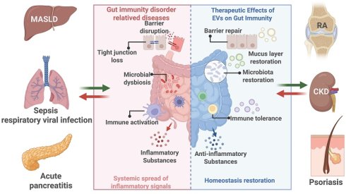
The intestine is the largest immune organ and plays a critical role in regulating local and systemic tissue homeostasis, while disruption of gut immunity can drive an array of diverse diseases both within and beyond the gut. However, current clinical therapeutics (mainly anti-inflammatory drugs) display limited efficacy in restoring gut immune homeostasis and often cause severe side effects. Therefore, new therapeutic strategies are needed to regulate gut immunity for disease treatment. In recent years, extracellular vesicle (EV)-based therapies have emerged as promising nanomedicines for treating gut immune disorder-related diseases by mediating multilayered signaling networks composed of immune cells, resident cells, and the gut microbiota. EVs from diverse sources (e.g., mammalian cells, food, plants and microbiota) may regulate gut immunity by modulating the phenotypes of diverse immune cells (e.g., macrophages, dendritic cells, and T cells), gut barrier integrity, and host–microbiota communication, with therapeutic effects reported mainly in preclinical models of gut immune disorders, such as inflammatory bowel disease, metabolic diseases, graft-versus-host disease, allergies, and colorectal cancer. EVs can be further engineered via cargo loading or surface modification to enhance their targeting ability and specific therapeutic efficacy. Overall, preclinical findings support further investigations of EV-based gut immunotherapy, but the clinical efficacy and long-term safety remain to be established.
Keywords: extracellular vesicles, gut immunity, nanomedicine, inflammation, host–microbe interaction, drug delivery
Introduction
The intestine is the largest immune organ in the human body and is composed of diverse types of immune cells, such as T cells, B cells, dendritic cells (DCs), and innate lymphoid cells, which play critical roles in regulating local and systemic tissue homeostasis via dynamic interactions with gut-resident cells, such as intestinal stem cells and epithelial cells (enterocytes, Paneth cells, goblet cells, tuft cells, etc.), as well as the gut microbiota [1,2]. Notably, disruption of intestinal immune homeostasis can drive an array of local and systemic diseases, such as inflammatory bowel disease, colorectal cancer (CRC), intestinal graft-versus-host disease (GvHD), chronic metabolic diseases, inflammatory disorders, and autoimmunity, via complex mechanisms, including microbiota-driven products (e.g., metabolites and toxic substrates) and immune cell-derived cytokines or the direct trafficking of immune cells [3]. However, current clinical treatments, such as corticosteroids and biologics, mainly relieve clinical symptoms by exerting overall immunosuppressive effects. Although these drugs have therapeutic effects in some patients, they still face some major issues, such as nonresponse, relapse, and severe off-target/side effects [3], which might be due to their limited efficacy in restoring the homeostasis of gut immunity and host–microbiota crosstalk. Therefore, novel biotherapeutics with high biosafety that fine-tune gut immunity for disease treatment are desired.
In recent years, extracellular vesicle (EV) therapeutics have been investigated for the treatment of gut immune disorders, with evidence derived mainly from preclinical studies. EVs are natural nanovesicles that are released by almost all types of cells and can exert therapeutic effects by transferring endogenous or exogenous payloads (e.g., RNAs, proteins, lipids, and metabolites) between cells [4]. Accumulating evidence has indicated that EVs can regulate crosstalk among gut epithelial, immune and microbial compartments [5,6]. For example, orally administered plant-derived vesicle-like nanoparticles and EVs derived from milk or probiotics can be taken up by innate immune cells (e.g., macrophages and DCs) in the gastrointestinal tract, and they can deliver bioactive cargoes to regulate gut immune responses [6-8], such as by inhibiting proinflammatory NF-κB and NLRP3 pathways in macrophages, reshaping DC and T-cell responses and reinforcing IgA- or Treg-associated tolerance [4]. Moreover, these EVs can be engineered via multiple strategies, such as cargo loading and surface modification, to further enhance their targeting ability and specific therapeutic efficacy [9-11]. For example, intestinal-homing ligand-modified EVs can improve their ability to target inflamed intestinal sites, whereas anti-inflammatory drug- or miRNA-loaded EVs can enhance both inflammation resolution and epithelial barrier repair [5,12].
While there are some previous reviews related to EV-mediated gut homeostasis, plant-derived nanoparticles, microbiota–host communication or engineered EVs and their applications [5-8], a comprehensive and critical evaluation on the roles of EVs in regulating gut immunity and associated diseases is still lacking. More importantly, it is necessary to focus on how disease-specific immune requirements guide the selection and engineering of different EV platforms. For instance, restoring mucosal tolerance and enhancing antitumor immunity impose different requirements on EV cargo, recipient cells and delivery. Connecting these requirements to source selection and engineering provides a basis for evaluating which platforms are appropriate for a given disease and where further validation is needed.
In this review, we examine EV-based therapies through the relationships among disease-specific immune requirements, source selection and engineering design. In intestinal and extraintestinal disorders, we compare mammalian EVs, plant-derived vesicle-like nanoparticles and microbial EVs in terms of their immune targets, mechanistic evidence and delivery constraints. We distinguish experimentally supported cargo functions from proposed effects and examine how the limitations of native preparations motivate cargo modification, targeting and formulation strategies. By connecting disease-specific comparisons with engineering approaches, this review identifies the biological rationale for platform selection, the problems that engineering addresses and the evidence still required for clinical translation.
1. Gut immunity as a critical regulator of tissue homeostasis
1.1 Composition and function of the gut immune system
Intestinal immune homeostasis is maintained by coordinated epithelial, immune, microbial and metabolic circuits. This balance is tightly regulated by dynamic communication among epithelial cells, innate and adaptive immune cells, the gut microbiota, microbial metabolites and microbiota-derived EVs [1] (Figure 1).
Gut immune homeostasis and dysregulation. In the healthy intestine, the epithelial barrier, mucus layer, commensal microbiota, antimicrobial peptides, and SIgA jointly maintain mucosal defense and cooperate with immune cells such as DCs, macrophages, Tregs, ILC3s, IELs, and IgA⁺ plasma cells to maintain intestinal immune tolerance and intestinal barrier homeostasis. In the dysregulated intestine, the intestinal epithelial barrier is damaged, and the intestinal mucus layer is reduced, which is accompanied by a disrupted microbial community, leading to abnormal antigen exposure and abnormal activation of immune cells, resulting in an imbalance of the Treg–Th17 axis, massive recruitment of neutrophils, and the release of proinflammatory cytokines, ultimately aggravating intestinal and systemic inflammation. Abbreviations: AMPs, antimicrobial peptides; CD, cluster of differentiation; DC, dendritic cell; Foxp3, forkhead box P3; GM-CSF, granulocyte–macrophage colony-stimulating factor; IEL, intraepithelial lymphocyte; IFN-γ, interferon-γ; IgA, immunoglobulin A; IL, interleukin; ILC1/2/3, group 1/2/3 innate lymphoid cells; M cells, microfold cells; M1/M2, classically/alternatively activated macrophages; MHC II, major histocompatibility complex class II; NET, neutrophil extracellular trap; ROS, reactive oxygen species; SIgA, secretory immunoglobulin A; TCR, T-cell receptor; Tfh, T follicular helper cell; TGF-β, transforming growth factor-β; Th1/Th17, T helper 1/17 cells; TNF-α, tumor necrosis factor-α; Treg, regulatory T cell.
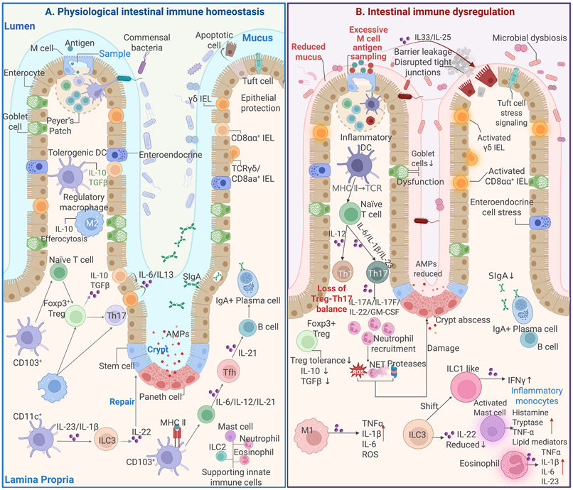
Goblet cell-derived mucin 2 (MUC2) forms a mucus layer that physically separates luminal microorganisms from the epithelium, whereas tight junction proteins, including zonula occludens-1 (ZO-1), occludin and claudins, regulate paracellular permeability [1]. This structural barrier is further reinforced by Paneth cell-derived antimicrobial peptides and secretory IgA, which constrain microbial adherence and overgrowth without inducing overt inflammation. Mucus barrier dysfunction can occur early in colitis, increasing microbial access to the epithelial surface [13,14]. Disruption of any of these barrier components allows microbial ligands, such as lipopolysaccharide (LPS), flagellin, peptidoglycan and bacterial DNA, to reach immune sensors in the epithelium and lamina propria. Their recognition by Toll-like receptors (TLRs), nucleotide-binding oligomerization domain (NOD)-like receptors and inflammasome pathways activates NF-κB/MAPK signaling and promotes the production of IL-1β, IL-6, TNF-α and IL-23, thereby converting barrier dysfunction into self-amplifying mucosal inflammation [2].
Under homeostatic conditions, intestinal macrophages clear invading microbes and apoptotic cells while maintaining an IL-10-producing, hyporesponsive phenotype toward commensal-derived signals [15]. Under inflammatory conditions, however, macrophages activate the TLR4/NF-κB and NLRP3/caspase-1 pathways and produce TNF-α, IL-1β and IL-6. DCs integrate luminal antigen sampling through M cells, goblet cell-associated antigen passages and transepithelial dendrite extension [16]. CD103+ DCs promote retinoic acid- and TGF-β-dependent Foxp3+ Treg differentiation, whereas proinflammatory DCs produce IL-6, IL-12 and IL-23 to drive Th1/Th17 responses [2]. ILC3s provide another supportive epithelial axis through the secretion of IL-22, which activates epithelial STAT3 signaling and promotes antimicrobial peptide production, mucus defense and tissue repair [17].
Adaptive immunity stabilizes long-term mucosal tolerance. The Treg/Th17 balance determines whether antigen exposure leads to tolerance or inflammation. TGF-β, retinoic acid and short-chain fatty acids promote Foxp3+ Treg differentiation and IL-10-dependent tolerance, whereas cytokines such as IL-6, IL-1β and IL-23 promote Th17 responses that support antimicrobial defense but can also drive tissue injury when they are excessive [2]. In addition to soluble metabolites such as short-chain fatty acids, bile acids and tryptophan metabolites, microbiota-derived EVs mediate host–microbe communication. Certain bacterial outer membrane vesicles (OMVs) can cross the intestinal epithelium and deliver microbial cargo to host cells [18]. OMV-associated peptidoglycan can activate epithelial NOD1 signaling [19]. OMVs containing polysaccharide A from Bacteroides fragilis promote regulatory immune responses through TLR2 signaling in DCs [20].
1.2 Gut immune disorder-related diseases
Although the manifestations of intestinal immune dysregulation differ among diseases, many conditions converge on defective epithelial–immune crosstalk. This disruption compromises barrier integrity, impairs immune surveillance and facilitates sustained inflammatory responses (Figure 1 and Table S1).
1.2.1 Intestinal diseases
Intestinal immune dysregulation initially presents as a mucosal disease. In inflammatory bowel disease (IBD), epithelial barrier injury facilitates the entry of microbial ligands (e.g., LPS, flagellin, peptidoglycan and bacterial DNA) into the intestinal lamina propria, resulting in the aberrant activation of intestinal innate immune cells. In epithelial cells, CX3CR1+ macrophages and DCs, microbial ligands engage TLR4, TLR5 and NOD2 while also activating inflammasome pathways. These events drive NF-κB/mitogen-activated protein kinase (MAPK) signaling and induce the production of TNF-α, IL-1β, IL-6 and IL-23 [15,21]. Following the activation of upstream inflammatory signaling, IL-23 supports the persistence of Th17 cells and ILC3 mediated IL-17A/IL-22 signaling, whereas IL-12 drives Th1-dependent IFN-γ production [2,22]. In parallel, excess TNF-α and IL-1β weaken epithelial barrier integrity through myosin light chain kinase (MLCK) activation, epithelial apoptosis and neutrophil recruitment, resulting in a self-amplifying cycle of barrier dysfunction and inflammation [15]. Moreover, immune tolerance is difficult to maintain when IL-10 signaling is disrupted, Foxp3+ Treg function is altered, and CD103+ DC regulatory activity is reduced [23].
CRC progression is shaped by both tumor-promoting inflammation and impaired antitumor immunity. In a mouse model of colorectal tumorigenesis, epithelial barrier defects allowed microbial products to activate tumor-associated myeloid cells and promote IL-23/IL-17-dependent tumor growth [24,25]. In mouse models of metastatic CRC, increased TGF-β signaling in the tumor microenvironment promoted T-cell exclusion and suppressed antitumor T-cell responses [26].
Intestinal GvHD represents another local intestinal disease driven by an immune imbalance, but its initiating event is alloimmune injury rather than chronic dysbiosis or epithelial transformation. Donor CD4+ and CD8+ T cells recognize host alloantigens and produce IFN-γ, TNF-α and GM-CSF, whereas inflammatory macrophages amplify the production of IL-1β, IL-6 and TNF-α [27]. These signals induce crypt epithelial apoptosis, disrupt Lgr5+ intestinal stem cell niches and damage Paneth cells, reducing the expression of antimicrobial peptides such as α-defensins and Reg3γ [28-30]. Barrier leakage and microbial translocation then further reinforce donor T-cell activation, whereas delayed Foxp3+ Treg recovery limits immune tolerance [31].
Intestinal immune dysregulation is also implicated in subsets of patients with irritable bowel syndrome (IBS). In diarrhea-predominant IBS, human biopsy analyses and complementary mouse experiments have revealed that mast-cell activation and LPS–TLR4 signaling are involved in epithelial barrier dysfunction [32]. Microbiome alterations differ across IBS subtypes, indicating that these disturbances are not uniform [33]. The term “preinflammatory bowel disease” (pre-IBD) has been used in selected studies to describe IBS-like presentations accompanied by low-grade intestinal inflammation. However, this research-defined phenotype should not be equated with IBS generally or interpreted as evidence of inevitable progression to IBD [34].
1.2.2 Extraintestinal organ diseases
Gut immune dysregulation also drives extraintestinal diseases, such as chronic liver and kidney diseases, cardiovascular diseases and pulmonary disorders, through direct (gut immune cell trafficking) or indirect (paracrine) effects.
1.2.2.1 Gut–kidney axis
The gut–kidney axis is also involved; as renal clearance decreases, urea accumulates in the intestinal lumen and is hydrolyzed by bacterial urease into ammonia and ammonium hydroxide, which disrupt ZO-1-, occludin- and claudin-dependent epithelial integrity [35]. Barrier leakage allows LPS, peptidoglycan, bacterial DNA and microbial vesicles to activate the TLR4, NOD and NLRP3 pathways in intestinal macrophages, DCs and circulating monocytes, promoting the production of IL-1β, IL-6, TNF-α and IL-23 [36]. Thus, gut-directed therapy for distal organ diseases should not be limited to reducing local intestinal inflammation [36]. More disease-relevant goals include restoring ZO-1/occludin-dependent barrier integrity, suppressing the activity of the urease- and indole/p-cresol-producing microbial pathways, reducing circulating indoxyl sulfate and p-cresyl sulfate levels, and interrupting TLR4/NF-κB/NLRP3-driven innate immune activation that links intestinal leakage to renal and systemic injury [36].
1.2.2.2 Gut–liver axis
The gut‒liver axis is shaped by the portal delivery of microbial and metabolic signals [37]. LPS activates TLR4 on Kupffer cells and hepatic stellate cells, inducing NF-κB-dependent TNF-α and IL-1β production and promoting fibrogenic TGF-β/alpha-smooth muscle actin (α-SMA)/collagen I signaling [38,39]. Thus, in individuals with metabolic disease and metabolic dysfunction-associated steatotic liver disease (MASLD), the intestine acts as an upstream inflammatory and metabolic checkpoint rather than a passive target organ.
1.2.2.3 Gut–heart axis
The gut microbial metabolism of dietary choline and L-carnitine generates trimethylamine, which is subsequently oxidized in the liver to trimethylamine N-oxide (TMAO) [40]. Increased TMAO signaling can promote macrophage foam-cell formation, platelet hyperreactivity and thrombotic risk, thereby linking intestinal microbial metabolism to atherosclerotic cardiovascular disease [41]. Emerging evidence further suggests that gut-derived bacterial EVs can cross a compromised intestinal barrier and amplify systemic and myocardial inflammation, although direct evidence supporting therapeutic EV-mediated regulation of the gut–heart axis remains limited [42-44].
1.2.2.4 Gut–lung axis
Intestinal dysbiosis and barrier dysfunction during respiratory viral infection can increase systemic exposure to microbial products and contribute to pulmonary immune activation. In severe COVID-19, circulating markers of disrupted intestinal barrier integrity and microbial translocation are associated with systemic inflammation [45,46]. EV-mediated gut–lung communication is also beginning to emerge: oral or gut commensal-derived bacterial EVs have been reported to suppress airway or pulmonary inflammation in animal models, although the contribution of intestinal immune-cell reprogramming has not yet been fully established [47].
1.2.3 Systemic immune disorders
Systemic immune disorders can be accompanied by an imbalance in intestinal immune homeostasis. For example, in food allergic reactions, impaired oral tolerance causes the body to develop abnormal type 2 immune responses to food antigens, which are characterized by the activation of Th2 cells and group 2 innate lymphoid cells (ILC2s), increased IgE production, mast cell degranulation, and allergic inflammatory responses [48,49]. In addition, in autoimmune diseases such as rheumatoid arthritis, gut microbiota dysbiosis and increased intestinal barrier permeability lead to excessive antigen uptake, thereby altering the function of DCs in the intestinal mucosa and changing the balance between Th17 and Treg cells [50]. Similarly, in individuals with psoriasis, changes in intestinal immune homeostasis increase the activity of the IL-23/IL-17 inflammatory axis and participate in the regulation of inflammatory changes in the skin [51,52].
In individuals with systemic inflammatory conditions, the injured intestine is an important amplification pathway for inflammatory dissemination [53]. In addition, EVs released by immune cells, endothelial cells, or injured tissues may carry proinflammatory, procoagulant, or immunosuppressive signals, further exacerbating systemic immune homeostasis imbalance [9,11]. In patients with severe acute pancreatitis, pancreas-derived inflammatory mediators and circulating EVs can act on intestinal immune cells and intestinal epithelial cells, promoting macrophage activation, enhancing NLRP3 inflammasome signaling, and inducing intestinal epithelial barrier injury [54].
1.3 Clinical treatments for regulating gut immunity
Biologics that target defined immune circuits have been used to treat IBD in the clinic [55-57]. Anti-TNF agents neutralize soluble and membrane-bound TNF-α produced mainly by macrophages, monocytes and activated T cells, thereby reducing tumor necrosis factor receptor 1/2 (TNFR1/TNFR2)-mediated NF-κB and MAPK activation in epithelial, endothelial and immune cells [3]. Vedolizumab is a gut-selective anti-α4β7 integrin antibody that blocks α4β7 integrin and mucosal addressin cell adhesion molecule-1 (MAdCAM-1) interactions and reduces gut-homing lymphocyte trafficking into the intestinal mucosa [58]. However, the clinical outcomes of these immunosuppressive drugs are restricted by several limitations, such as primary nonresponse, loss of response, and infection risk [59-61].
Recently, microbiota-based therapies, which act via gut ecological and metabolic reconstruction rather than direct immunosuppression, have received increasing attention from researchers and clinicians [62,63]. However, the efficacy of microbiota-based therapies depends on donor strain engraftment and the recipient’s baseline microbiota, mucosal inflammation and diet [64]. Table S2 compares current therapies with EV-based approaches in terms of therapeutic targets, mechanisms, benefits and limitations. EVs offer opportunities for therapeutic cargo delivery, as illustrated by oral delivery of IL-10 to inflammatory macrophages in murine colitis [65]. However, comparative clinical benefits over established treatments remain unproven.
2. EV biology
2.1 Major EV biogenesis pathways
EVs are a group of heterogeneous lipid bilayer nanovesicles released by almost all types of mammalian cells and carry diverse types of bioactive cargos, such as proteins, lipids, mRNAs and noncoding RNAs [9,66]. Following the Minimal Information for Studies of Extracellular Vesicles 2023 (MISEV2023) recommendations, we use EVs with source-based descriptors for preparations of unverified biogenesis and reserve exosomes and ectosomes for vesicles of established endosomal and plasma membrane origin, respectively [66]. Several major EV biogenesis routes, such as exosome (~30–200 nm) formation through the endosomal pathway, ectosome (MV, ~200–1000 nm) shedding from the plasma membrane, and apoptotic body (~800–5000 nm) formation, have been reported (Figure 2).
EV biogenesis pathway, production, and quality control. EVs arise through distinct biogenesis pathways, giving rise to exosomes, ectosomes and apoptotic bodies. Exosomes originate from the intracellular endosomal pathway, ectosomes form through outward budding of the plasma membrane, and apoptotic bodies are generated during apoptosis and fragmentation. The plant panel illustrates PDNV preparation by tissue homogenization and a schematic vesicular component. The recovered preparation may also contain intracellular vesicles, reassembled membrane fragments and nonvesicular material. The schematic does not establish an extracellular origin or a universal marker profile. EVs from different sources can be isolated and purified using ultracentrifugation, size exclusion chromatography and density gradient centrifugation. In addition, tangential flow filtration is suitable for larger-scale EV preparation. Standardized quality control evaluates purity, integrity and stability, safety, functional potency, and batch consistency. Abbreviations: EVs, extracellular vesicles; NTA, nanoparticle tracking analysis; PDNVs, plant-derived vesicle-like nanoparticles; RNA, ribonucleic acid; SEC, size exclusion chromatography; TEM, transmission electron microscopy.
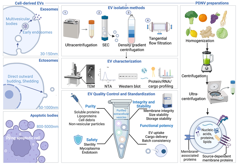
Exosomes originate from the endosomal system. After endocytosis, early endosomes mature into late endosomes and multivesicular bodies (MVBs), where intraluminal vesicles (ILVs) are generated by the inward budding of the endosomal membrane. ILV formation involves both endosomal sorting complex required for transport (ESCRT)-dependent and ESCRT-independent mechanisms [67]. In the canonical ESCRT-dependent pathway, ESCRT 0 recognizes and clusters ubiquitinated cargo on endosomal membranes, ESCRT I and ESCRT II facilitate cargo sorting and membrane deformation, and ESCRT III drives membrane neck constriction and scission, followed by VPS4-mediated disassembly and recycling of the ESCRT machinery [67,68]. ESCRT-independent cargo sorting and ILV formation can involve tetraspanin-enriched microdomains and ceramide-mediated membrane curvature. Mature MVBs are either delivered to lysosomes for degradation or transported to the plasma membrane for exosome secretion [67]. Their fusion with the plasma membrane is regulated by Rab GTPases, including Rab27a, Rab27b, Rab11 and Rab35, together with the SNARE-associated membrane fusion machinery [67]. Once released extracellularly, ILVs are referred to as exosomes.
Ectosomes are generated by outward budding and shedding of the plasma membrane [68]. Increased intracellular calcium levels activate scramblases and inhibit flippases, which promote phosphatidylserine exposure and disrupt membrane lipid asymmetry. Together with actomyosin contraction and small GTPase-mediated cytoskeletal remodeling, these changes drive membrane blebbing and EV release [69]. ARF6, RhoA, ROCK, Rac1 and myosin light chain kinase contribute to this process by regulating actin organization, cell contractility and membrane–cytoskeleton detachment [69]. Apoptotic bodies are released from dying cells and are usually larger and more heterogeneous than exosomes or ectosomes [70]. During apoptosis, caspase activation cleaves cytoskeletal and nuclear structural proteins, leading to membrane blebbing, nuclear fragmentation and cellular disassembly [70]. In addition, apoptotic body formation and release require the involvement of processes such as ROCK1 activation, actomyosin contraction and PANNEXIN1-associated changes in membrane permeability, allowing cells to fragment into bodies enclosed by a membrane bilayer. Phagocytes, such as macrophages, recognize and clear vesicles containing nuclear fragments, histones, mitochondria and other cellular components. It is evident that the timely clearance of apoptotic bodies through efferocytosis helps maintain tissue immune homeostasis, whereas delayed clearance of these EVs can lead to inflammation and other problems [71].
Milk-derived EVs and plant-derived vesicle-like nanoparticles (PDNVs) have considerable potential for treating diseases associated with gut immune dysregulation. These preparations can be isolated from various foods or medicinal plants, including milk [72], garlic [73], broccoli [74], grapefruit [75] and Pueraria [76]. Moreover, the lipid-rich membranes of these food- or plant-derived vesicles help these vesicles withstand gastric acid, bile acids and digestive enzymes, making them suitable oral delivery carriers for managing diseases associated with gut immune dysregulation. Compared with mammalian EVs, PDNVs offer more abundant raw materials, lower production costs and better compatibility with food-grade manufacturing. However, their biogenesis pathways and marker systems remain elusive compared with those of mammalian EVs. PDNVs are vesicle-like preparations obtained from processed plant tissues when natural EVs released from plants have yet to be well established [77]. However, mechanical homogenization can mix naturally released EVs with other types of intracellular vesicles and disrupt membranes that may reassemble them into vesicles and nonvesicular components [77,78]. Therefore, the particle size and vesicular morphology of PDNVs cannot be assumed to reflect structures naturally released into the extracellular space [66,77]. Moreover, PDNV components, such as lipids, proteins, RNAs and phytochemicals, detected in these preparations should be distinguished from EV components whose contribution to biological activity has been functionally demonstrated [66].
2.2 EV production and characterization
Differential ultracentrifugation and density gradients are used to enrich EVs by sedimentation and buoyant density, whereas size exclusion chromatography (SEC) separates particles by size [66]. In platelet-free supernatants from human platelet concentrates, pooling SEC fractions 9-12 increased CD61-positive vesicle recovery from 14 ± 9% in fraction 9 alone to 43 ± 23% but reduced vesicle-to-protein enrichment relative to the starting material from 562 ± 337-fold to 70 ± 19-fold [79]. In rat plasma, the SEC and ultracentrifugation preparations induced similar endothelial cell migration at matched particle doses, although the CD81 signal in the SEC preparations was approximately one-eleventh that in the ultracentrifugation preparations. SEC preparations also contained more APOB-positive lipoproteins, suggesting that coisolated components could contribute to the observed activity [80]. These findings highlight the need to standardize fraction collection and dose normalization and to assess nonvesicular contributions when comparing biological activity.
When a hollow-fiber bioreactor is used, the MSC-EV yield increases to approximately 1.4 × 10⁹ particles per 10⁵ cells, which is greater than the 4.7 × 10⁸ yield reported in flask cultures [81]. Additionally, compared with conventional 2D culture, microcarrier-based 3D culture increased the yield of MSC-EVs approximately 20-fold when both preparations were isolated by ultracentrifugation. Replacing ultracentrifugation with tangential flow filtration (TFF) for 3D cultures further increased recovery sevenfold [82]. Functionally, siRNA-loaded EVs from 3D culture and TFF may have enhanced bioactive properties, and they achieved half-maximal Huntingtin silencing in primary neurons at ~13 nM siRNA, compared with ~63 nM for EVs from 2D culture and ultracentrifugation [82]. However, proteomic analysis revealed enrichment of secreted proteins in TFF preparations, indicating persistent coisolation despite improved recovery and delivery activity [82]. Scale-up therefore requires functional testing alongside the assessment of coisolated proteins.
The standard framework for EV characterization has been established in the MISEV guidelines [66]. MISEV2023 guidelines recommend that mammalian cell-derived EVs should be characterized by complementary approaches, including particle size and concentration analyses, such as nanoparticle tracking analysis (NTA), tunable resistive pulse sensing (TRPS) or high-resolution flow cytometry; morphological assessments by transmission electron microscopy (TEM), cryo-electron microscopy (cryo-EM) or atomic force microscopy (AFM) to confirm lipid bilayer-enclosed vesicular structures; and the expression of EV markers, including transmembrane or membrane-associated markers such as CD9, CD63 and CD81; and cytosolic EV-associated proteins such as TSG101, ALIX, HSP70 and syntenin [66]. Negative EV markers, such as calnexin, GM130, ApoA1/ApoB or albumin, should also be detected to evaluate contamination from intracellular organelles, lipoproteins or soluble proteins (Figure 2). The particle/protein ratio alone cannot be used to establish purity. Batch comparisons should combine physical characterization with a stated dose basis and functional assays, such as epithelial barrier recovery or macrophage cytokine modulation. Matched nonvesicular fractions help assess whether coisolates contribute to the observed activity [66].
Furthermore, quality control should address source-specific contaminants. For mammalian cell-derived EVs, identically processed unconditioned medium provides a control for medium-derived vesicles and proteins [66]. In bovine milk, the addition of SEC after ultracentrifugation reduced the recovered total protein concentration from 125 to 10.6 μg per mL of starting milk, whereas the measured particle recovery decreased from 1.47 × 10¹² to 2.19 × 10¹¹ particles per mL. Proteomic analysis also revealed depletion of caseins and whey proteins [83]. Milk EV purification should therefore monitor contaminant removal alongside particle recovery. In Arabidopsis, compared with PDNVs isolated from disrupted leaves, apoplastic EVs exhibit approximately threefold higher uptake by cancer cells [84]. The isolation of plant EVs from apoplastic wash fluid should include assessment of cell viability in the source tissue and of intracellular contamination in the collected fluid [78,85]. In Arabidopsis, differential centrifugation has been combined with density gradients or immunoaffinity capture to purify EV subclasses [86]. Reproducible comparisons require defined tissue sources, extraction conditions and collected fractions [78,86]. Purity assessment should compare EV marker enrichment and depletion of source-relevant non-EV components against the starting material [66]. RuBisCO-containing nonvesicular complexes are relevant potential coisolates in leaf preparations [78]. Such negative purity controls require validation for each source and cannot be inferred from particle morphology alone [66,78].
With respect to meningococcal OMVs, detergent extraction depleted the lipoprotein fHbp and reduced both TLR4 and TLR2 activation, whereas lpxL1 deletion reduced TLR4 activation while retaining TLR2 signaling [87]. Thus, OMV quality control should assess inflammatory activity and the retention of immunologically active components, along with testing for residual viable bacteria. Due to the strong pro-inflammatory activity of OMVs, it is critical to distinguish EV-associated components from nonvesicular material. Detection of LPS alone cannot make this distinction, because it may be present in nonvesicular micelles and protein complexes [66]. Protein markers likewise require careful interpretation. GroEL and flagellin cannot serve as universal negative markers, as both have been reported as enriched or depleted in bacterial EV preparations [88]. Accordingly, OMV-specific pro-inflammatory activity should be established using source-validated purity controls and functional comparisons with matched nonvesicular fractions [66,88].
2.3 Major EV sources for regulating gut immunity
EVs that regulate gut immunity can be classified according to their biological origin because the source strongly affects the cargo composition, delivery route, immune target and translational feasibility (Table S3).
2.3.1 Mammalian cell-derived EVs
Mammalian cell-derived EVs used for gut immune regulation are obtained mainly from mesenchymal stem cells (MSCs), intestinal epithelial cells and immune cells [4]. Their biological functions are largely determined by the identity and activation state of the parental cells. MSC-derived EVs (MSC-EVs) can regulate intestinal immunity through specific bioactive cargoes. For example, miR-216a-5p delivered by EVs from hypoxia-preconditioned adipose-derived stromal cells targets HMGB1 and suppresses TLR4/NF-κB signaling, promoting macrophage M2-like polarization in experimental colitis [89,90]. In an engineered mammalian EV platform, the delivery of IL-10 to inflammatory macrophages reduced reactive oxygen species production and proinflammatory cytokine expression [65]. Intestinal epithelial cell-derived EVs (IEC-EVs) are closely linked to mucosal communication, such as epithelial–immune crosstalk and antigen handling, because they carry epithelial antigens, major histocompatibility complex (MHC)-related molecules, heat shock proteins, trefoil factors and epithelial miRNAs [91].
The functions of immune cell-derived EVs are more context dependent [6]. Although direct evidence from tissue-resident intestinal macrophage-derived EVs remains limited, studies of colitis and intestinal injury models have suggested that macrophage-derived EVs can transfer polarization-associated miRNAs, long noncoding RNAs (lncRNAs) and WNT-related signals to epithelial and immune cells [15,92,93]. Treg cell-derived EVs may suppress local inflammatory responses by transferring inhibitory molecules or regulatory signals, including cytotoxic T lymphocyte-associated protein 4 (CTLA-4), molecules involved in CD39/CD73-related adenosine pathways, TGF-β/IL-10-associated signals and regulatory miRNAs [94,95]. DC-derived EVs can transmit antigen-presenting signals through MHC–peptide complexes, although their functional effects depend on the state of the parental DC [4,96]. EVs from mature DCs carrying CD80 and CD86 may support T-cell activation, whereas tolerogenic dendritic cell-derived EVs (DC-EVs) enriched in PD-L1 or Fas ligand (FasL) may favor immune restraint [97,98]. Thus, the therapeutic use of these EVs requires disease-matched control of parental cell identity, activation state, cargo composition and functional potency [9,67].
2.3.2 Food- or plant-derived vesicle-like nanoparticles
Milk-derived EVs and plant-derived vesicle-like nanoparticles have been investigated for intestinal therapy because their biological properties depend on their source [7,99,100]. For example, ginger-derived nanoparticles have been investigated as orally stable vesicle-like carriers for intestinal inflammation [101]. In ginger-derived vesicle-like nanoparticles, phosphatidic acid contributes to uptake by Lactobacillus rhamnosus GG, whereas mdo-miR7267-3p targets bacterial ycnE, increasing indole-3-carboxaldehyde production and promoting AhR-dependent IL-22 responses and intestinal protection [102]. In this mechanism, the metabolite is a downstream microbial product rather than a directly delivered EV cargo. Evidence for intravesicular RNA delivery also requires more than RNA detection. In apoplastic Arabidopsis preparations, combined protease and RNase analyses revealed that much of the extracellular RNA was associated with proteins outside EVs [103]. To standardize RNA localization in plant-derived preparations, future studies should compare untreated aliquots with RNase alone and sequential proteinase K–RNase treatment, with and without detergent-mediated membrane disruption. RNA resistance to proteinase K–RNase treatment, followed by degradation after membrane disruption, supports membrane encapsulation, provided vesicle integrity and enzyme activity are verified [86,103]. Because nonvesicular ribonucleoprotein or lipid complexes may co-isolate, these assays should be combined with fractionation and nonvesicular controls before attributing activity to vesicular RNA; cargo-specific inhibition or depletion remains necessary to establish its functional contribution [66,78,103]. Grapefruit and citrus-derived nanovesicles have been used as scalable lipid nanoplatforms for the oral delivery of small molecules or nucleic acids for targeted gut therapy [75]. Milk-derived EVs contain miRNAs, proteins, lipids and milk-associated bioactive molecules and are being investigated for oral delivery [104-106]. However, the clinical translation of food- and plant-derived vesicle preparations is still limited by multiple factors, such as source variability, coisolated nonvesicular components, heterogeneous cargo composition, uncertain uptake specificity and insufficient batch-to-batch reproducibility.
2.3.3 Microbe-derived EVs
Microbe-derived EVs, such as outer membrane vesicles (OMVs) from gram-negative bacteria, are vital mediators of host–microbiota communication [6,8]. They are released by commensals, probiotics and pathobionts and contain diverse cargoes, such as LPS, peptidoglycan, lipoproteins, outer membrane proteins, adhesins, bacterial DNA/RNA and metabolites [8]. The biological effects of microbial EVs are highly dependent on the source and strain [6,8]. EVs from commensal or probiotic bacteria may carry tolerogenic or barrier-supportive signals, whereas EVs from pathobionts may deliver virulence-associated proteins, inflammatory LPS species or adhesins that amplify mucosal inflammation or tumor-promoting immune responses [8,107]. For example, polysaccharide A (PSA) carried by Bacteroides fragilis OMVs is sensed through TLR2 on DCs, promoting IL-10-producing regulatory T (Treg) responses and protection against experimental colitis [20]. Similarly, OMVs from E. coli Nissle 1917 ameliorate DSS-induced colitis in mice [108] and strengthen epithelial barrier function in vitro by regulating tight-junction proteins [109].
Moreover, fungal EVs in the gut system are emerging as potential mediators of fungal–host communication, although direct evidence in intestinal settings remains more limited than that for bacterial or mammalian EVs [110]. Studies of pathogenic fungi, particularly Candida albicans, have indicated that fungal EVs can carry immunologically active cargo, including mannans, β-glucans, chitin-related structures, stress-response proteins, virulence-associated proteins and fungal RNAs [111]. These EV-associated molecules may influence host innate immune responses through pattern recognition pathways, especially TLR4 and Dectin-1 signaling. With respect to EV-associated mRNAs, the proposed immune effects mediated by encoded proteins require evidence of translation in recipient cells [112]. More generally, cargo profiling alone does not establish causality; attribution to individual EV components requires functional perturbation and controls for coisolated nonvesicular material [66].
3. EV-based therapies for regulating gut immunity
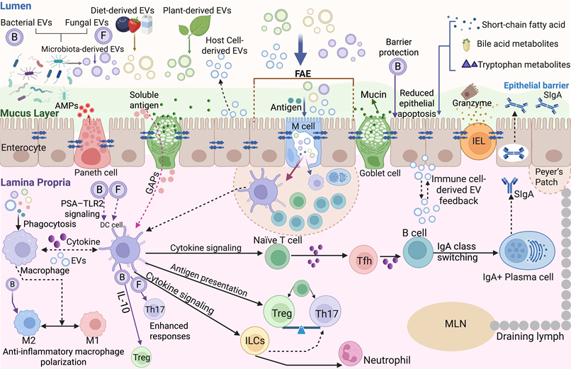
Research on the biological properties of native EVs provides a basis for selecting appropriate EV sources for treating specific diseases. On this basis, EV engineering further demonstrates how approaches such as cargo modification or delivery control can address specific therapeutic limitations (Figure 3 and Table S4).
Regulatory effects of EVs on gut immunity. EVs can interact with the intestinal mucus layer, intestinal epithelial cells, and M cells, regulating intestinal antigen sampling, mucus secretion, antimicrobial peptide release, SIgA production, and barrier function. In the intestinal lamina propria, EVs can regulate macrophages, DCs, T cells, B cells, ILCs, and neutrophils, participating in antigen presentation, cytokine signaling, Treg–Th17 balance, IgA class switching, and the maintenance of mucosal immune homeostasis. B and F denote bacterial and fungal extracellular vesicles (EVs), respectively. Bacterial EVs include outer membrane vesicles (OMVs) released by gram-negative bacteria and membrane vesicles released by gram-positive bacteria. Abbreviations: AMPs, antimicrobial peptides; DC, dendritic cell; EVs, extracellular vesicles; FAE, follicle-associated epithelium; GAPs, goblet cell-associated antigen passages; IEL, intraepithelial lymphocyte; IgA, immunoglobulin A; IL-10, interleukin-10; ILCs, innate lymphoid cells; M cells, microfold cells; M1/M2, classically/alternatively activated macrophages; MLN, mesenteric lymph node; OMVs, outer membrane vesicles; PSA, polysaccharide A; SIgA, secretory immunoglobulin A; Tfh, T follicular helper cell; Th17, T helper 17 cell; TLR2, Toll-like receptor 2; Treg, regulatory T cell.
3.1 Inflammatory bowel disease
Inflammatory bowel disease (IBD) primarily includes Crohn’s disease and ulcerative colitis. Among intestinal diseases, EV-based therapy has been most extensively studied in IBD. Its major pathological processes include epithelial barrier failure, TLR/NOD-driven innate immune activation, inflammatory macrophage polarization, Th17/Treg imbalance and dysbiosis. Therapeutic EVs have been investigated across these interconnected circuits, although most evidence remains limited to DSS-, TNBS- and IL-10-deficient colitis models.
At the epithelial level, MSC-EVs reduce apoptosis, oxidative stress and mucosal injury while restoring the expression of tight junction proteins [113,114]. TSG-6 carried by bone marrow mesenchymal stem cell-derived EVs (BMSC-EVs) inhibited intestinal epithelial pyroptosis through the NLRP3/caspase-1/GSDMD pathway, restored ZO-1 and occludin expression and alleviated DSS-induced colitis. TSG-6 knockdown weakened these effects, supporting a cargo-dependent mechanism [115]. In another study, EV-delivered circHECTD1 bound to CTCF and suppressed METTL3 transcription, thereby reducing the m6A modification of claudin-1 mRNA and restoring claudin-1 expression [116]. Orally administered milk-derived EVs also improved epithelial integrity while suppressing TLR4/NF-κB and NLRP3 signaling and partially restoring the Th17/Treg balance [117]. Aloe-derived nanovesicles also alleviated DSS-induced colitis and increased tight junction protein expression [118]. Oral pretreatment with L. plantarum Q7-derived EVs alleviated DSS-induced colitis and altered the gut microbiota composition [119]. Lactiplantibacillus plantarum EVs reduced epithelial apoptosis through the mPTP/CytC/caspase-9/caspase-3 pathway [120], whereas Roseburia intestinalis EVs inhibited DPP4 through an IPI peptide cargo, increased the level of active GLP-1 and attenuated inflammatory NF-κB/STAT3 signaling [121]. In addition, Akkermansia muciniphila-derived EVs increased MUC2 expression, improved mucus integrity, and reduced intestinal permeability in mice with colitis [122].
Macrophages represent an important recipient population for EV-mediated intestinal immunoregulation. Human BMSC-EVs preferentially accumulate in inflamed colon tissue and are predominantly taken up by F4/80⁺CD11b⁺ macrophages, with macrophage depletion weakening their therapeutic activity [123]. miR-146a-enriched BMSC-EVs suppressed IRAK1/TRAF6/NF-κB signaling and reduced TNF-α, IL-6 and IL-1β levels in TNBS-induced colitis [124]. Synthetic lipid nanoparticles inspired by ginger-derived PDNVs loaded with osa-miR164d promoted reparative macrophage polarization by targeting TAB1 and inhibiting TNF signaling [125]. Similarly, Prunus mume-derived vesicle-like nanoparticles were preferentially internalized by colonic macrophages and delivered miR-159, which disrupted NEK7–NLRP3 binding and inhibited inflammasome activation [126]. Lactobacillus johnsonii EV-associated GAPDH also alleviated colitis in a macrophage-dependent manner by suppressing MAPK–STAT3 signaling [127]. In addition to conventional colitis models, orally administered milk-derived EVs attenuated mucosal inflammation in a murine pre-IBD model by increasing Lactobacillus murinus and reducing lysophosphatidylcholine accumulation, thereby modulating TLR2-dependent macrophage polarization [128]. These findings remain preclinical and do not establish therapeutic efficacy in patients with IBS.
Microbial EVs can additionally reshape adaptive mucosal immunity. EVs from B. longum NSP001 alleviate DSS-induced colitis even after microbiota depletion, reducing macrophage infiltration and the expression of inflammatory cytokines while increasing the levels of IL-10 and TGF-β and shifting the Th17/Treg balance toward Treg responses [129]. L. johnsonii EVs restored taurine-associated immune regulation, reduced Th17 responses, increased the number of Tregs and corrected abnormal pIgR/FcRn expression and immunoglobulin-coated bacterial populations [130]. Source selection in IBD involves overlapping biological functions across different sources. For example, both MSC-EVs and Prunus mume-derived particles act on macrophages, whereas both milk-derived EVs and B. longum-derived EVs affect barrier and adaptive immune responses [117,123,126,129]. Macrophage depletion and cargo perturbation support specific mechanisms in selected studies, whereas reduced cytokine levels alone provide weaker mechanistic evidence. Engineering can combine these biological activities with controlled delivery. For instance, galactose-modified EVs carrying IL-10 use a chitosan/alginate coating for oral protection and macrophage-directed delivery [65] (Sections 4.1 and 4.4–4.6). Differences in models, routes and dose definitions currently limit efficacy comparisons between sources.
3.2 Anticolorectal cancer immunity
Colorectal cancer (CRC) is characterized by the use of EVs to remodel both mucosal and tumor immunity. Approximately 85% of CRCs are microsatellite stable (MSS) and respond poorly to immune checkpoint blockade, owing partly to insufficient antigen presentation, limited cytotoxic T-cell infiltration and suppressive myeloid and stromal networks. EV-based platforms may address these defects by delivering tumor antigens and innate agonists, restoring T-cell costimulation or promoting immunogenic tumor-cell death.
Oral EV administration provides a direct strategy for engaging intestinal immunity. Orally delivered tumor cell-derived microparticles were taken up by ileal epithelial cells, where they activated NOD2–MAPK–NF-κB signaling and induced CCL2 production. Consequently, CD103+CD11c+ DCs were recruited, while epithelial transcytosis transferred EV-associated tumor antigens to these cells for cross-presentation and subsequent antitumor T-cell activation in CT26-bearing mice [131]. A related strategy involved the oral administration of engineered Escherichia coli to produce antigen-bearing OMVs in situ. These OMVs cross the intestinal epithelium, enter the lamina propria and promote DC maturation, thereby suppressing colon tumor growth and generating protective immune memory [132]. Together, these studies show that intestinal epithelial cells can function as an entry point through which orally delivered EVs connect mucosal antigen sampling with systemic antitumor immunity.
Bacterial vesicles can also provide antigen-independent innate stimulation or serve as modular tumor vaccines. Systemic administration of detoxified E. coli OMVs induced IFN-γ-dependent tumor regression and long-term immune memory in CT26 and MC38 models [133]. More selective immunity was achieved using a plug-and-display platform in which the MC38 neoantigen Adpgk was displayed on engineered OMVs. Treatment with these OMVs increased the numbers of tumor-infiltrating DCs, CD4+ and CD8+ T cells and activated neutrophils; reduced the number of Foxp3+ Tregs; and resulted in complete tumor regression in 60% of the mice [134]. OMVs from the intestinal commensal Parabacteroides distasonis increased CXCL10 expression and CD8+ T-cell infiltration in CT26 tumors, and CXCL10 neutralization weakened their antitumor activity [135]. Synthetic bacterial vesicles loaded with STING agonists similarly enhanced DC activation and IFN-β production, increased intratumoral T-cell infiltration and suppressed colon tumor growth [136]. Immunologically cold MSS CRC remains a clinical challenge because T-cell exclusion limits responses to immune checkpoint blockade [26]. In stroma-rich tumors, the dense desmoplastic matrix restricts therapeutic penetration and immune-cell infiltration, while hypoxia-associated CD73–adenosine signaling reinforces metabolic immunosuppression [137]. Engineered EVs may address both barriers: hyaluronidase-expressing OMVs carrying CD73 siRNA combine matrix degradation with suppression of adenosine-mediated immunosuppression [137]. However, the applicability of this approach to cold MSS CRC still requires further validation in immunocompetent orthotopic models with defined stromal and immune features.
Mammalian EVs offer complementary strategies for restoring adaptive immunity. Native CRC-derived EVs containing miR-424 suppressed CD28 expression in tumor-infiltrating T cells and CD80/CD86 expression in DCs, thereby weakening costimulation and promoting resistance to immune checkpoint blockade. Removing miR-424 or its murine homolog, miR-322, converted these vesicles into immunogenic antigen carriers that restored CD8+ T-cell responses and improved checkpoint blockade efficacy in CRC models [138]. M1 macrophage-derived EVs loaded with zinc phthalocyanine provide another approach for inducing photodynamic immunogenic cell death, release of damage-associated molecular pattern (DAMP), DC maturation and protective immune memory in MC38 tumor-bearing mice [139]. GEMINI-engineered EVs further combined αCD3 and αEGFR to redirect cytotoxic T cells toward EGFR-positive CRC cells, while surface PD-1 and OX40L promoted checkpoint inhibition and costimulation. Bacterial and tumor-derived platforms require different cargo strategies [140]. OMV antigen display and STING agonist loading into synthetic bacterial vesicles strengthen immune stimulation [134,136], whereas the depletion of miR-424 removes an immunosuppressive component from tumor-derived EVs [138]. The latter provides causal support for cargo removal, rather than assuming that tumor antigen carriage is sufficient for vaccination. These examples connect source selection to antigen display, payload loading or cargo depletion (Section 4.6), but their different models and delivery routes preclude simple efficacy rankings.
3.3 Intestinal graft-versus-host disease
After allogeneic hematopoietic stem cell transplantation, conditioning damages the crypt epithelium and Paneth cells, allowing microbial products and danger signals to activate host antigen-presenting cells. Activated donor CD4+ and CD8+ T cells produce IFN-γ and TNF-α, whereas inflammatory macrophages release TNF-α, IL-1β and IL-6. These responses promote epithelial apoptosis, microbial translocation and alloimmune activation. EV-based therapy for intestinal GvHD should therefore suppress local antigen presentation, T-cell activation or macrophage inflammation without abolishing graft-versus-leukemia (GvL) activity.
Direct evidence that EVs repair the intestinal epithelium in patients with GvHD remains limited. However, systemic MSC-EVs can attenuate immune pathways that drive intestinal injury [141]. Prophylactic administration of hUC-MSC-derived EVs reduced acute GvHD severity and colonic injury in mice [142]. In an allogeneic HSCT mouse model, intravenous administration of 300 µg of BMSC-EVs at transplantation reduced clinical GvHD and small intestinal histological injury. These EVs decreased the expression of CD40, CD80 and CD86; reduced the expression of IL-6 and IL-12p70; and increased the expression of IL-10 in DCs. They also suppressed T-cell proliferation, promoted CD4+CD25+Foxp3+ Treg differentiation and shifted macrophages toward a CD206+ M2-like phenotype. Importantly, CD8+ T cells retained antileukemia cytotoxicity, suggesting that these immunoregulatory effects did not completely eliminate GvL activity [143].
A more intestine-directed strategy uses microbial outer-membrane vesicles for local cargo delivery. In a murine allo-HSCT model, engineered E. coli Nissle 1917 produced OMVs with low endotoxicity that displayed ClyA-fused BigLEN. Following oral administration, OMVs delivered BigLEN to intestinal lamina propria macrophages and inhibited NLRP3 inflammasome activation and GSDMD-associated pyroptosis. This treatment reduced intestinal inflammation and GvHD severity while preserving the control of A20 lymphoma cells [144]. Intravenously administered MSC-EVs modulate DCs, macrophages and T cells [143], whereas orally administered engineered E. coli Nissle produces BigLEN-bearing OMVs to locally inhibit macrophage pyroptosis [144]. Both approaches preserved GvL activity in the tested mouse models, but this does not establish equivalent safety or efficacy. For GvHD, the source and delivery engineering must therefore be assessed against two linked outcomes: intestinal protection and the preservation of antileukemia immunity.
3.4 Metabolic diseases
Obesity, type 2 diabetes and metabolic dysfunction-associated steatohepatitis are accompanied by chronic low-grade inflammation that is partly sustained by intestinal dysbiosis and barrier failure. Increased translocation of LPS, bacterial DNA and microbial vesicles activates inflammatory pathways in intestinal macrophages, adipose-tissue macrophages and Kupffer cells, thereby exacerbating metabolic injury [145,146]. The most direct evidence comes from Akkermansia muciniphila-derived EVs. Oral administration of these EVs to HFD-induced diabetic mice reduced intestinal permeability and body weight gain, improved glucose tolerance and restored the expression of tight junction proteins, including occludin, ZO-1 and claudins [147]. In Caco-2 cells, the induction of occludin expression and resistance to LPS-induced permeability involved AMPK activation, although the requirement for AMPK was not established in vivo [147]. A subsequent study revealed that A. muciniphila EVs reduced adiposity and plasma glucose levels; suppressed inflammatory changes in the colon, adipose tissue and liver; and partially restored HFD-disrupted microbial communities. In the preventive HFD setting, these effects were associated with increased colonic TLR2 and IL-10 expression and reduced TLR4 and TNF-α expression [148]. However, the specific intestinal immune cells that directly respond to these vesicles remain undefined.
Food-derived EVs may act through related interorgan pathways. In mice concurrently fed a NASH-inducing diet, oral administration of bovine colostrum-derived EVs reduced hepatic steatosis, fibrosis and inflammation. These changes were accompanied by increased intestinal Akkermansia abundance; increased Muc2, claudin-1 and occludin expression; reduced intestinal TNF-α and IL-6 levels; and improved barrier integrity, supporting a gut–liver mechanism [72]. However, microbiota depletion and immune cell-specific experiments were not performed; therefore, the contributions of individual microbial or immune pathways remain unresolved. Plant-derived nanoparticles can also modify the vesicles produced by resident bacteria. PDNVs derived from garlic reprogrammed A. muciniphila to release OMVs enriched in Amuc_1100, P9 and phosphatidylcholines. Oral administration of these OMVs increased GLP-1 signaling, inhibited cGAS–STING-associated inflammation in brain microglia and improved insulin resistance in HFD-fed mice. This pathway represents a microbiota–gut–brain mechanism rather than the direct regulation of intestinal immune cells [149].
A. muciniphila EVs and milk EVs support oral strategies for barrier protection [72,147], whereas garlic-derived nanoparticles represent an indirect approach through the modification of bacterial OMV production [149]. These platforms act at different points in host–microbiota communication. Improved permeability and metabolic outcomes do not by themselves establish which intestinal response mediates systemic benefit. Engineering should therefore distinguish delivery to host cells from reprogramming bacterial vesicle production, with active cargo and the contribution of intestinal versus peripheral actions remaining priorities for validation.
3.5 Allergic diseases
Food allergy reflects a failure of oral tolerance to dietary antigens. EV-based therapy should therefore restore antigen-specific immune tolerance rather than impose broad immunosuppression. Under homeostatic conditions, intestinal CD103+ DCs generate retinoic acid- and TGF-β-dependent signals that promote Foxp3+ Treg differentiation in mesenteric lymph nodes. This pathway has been reproduced using engineered DC-derived EVs. EVs displaying IL-2 and OVA peptide–MHC II complexes bound OVA-specific CD4+ T cells, induced intestinal Tregs and suppressed food allergy in an experimental model [150,151]. A complementary construct carrying OVA peptide–MHC II complexes and cleaved caspase-3 selectively induced the apoptosis of OVA-specific Th2 cells, increased intestinal Treg numbers and attenuated allergic responses [151]. These studies support the use of antigen-coupled EVs as a strategy for increasing the number of allergen-specific Tregs or eliminating pathogenic Th2 cells.
Milk-derived EVs provide a distinct oral strategy that acts on epithelial and immune responses. In OVA-induced food allergies, milk EVs reduced hypothermia and serum IgE, IgG1 and MCPT-1 levels; decreased the levels of Th2 cytokines and numbers of Th2/Th17 cells; and limited jejunal injury and mast cell infiltration. Treatment also increased the number of tight junction-related transcripts and partially restored the diversity of microbes, including Firmicutes and Lactobacillus. A comparative study revealed that EVs derived from cow, buffalo and yak milk reduced OVA-specific IgE/IgG1 and MCPT-1 levels, whereas EVs derived from yak milk produced the strongest barrier protection; increased ZO-1, occludin and claudin-1 expression; suppressed IL-4, IL-5 and IL-13 expression; and partially restored Lactobacillus, Ligilactobacillus and Akkermansia abundances [152]. Thus, engineered EVs can directly target allergen-specific T cells, whereas milk-derived EVs act across epithelial, Th2/Th17, mast cell and microbiota-associated pathways. However, these findings remain limited to murine models, and the active milk-derived EV cargoes and causal contributions of changes in the microbiota remain unresolved. Engineered DC-EVs couple antigen recognition to Treg induction or Th2-cell elimination [150], whereas milk-derived EVs improve barrier and immune readouts without an engineered antigen-recognition module [152]. The former offers a defined strategy for antigen-specific intervention; the latter still requires identification of the components responsible for protection. OVA-based studies do not establish efficacy across different food allergens. Accordingly, antigen selection is central to DC-EV engineering, but the mechanism-guided development of milk EVs first requires cargo validation.
3.6 Chronic inflammatory diseases
Chronic inflammatory diseases involving the kidney, joints and skin may be influenced by gut-derived immune and vesicular signals, but the strength and nature of the evidence differ across disease contexts. In these disorders, EVs may act as bidirectional regulators, with tolerogenic host cell-derived EVs restoring immune balance and dysbiotic microbial EVs propagating gut-derived inflammatory signals to distant tissues [153].
3.6.1 Chronic kidney disease (CKD)
In individuals with CKD and diabetic kidney disease (DKD), targeting gut-derived metabolic, barrier and immune disturbances may help slow renal injury, although the potential of EV-based approaches remains underinvestigated. Uremia increases luminal urea levels, and bacterial urease converts urea into ammonia and ammonium hydroxide, which disrupt epithelial tight junctions and increase intestinal permeability [35]. Barrier leakage facilitates systemic exposure to microbial ligands and indole- or p-cresol-derived metabolites, which are converted into indoxyl sulfate and p-cresyl sulfate. Once absorbed, gut-derived uremic toxins and microbial vesicles can convert barrier leakage into renal and vascular inflammation. These uremic toxins activate AhR, ROS generation, and NF-κB and TGF-β/Smad signaling in endothelial cells, renal tubular epithelial cells and macrophages, thereby amplifying vascular inflammation, tubular injury and renal fibrosis [154].
EV-based therapeutic strategies may act either by blocking pathogenic signals or by reshaping gut microbiota metabolism. Plant-derived EV-like nanoparticles have emerged as an additional therapeutic modality for the gut. In DKD, orally administered Salvia miltiorrhiza-derived vesicle-like nanoparticles have been reported to protect against kidney injury through gut microbiota and metabolomic remodeling. This study is particularly relevant because it links oral administration of PDNVs to microbiota-derived metabolic regulation rather than direct kidney targeting [155]. Oral Salvia preparation links renal improvement to changes in the gut microbiota and metabolites, but the causal contribution of these changes remains unresolved [155]. This evidence does not establish superiority over mammalian EVs. Before a gut-targeted engineering strategy is selected, the relevant microbial or host target needs to be defined and distinguished from the possible systemic effects of the administered preparation. In IgA nephropathy mice, orange-derived EVs carrying dexamethasone phosphate targeted intestinal lymphocytes and reduced renal IgA deposition [156].
3.6.2 Rheumatoid arthritis (RA)
Rheumatoid arthritis (RA) is increasingly recognized as an extraintestinal autoimmune disease in which intestinal dysbiosis, barrier dysfunction and mucosal immune activation may sustain joint inflammation. In RA, gut dysbiosis and impaired intestinal barrier function may promote systemic autoimmunity by reshaping mucosal antigen presentation and T-cell polarization. A recent fate mapping study further revealed that gut TH17 cells can acquire a TFH-like program in Peyer’s patches and migrate to the spleen, thereby promoting germinal center B-cell responses and arthritis. A related transcriptional signature was detected in patients with RA [157]. The enrichment of Prevotella-related taxa is associated with increased intestinal permeability, mucosal DC activation and Th17-skewed immunity, which may contribute to anti-citrullinated protein antibody-associated autoimmune responses. Immune cell-derived EVs may restore immune tolerance in inflamed states. Treg-derived EVs represent a more defined tolerance-restoring strategy. These EVs may carry CTLA-4, CD39/CD73, TGF-β, IL-10 and regulatory miRNAs, allowing them to suppress effector T-cell activation and antigen-presenting-cell costimulation without transferring whole cells. In collagen-induced arthritis models, Treg-derived EVs have been reported to reduce the expression of IL-17A, TNF-α and IFN-γ while increasing the expression of IL-10, partly through the miRNA-mediated regulation of Notch signaling [158].
The gut–joint axis can be therapeutically targeted at the microbial, microbe-EV and host-EV levels. PDNVs derived from Pueraria lobata were preferentially taken up by the RA-enriched taxon Ruminococcus gnavus and reduced its production of phenylethylamine. The intestinal accumulation of gma-miR4412 suppressed bacterial phenylalanine decarboxylase expression and attenuated the PEA–BTK–NET axis, thereby limiting the aggravation of arthritis [159]. EV-mediated signaling may also be modulated indirectly. In mice with collagen-induced arthritis (CIA), Guizhi-Shaoyao-Zhimu decoction increased the abundance of Parabacteroides goldsteinii, whose outer membrane vesicles entered joints and reduced NET formation through Cav-1–Nrf2 signaling [160]. At the host-EV level, human umbilical cord mesenchymal stem cell-derived EVs (hUC-MSC-EVs) combined with the ginsenoside Rh2 reduced the severity of arthritis in CIA rats and induced changes in the gut microbial composition and fecal metabolite profiles [161]. Finally, a non-peer-reviewed study revealed that EVs from P. goldsteinii ASF519 activated IL-10-producing myeloid cells through pannexin1/CD39/CD73/A2a signaling and attenuated arthritis in a murine model, identifying a bacterial EV strategy that requires independent validation [162]. Treg-EVs regulate host T-cell signaling through miR-449a/Notch1 [158], whereas Pueraria-derived particles act on bacterial phenylethylamine production [159]. The former supports a host immune mechanism but does not establish an intestinal site of action; the latter directly links a microbial target to arthritis. This distinction is important for engineering: joint-directed delivery and retention of microbiota-active cargo in the intestine involve different therapeutic mechanisms.
3.6.3 Psoriasis
In psoriasis, the OMVs of Parabacteroides goldsteinii illustrate how the administration route affects the use of the same microbial source. Orally administered OMVs reach inflamed skin after intestinal transit, whereas subcutaneous delivery of OMVs in a thermosensitive PF-127 hydrogel results in sustained local release; both of these processes reduce psoriasis-like inflammation in mice [163]. Thus, this study supports both gut–skin delivery and local biomaterial-assisted treatment (Section 4.5). It does not establish that mammalian EVs reproduce the same distribution or that one route is superior. The relative contributions of intestinal immune modulation and direct skin exposure remain important to distinguish.
3.7 Infectious diseases and critical illness
In individuals with sepsis and acute pancreatitis, EVs act as mediators of gut barrier failure, thromboinflammation and immune paralysis. Macrophage-derived EVs enriched in miR-146a can inhibit IRAK1/TRAF6-NF-κB signaling and reduce excessive cytokine release, but persistent transfer of this negative-feedback miRNA may also contribute to late immunosuppression by reducing monocyte responsiveness. Therefore, therapeutic studies target systemic antimicrobial competence, the gut vascular–epithelial barrier and pathological EV cargo. In rats with sepsis induced by cecal ligation and puncture (CLP), MSC-EVs transferred Mfn2, PGC-1α and functional mitochondria to intestinal epithelial cells. These cargos restored mitochondrial fusion and biogenesis and attenuated barrier failure [164]. Pericyte-derived EVs further protected the gut vascular barrier through Angpt1-dependent endothelial repair and pericyte recruitment [165]. In mice with taurocholate-induced acute pancreatitis, hUC-MSC-derived EVs loaded with emodin inhibited inflammasome activation, restored occludin and ZO-1 expression and promoted epithelial regeneration [166].
Conversely, circulating EVs from rats with severe acute pancreatitis transfer miR-155-5p to intestinal epithelial cells, suppress expression and promote NLRP3 inflammasome-mediated pyroptosis and barrier injury [54]. This mechanism explains why the nonselective inhibition of EV release can attenuate intestinal injury while also underscoring the need to preserve protective EV populations. In cell and mouse models, small EV-enriched serum preparations from healthy donors restored the expression of tight junction proteins and reduced pyroptosis through the targeting of annexin A3 (ANXA3) by miR-579-3p, whereas preparations from patients with acute pancreatitis aggravated barrier dysfunction [167]. These studies distinguish administered MSC-derived products from heterogeneous circulating EV populations whose effects depend on donor health and cargo. Emodin-loaded MSC-EVs provide a defined delivery strategy for suppressing intestinal pyroptosis in experimental pancreatitis [166], but serum studies have revealed protective or injurious signaling that should not be generalized to all circulating EVs [167]. Engineering should be evaluated for added benefit over unloaded EVs and free cargo, with infection control assessed separately from reduced inflammation in sepsis.
4. Engineering EVs to increase their therapeutic capacity
Although native EVs can transfer bioactive cargoes, their therapeutic application is limited by manufacturing variability, biological safety concerns and delivery barriers. Limited or variable production yields, heterogeneous compositions and batch-to-batch variability complicate reproducible dosing. Differences in purification and storage can affect product purity, stability and functional potency [66]. Route-dependent pharmacokinetics and biodistribution may restrict target-tissue exposure, while potential immunogenicity requires source- and formulation-specific assessment. Together, these issues complicate product comparability, safety evaluation and regulatory assessment. Orally administered EVs must survive gastric acid, bile salts and digestive enzymes; penetrate the mucus layer; and remain at the inflamed mucosa long enough to be taken up by epithelial cells or immune cells [6]. Systemically administered EVs are rapidly cleared by liver, spleen and lung macrophages, which limits intestinal delivery [5]. Even after the target tissue is reached, efficacy depends on sufficient therapeutic cargo loading, protection and release in the correct recipient cells. Therefore, different disease states may require engineered EVs with specific properties, such as protecting orally delivered immunoregulatory cargo in IBD, controlling stimulatory and suppressive cargo in CRC, and coupling antigen recognition to tolerogenic signals in food allergies. These constraints and disease-specific objectives motivate the targeting, donor-cell modification, oral protection, biomaterial integration and payload‒loading strategies discussed below (Figure 4 and Table S5). Moreover, engineered EVs must be accompanied by standardized manufacturing, validated quality control and source-specific safety assessment.
EV engineering strategies for advancing gut immune regulation. Multiple EV engineering strategies, such as donor-cell programming, exogenous cargo loading, surface modification, and EV–liposome hybridization, have been reported. Platform engineering strategies include layer-by-layer coatings for cargo protection and responsive release, EV–nanomaterial composites for enhanced payload loading and stimulus-responsive release, and bioadhesive hydrogels for mucosal retention and sustained delivery. In situ approaches seek to modulate the gut microbiota and its production of bioactive EVs to influence intestinal immunity. Abbreviations: EVs, extracellular vesicles; GI, gastrointestinal; miRNA, microRNA; mRNA, messenger RNA; siRNA, small interfering RNA.
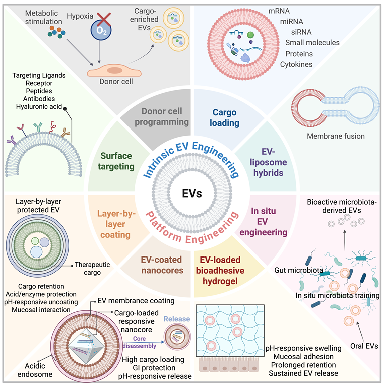
4.1 Target selection for intestinal EV delivery
The surface engineering of EVs should first be guided by disease-enriched receptors and by evidence from receptor-blocking or biodistribution studies. In intestinal inflammation, α4β7–MAdCAM-1 signaling is a clinically established therapeutic target, as α4β7 blockade with vedolizumab is effective in individuals with ulcerative colitis [58]. Anti-MAdCAM-1 antibody-conjugated milk EVs carrying cyanidin-3-O-glucoside increased intestinal accumulation and therapeutic activity in a murine microplastic-induced colitis model [168]. However, MAdCAM-1-oriented EV targeting remains at an early preclinical stage, and increased tissue accumulation alone does not establish receptor-dependent delivery. Similarly, RGD-containing cyclic peptides such as iRGD can bind αvβ3/αvβ5 integrins and support tumor-targeted nanoparticle delivery [169]. However, these findings cannot be directly extrapolated to EV delivery to inflamed intestinal tissue.
Because several trafficking axes remain incompletely validated for intestinal EV delivery, near-term targeting should prioritize receptors enriched on inflamed mucosal immune cells and the injured epithelium. CD44 is upregulated on activated immune cells and injured epithelium, providing a rationale for the use of hyaluronic acid-functionalized EVs or EV-mimetic nanoparticles to improve retention in the inflamed colon [170]. Sugar-ligand modification offers another approach to immune-cell targeting. For example, galactose-modified, IL-10-carrying EVs were combined with macrophage-directed delivery and hydrogel-mediated oral protection in experimental colitis [65].
4.2 Surface engineering for active targeting and mucosal delivery
Postisolation surface engineering can introduce these targeting functions through covalent conjugation or lipid insertion. Click chemistry enables covalent surface functionalization of EVs without modifying donor cells [171], although the membrane integrity and removal of residual reagents require verification. Alternatively, ligand-bearing 1,2-distearoyl-sn-glycero-3-phosphoethanolamine–polyethylene glycol (DSPE–PEG) can be anchored through noncovalent insertion of its lipid tail into the vesicle membrane. In a direct intestinal example, DSPE–PEG–hyaluronic acid insertion into red cabbage-derived EV-like nanoparticles enhanced epithelial and macrophage uptake, with CD44-blocking experiments supporting receptor involvement [170]. Unlike covalent surface conjugation, lipid insertion does not require direct chemical modification of native membrane proteins. Nevertheless, both approaches require assessment of ligand density, surface retention, accessibility and preservation of EV biological activity.
By fusing targeting molecules (e.g. peptides) to EV-associated membrane proteins, such as lysosome-associated membrane protein 2b (Lamp2b), donor-cell engineering provides a complementary strategy. Engineered glycosylation of Lamp2b fusion proteins protected the displayed peptides from proteolytic degradation and improved functional surface display [172]. This differs from the binding of sugar ligands to isolated EVs: glycosylation here stabilizes the displayed protein rather than simply providing a receptor-binding sugar. In contrast to postisolation modification, this approach requires genetically tractable producer cells and control of fusion protein expression and processing. These findings provide a technical precedent but do not establish efficacy in intestinal disease.
For oral intestinal delivery, surface engineering should also balance mucus transport with receptor accessibility. Increased mucosal retention should not be equated with enhanced mucus penetration, and greater tissue fluorescence alone cannot establish active targeting. Evaluation should therefore distinguish mucus transport, receptor-dependent uptake and functional cargo delivery while accounting for ligand stability during gastrointestinal transit.
4.3 Preconditioning of donor cells
By changing the cargo loaded during EV biogenesis, donor-cell preconditioning is used to improve EV potency. For intestinal immune diseases, the key issue is not only whether preconditioning increases therapeutic activity but also whether the induced changes in the cargo match the intended barrier, macrophage or T-cell mechanism. One of the major strategies is to use environmental stressors, such as hypoxia, to redirect EV cargo toward epithelial protection and immune repair [113]. Donor cell preconditioning improves EV function by reshaping cargo sorting during EV biogenesis, and EV function depends on the donor cell source, stimulation dose and exposure time [113,173]. In intestinal inflammation, the most convincing examples are hypoxia and metabolite-licensed MSC-EVs. EVs derived from hypoxia-preconditioned BMSCs were recently shown to protect against DSS-induced colitis through a defined HIF-1α/BNIP3 mitophagy pathway [113]. These EVs reduced epithelial ROS accumulation, DNA damage and apoptosis; restored barrier integrity; decreased the DAI and histological injury; and partially restored colon length [113]. HIF-1α interference or autophagy blockade with 3-MA weakened these effects [113]. Hypoxia enriched adipose-derived stem cell-derived EVs (ADSC-EVs) with miR-216a-5p, which targeted HMGB1 and suppressed the HMGB1/TLR4/NF-κB axis, thereby promoting macrophage M2 polarization and attenuating experimental colitis [89].
In addition to hypoxic stress, metabolic licensing directly affects macrophage-mediated immunometabolism, a central driver of intestinal inflammation. EVs from Rg1-preconditioned adipose-derived stem cells (ADSCs) were reported to suppress glycolysis in macrophages in a DSS-induced colitis model, partly through the activity of candidate miRNA cargoes such as miR-574-3p and the inhibition of RAS/RAF/MEK/ERK-HIF-1α signaling [173]. These factors reduce the expression of glycolytic markers, including GLUT1, LDHA and PKM2; decrease M1 macrophage polarization; promote M2 macrophage-associated repair; and restore epithelial ZO-1/occludin expression [173]. More intensive licensing or genetic modification may further increase EV potency, but it also increases the need for cargo level and function level safety control. BMI1 overexpression combined with hypoxic preconditioning in Wharton’s jelly derived MSCs (WJ-MSCs) has been used to increase the immunoregulatory potency of EVs and was further linked to PD-L1/miR-27a-3p-engineered EVs that target PD-1 and PHB1 signaling in pathogenic T cells [12]. Physiological hypoxic preconditioning should be distinguished from intense cytokine licensing, such as IFN-γ/TNF-α priming. IFN-γ or TNF-α stimulation can increase the expression of immunoregulatory signals such as IDO-related molecules and PGE2/TSG-6, TGF-β and miR-146a-like NF-κB regulators but may also introduce inflammatory or donor cell-specific cargo shifts [174,175]. Therefore, for cytokine-licensed EV preparations, cargo profiling and functional potency assays should be complemented by an assessment of the potential risks of transferring trace pro-inflammatory proteins and eliciting variable host immune sensitization upon repeated dosing.
4.4 Improving the stability of oral EVs
Improving oral EV stability should not be understood simply as keeping EVs intact after gastric and bile exposure. For intestinal therapy, stability is useful only when it preserves cargo activity, mucosal delivery and pathway-level biological effects [7]. A practical starting point is to explore the oral delivery potential of food- and plant-derived EV-like nanoparticles [7]. PDNVs from edible or medicinal plants are being explored for oral delivery, with gastrointestinal tolerance and interactions with intestinal epithelial cells, macrophages and the gut microbiota reported for specific preparations. Garlic-derived PDNVs, with a mean size of approximately 79.6 nm, were reported to remain stable in the gut and alleviate DSS-induced colitis [176]. Mechanistically, their peu-MIR2916-p3 cargo promoted the growth of the anticolitic bacterium Bacteroides thetaiotaomicron, linking EV stability to microbiota-directed therapy [176]. PDNVs derived from Prunus mume selectively accumulated in inflamed colon tissue and were taken up mainly by macrophages; their RNA cargo, especially miR159, disrupted the NEK7–NLRP3 interaction, thereby blocking NLRP3 inflammasome assembly, caspase-1 cleavage and IL-1β secretion [126].
Biomaterials can also protect EVs from upper gastrointestinal degradation and deliver them to the colon in an active form [177]. In intestinal therapy, biomaterials can help EVs overcome barriers that native EVs do not fully withstand, including gastric acid, bile salts, digestive enzymes, mucus and rapid luminal clearance [177]. This stability is important even for plant-derived EVs, which are often assumed to be naturally stable after oral administration [178]. In a recent cabbage PDNV study, approximately 82% of EVs were disrupted under simulated upper gastrointestinal conditions [178]. Encapsulating lyophilized cabbage PDNVs in Eudragit S100-coated capsules improved oral protection, colonic release and therapeutic efficacy in a DSS-induced colitis model. Similarly, MSC-EVs encapsulated in sodium alginate hydrogel microspheres, which were protected by gelatin and coated with Eudragit FS30D, retained bioactivity during storage and gastrointestinal transit and reduced inflammatory macrophage activation in a colitis model [179]. These studies show that enteric materials can convert fragile EV preparations into colon-deliverable oral formulations.
4.5 Integrating EVs with functional biomaterials
Integrating EVs with biomaterials aims to convert biologically active but formulation-sensitive EVs into controllable intestinal therapeutics [180,181]. In oral intestinal therapy, the key design logic is to protect EV integrity, guide colonic release, improve mucosal interactions and preserve pathway-level potency [177]. Layer-by-layer (LbL) and ligand coatings can add modular targeting and cargo delivery functions. MSC-EVs coated with quaternized chitosan and oxidized konjac glucomannan retained an EV-like morphology, gained gastrointestinal protection and achieved efficacy in treating colitis with only half the dose of EVs required for intravenous administration [182]. Mechanistically, these LbL-EVs were taken up by macrophages and intestinal epithelial cells and suppressed MAPK/NF-κB signaling [182]. Chitosan/alginate hydrogel-coated Gal-IL10-EVs integrated hydrogel-mediated gastrointestinal protection, galactose-directed uptake by inflammatory macrophages and IL-10 delivery [65]. This system reduced macrophage ROS production and proinflammatory cytokine expression in a colitis model [65]. Chitosan/hyaluronic acid-coated milk-derived EVs extend the same logic to food EVs, improving their stability, controlled release and mucosal delivery while repairing physical, mucus and immune barriers partly through reduced epithelial apoptosis and PI3K/AKT inhibition [183].
Bioactive biomaterials may function in increasing mucus penetration, antioxidant activity or epithelial repair [184,185]. Marine- and plant-derived polysaccharides can also serve as functional coatings rather than passive protection [186]. Fucoidan-coated milk EVs carrying anthocyanins formed nanoparticles; delayed anthocyanin degradation during processing, storage and digestion; improved artificial mucus penetration; and reduced inflammatory cytokine levels and apoptosis in macrophages [186]. PDNVs derived from bananas loaded with curcumin and incorporated into sodium alginate/banana pectin hydrogel beads showed higher curcumin bioaccessibility than free curcumin crystals did, as well as neutral pH-responsive swelling and delayed release [187]. PDNVs derived from mulberry leaves loaded with resveratrol nanocrystals and embedded in fluorinated Pluronic F127/chitosan/alginate hydrogels improved accumulation in the inflamed mucosa, enhanced mucus penetration, promoted epithelial barrier reconstruction and shifted macrophages toward an M2-like phenotype in UC models [188].
The role of biomaterials can also extend upstream, from protecting the administered EVs to regulating EV generation or microbiota-derived EV production in situ. A mechanoelectronic hydrogel system embedded macrophages and used wireless mechanical stimulation to increase therapeutic EV biosynthesis by up to 20-fold, thereby modulating the gut microbiota and alleviating colitis [189]. Nanomedicine-trained microbiota-derived EVs represent another conceptual shift [190]. Compared with FMT or untrained EVs, tea leaf lipid/Pluronic F127-coated curcumin nanocrystals trained the intestinal microbiota to produce gut microbiota-derived EVs with stronger therapeutic activity, improving colonic inflammation, mucosal repair, microbiota balance and purine metabolism [190]. Oral microcapsules containing endometrial regenerative cell-derived EVs further suggest that biomaterial protection is not limited to MSC-EVs and may be extended to other tissue-repair EV sources [191].
4.6 Loading EVs with therapeutic payloads
For gut immunotherapy, effective EV design requires matching the EV source, therapeutic cargo and release profile to the specific mucosal pathway being targeted [192]. Loading therapeutic payloads into EVs aims to convert natural EVs into programmable delivery systems with defined cargo, target cells and pharmacodynamic readouts. Endogenous payload loading involves the use of producer-cell engineering to package therapeutic RNAs or proteins during EV biogenesis. RNA loading systems based on RNA-binding proteins, such as MS2 coat protein fused to EV-associated proteins, can enrich engineered RNAs in EVs [193]. In the MS2-mediated targeted and modular EV loading (TAMEL) system, MS2-mediated loading increased cargo RNA incorporation by up to 6-fold, and vesicular stomatitis virus glycoprotein (VSVG)-containing EVs achieved approximately 40-fold RNA enrichment [193]. However, much of the cargo RNA is degraded after EV uptake by recipient cells, indicating that efficient loading does not necessarily ensure functional intracellular delivery [193]. More recent platforms use CD63 RNA binding domain fusion or scaffold proteins such as prostaglandin F2 receptor inhibitor (PTGFRN) and brain abundant membrane attached signal protein 1 (BASP1) to increase mRNA, cytokine, antibody fragment, vaccine antigen or Cas9 loading [194]. Modular systems such as safeEXO may serve as cleaner EV chassis with a reduced endogenous RNA background [195]. These platforms could be adapted for IL-10, IL-22, antigenic mRNA or macrophage-reprogramming proteins, but their intestinal validation is still limited.
Exogenous payload loading is currently the most directly supported approach to gut-directed anti-inflammatory therapy [192,196]. Milk-derived EVs loaded with TNF-α siRNA protected the siRNA during oral delivery and allowed it to reach the colonic tissue and suppress the expression of TNF-α and related inflammatory cytokines in a colitis model. Ginger EV-coated ZIF-8 nanoparticles carrying TNF-α siRNA represent a more engineered design [197]. The EV membrane provides gastric protection and affinity for colon and inflammatory macrophages, whereas the ZIF-8 core increases siRNA loading and enables acid-responsive endolysosomal release. Galactose-modified IL-10-loaded EVs protected by a chitosan/alginate hydrogel further combined cytokine loading, macrophage targeting and oral protection; the delivered IL-10 protein reduced ROS production and proinflammatory cytokine expression in inflammatory macrophages [65]. Similarly, Paeonia lactiflora-derived vesicle-like nanoparticles loaded with cyclized ppTCPTP reduced inflammation and improved intestinal barrier integrity in experimental colitis [198]. In addition to RNA and cytokines, milk-derived EVs functionalized with anti-TNF-α nanobodies and LL37 can also orally deliver biologics, combining TNF-α neutralization, antimicrobial activity, barrier repair and microbiota modulation [199].
Oral payload delivery can be further improved by the use of food-derived, plant-derived or hybrid EV platforms that combine gastrointestinal compatibility with engineered loading capacity [197]. Food- and plant-derived EVs represent additional oral payload platforms. Oral delivery of CX5461 using Sophora flavescens-derived vesicle-like nanoparticles alleviated DSS-induced colitis in mice [200]. As discussed above, food-grade EV biomaterial systems can protect dietary payloads [201]. PDNVs derived from Coptis chinensis loaded with a CA1 siRNA further support the use of PDNVs as stabilizing carriers for exogenous RNA delivery [202]. Milk-derived EV–liposome hybrids are not yet IBD specific, but they illustrate how hybrid systems can combine milk-derived EV-associated oral compatibility with increased payload loading, mucus penetration and epithelial uptake. Hybrid EV–liposome systems may help address loading and release barriers [203]. DNA zipper-mediated membrane fusion has been shown to generate siRNA-loaded EV–liposome hybrid vesicles that combine the liposomal loading capacity with EV membrane tropism, supporting the future intestinal delivery of siRNAs against TNF-α, NLRP3, RELA, STAT3 or chemokine axes [204].
In addition to their roles in inflammatory bowel disease, EVs containing a payload can also be designed to remodel antitumor immunity in CRC. In CRC and antitumor immunity, EV payload engineering is shifting from cytotoxic drug delivery toward immune reprogramming. EVs carrying PD-L1 siRNA and CTLA-4 siRNA have been reported to suppress CRC progression by reducing checkpoint-mediated immune escape [205]. Modified allogeneic tumor-derived EVs lacking the immunosuppressive cargo miR-424 can be taken up by DCs and induce stronger CD8+ T-cell responses, suggesting that cargo depletion can convert tumor-derived EVs into immunogenic vesicular vaccines [206]. STING agonist loading is another important strategy. ExoSTING established the principle that EV-loaded cyclic dinucleotides can activate antigen-presenting cells, enhance Th1 responses and recruit CD8+ T cells without strong systemic cytokine induction [207]. More recently, synthetic bacterial vesicles loaded with STING agonists have been shown to increase IFN-β production in DCs and inhibit colon tumor growth by increasing T-cell infiltration, supporting further investigations of this approach in CRC [136]. Its applicability to MSS CRC requires further validation in appropriately characterized models.
Engineering strategies should be selected according to the dominant therapeutic barrier. Donor-cell programming is useful when coordinated changes in endogenous cargo are desired, but unintended cargo shifts and culture-dependent variability complicate quality control. Cargo loading enables the delivery of defined therapeutic effectors, although loading efficiency does not guarantee functional intracellular delivery. Surface targeting may improve uptake by selected recipient cells but requires validation of receptor accessibility and ligand-dependent targeting. For oral EV therapy, layer-by-layer coatings primarily provide gastrointestinal protection, whereas bioadhesive hydrogels favor mucosal retention and sustained release; both require release profiles compatible with uptake at the intended site. EV–liposome hybrids and EV-coated nanocores can integrate additional loading capacity or material functions but introduce manufacturing complexity and require controls that distinguish vesicle-mediated effects from material-mediated effects. In situ engineering enables local or sustained EV production while requiring monitoring of producer-cell behavior, EV output and composition. These approaches are therefore complementary rather than universally superior to one another. Combining modifications is justified when each component addresses a demonstrated barrier and provides measurable benefit over the simpler formulation.
5. Clinical studies of EV-based therapies
The clinical translation of EV-based intestinal immunotherapy is still at an early stage, and current trials mainly test its feasibility, safety, route of administration and preliminary pharmacodynamic signals rather than its definitive efficacy. The available studies include oral plant/food-derived EVs for mucosal protection, systemic MSC-EVs as treatments for active ulcerative colitis, and local MSC-EV delivery as a treatment for fistulizing Crohn’s disease [208,209]. The earliest clinical attempt relevant to mucosal EV delivery was NCT01668849, a phase 1 study in which grape-derived exosomes were administered in the form of a powder to prevent chemoradiotherapy-induced oral mucositis in patients with head and neck cancer [210]. Although this trial was not designed for intestinal disease, it is informative for the translation of orally delivered EVs because it tested a dietary EV-based product in a human mucosal injury setting [210]. The primary endpoint was mucositis-related pain over six months, with a secondary assessment of immune biomarkers in blood and mucosal tissue. Its value for this review is therefore not direct IBD efficacy but proof that edible plant-derived EV preparations can enter early clinical testing after administration through an oral/mucosal route.
Beyond NCT01668849, several additional food-derived vesicle preparations have entered early clinical testing, although publicly available outcome data remain extremely limited. Preliminary results have been reported for NCT04698447, in which 20 healthy adults received 1,000 mg/day of CitraVes, a supplement containing Citrus limon-derived nanovesicles, for 12 weeks. Nineteen participants were included in the final analysis. Waist circumference decreased significantly in women but not in men, while LDL cholesterol was lower at week 12 than at week 4 [211]. However, this cohort was open-label and uncontrolled, and the formulation contained other lemon-derived components; therefore, the observed effects cannot be attributed specifically to the vesicle fraction [211]. Other registered studies have not yet yielded publicly available clinical outcomes. NCT04879810 evaluated orally administered ginger-derived nanoparticles, curcumin, or their combination in patients with IBD and is listed as completed, but no results have been published [208]. NCT01294072 investigates plant-derived vesicle preparations as carriers for curcumin delivery to normal and malignant colon tissues, but its current status is unknown [212]. A completed study of a whey protein milk-derived EV-enriched supplement in 13 healthy adults (NCT06742203) has also not reported its findings [213], while a subsequent open-label study of a related formulation in patients with ulcerative colitis (NCT06755021) has not yet begun recruitment [214]. These studies therefore indicate growing clinical interest and preliminary feasibility rather than established therapeutic efficacy.
Studies of patients with perianal fistulizing Crohn’s disease have reported early clinical outcomes after local EV therapy. In a phase I study, a local injection of umbilical cord MSC-derived EVs in five patients with refractory perianal Crohn’s fistulas was followed by complete healing in three patients and partial clinical improvement in one patient, with no treatment-related adverse events reported during follow-up [215]. A subsequent single-center, nonrandomized phase II study included 23 patients, 20 of whom completed treatment. Following three intratract injections of MSC-EVs, 12 patients achieved complete fistula closure, and 30 of 43 treated fistula tracts closed at six months after the final injection [216]. These findings provide an encouraging proof of concept for local EV therapy, but the absence of a control group, the small sample size and the limited product characterization data preclude conclusions regarding clinical efficacy. Ongoing studies, including the recruiting phase II trial of DB-3Q (NCT06918808), may extend this evidence base [217]. Overall, EV-based therapy for gut-related immune disease still lacks completed, adequately powered and controlled trials with standardized EV characterization, dose justification, potency assays, biodistribution data and mechanism-linked biomarkers.
6. Conclusions and future perspectives
Many preclinical studies have shown that MSC-derived EVs, PDNVs, milk-derived EVs or microbial OMVs can beneficially modulate intestinal barrier function and immune responses. However, it remains unclear which EV cargoes or molecular features mediate therapeutic effects, which recipient cells take up EVs, and at which disease stages EV-based intervention is most effective. Future studies of EV-based gut immunotherapy should therefore move beyond source-based selection and instead choose or engineer EVs according to the key pathological circuits that drive intestinal immune dysregulation. In UC, the relevant circuit may involve epithelial barrier leakage, macrophage NF-κB/NLRP3 activation and defective IL-22-associated repair [101,117]. In fistulizing Crohn’s disease or intestinal GvHD, tissue repair, stromal remodeling and local immune tolerance may be more decisive. In CRC, EV engineering needs to address antigen presentation, myeloid suppression, STING activation and cytotoxic T-cell recruitment rather than simply delivering cytotoxic drugs [136]. In diseases involving communication between the gut and distant organs, therapeutic targets may interrupt barrier disruption, microbial vesicle signaling, uremic toxins, bile acid imbalance or systemic inflammatory signal transmission [145,155].
Furthermore, pathological EVs may disrupt intestinal homeostasis through specific cargo-dependent mechanisms. For example, circulating EV-associated miR-155-5p suppressed SOCS1 expression in intestinal epithelial cells and promoted NLRP3 inflammasome-mediated pyroptosis and barrier injury in severe acute pancreatitis. The inhibition of miR-155-5p or the restoration of SOCS1 expression partially reverses epithelial damage in vitro [54]. In CRC, tumor-derived EVs containing miR-424 suppress CD28–CD80/CD86 costimulatory signaling in T cells and DCs, weakening antitumor immunity and contributing to immune checkpoint blockade resistance. The knockdown of miR-424 enhances the immunotherapeutic activity of these vesicles in CRC models [138]. These findings demonstrate the pathogenic functions of disease-associated EVs and should be distinguished from adverse events following therapeutic EV administration. These findings support the evaluation of candidate EV preparations for epithelial toxicity and unintended immune suppression alongside their intended therapeutic activity.
Future studies should prove that EV therapy works through defined cargo–cell–pathway relationships rather than through nonspecific anti-inflammatory effects. Improved histology, reduced cytokine levels or lower disease scores are not sufficient unless they can be linked to EV uptake by defined recipient cells, functional cargo delivery and the engagement of disease-relevant pathways [123]. In the context of immune diseases of the gut, determining whether the therapeutic effect is mediated by epithelial cells, macrophages, DCs, T cells, the microbiota or distant organ targets is important. Organoids, microbiota-depleted models, cell-specific knockout systems, single-cell and spatial transcriptomics, and EV tracing approaches are needed to distinguish causal mechanisms from secondary phenotypic changes.
Model rigor is essential for interpreting therapeutic evidence. Much of the IBD evidence reviewed here comes from acute DSS- or TNBS-induced colitis. Reduced disease activity and mucosal injury in these settings do not establish sustained control of relapsing inflammation or the reversal of established fibrosis in patients with Crohn’s disease. IL-10-deficient mice and CD4⁺CD45RBhigh T-cell transfer models provide complementary settings for evaluating chronic immune-mediated inflammation [218,219]. However, efficacy in chronic inflammatory models should also be distinguished from antifibrotic activity, which requires direct assessment of collagen deposition and bowel wall remodeling. Evidence supporting EV therapy for chronic fibrotic intestinal disease remains limited.
With respect to CRC, subcutaneous tumor models do not reproduce the intestinal immune microenvironment. Direct comparisons have shown that tumor location alters immune cell infiltration and sensitivity to immune checkpoint blockade [220]. Microsatellite instability-high (MSI-H) tumors frequently exhibit greater T-cell infiltration, whereas many MSS tumors are less inflamed [221]. These patterns are heterogeneous, so the microsatellite status alone should not define a tumor as immune hot or cold. The applicability of OMV-based strategies and bacterial vesicle delivery of STING agonists therefore requires evaluation in immunocompetent orthotopic models with defined microsatellite status and baseline immune infiltration. The efficacy of tumor suppression in a single transplant model cannot be established across these subtypes, limiting predictions of clinical benefit.
These mechanistic requirements also reflect several major translational bottlenecks in the EV field. First, native EVs remain difficult to manufacture on a large scale because of their low yield, variable composition and inconsistent potency. Second, EV cargos are highly complex, and the effective components responsible for therapeutic activity are often not clearly distinguished from bystander proteins, lipids or RNAs, which limits mechanism-based optimization and regulatory evaluations [67]. Third, efficient delivery remains a major unresolved issue. Intravenously administered EVs are frequently enriched in the liver, spleen and lung, whereas orally administered food- or plant-derived EVs must survive gastric acid, bile salts and digestive enzymes before being taken up by intestinal epithelial or immune cells [5,177]. For oral EV products, determining how much functional cargo remains intact after gastrointestinal transit, which cells take up the EVs, and whether this uptake is sufficient to engage the intended mucosal pathway are essential.
A major engineering direction is therefore to replace poorly defined native EV preparations with biomimetic and modular EV-inspired nanoplatforms. Lipid-reassembled or synthetic PDNV-inspired nanoparticles may preserve the oral stability, mucus penetration and intestinal tropism of food- or plant-derived EVs while enabling the controlled loading of defined cargos. Potential payloads include plant miRNAs, mammalian miRNA mimics, siRNAs, mRNAs and small immunoregulatory molecules [197]. In addition to classical EVs, naturally sourced biomimetic nanomaterials, including plant- or probiotic-derived carbon dots and engineered postbiotic nanoparticles, may represent complementary strategies for oral gut immunotherapy. Although these systems do not retain a native vesicular structure, they are designed to solve many of the same translational problems, including gastrointestinal stability, scalable production, redox control, microbiota interaction and batch reproducibility.
For clinical translation, EV preparations need to maintain consistent quality and reproducible biological activity across batches. During preparation, researchers need to control the source materials, production conditions and purification methods and establish appropriate release criteria on the basis of product characteristics. These criteria should cover product identity, purity, quantity, microbial safety and potency. The particle counts and EV marker detection results are insufficient to demonstrate that a preparation has the intended biological activity. Therefore, researchers also need to assess its effects through functional assays, such as testing its ability to suppress macrophage inflammatory signaling or reduce epithelial permeability. In addition, researchers should evaluate the stability of EV preparations during storage and compare product quality and biological activity before and after changes to the manufacturing process.
The practical difficulties vary with the source. MSC-EVs have the potential for immunomodulation and tissue repair, but their development requires the use of qualified cell banks, controlled donor-cell states and the removal of culture-derived contaminants. Plant-derived EV-like nanoparticles have potential for large-scale production and oral delivery. However, differences in plant source materials and extraction methods, together with residual nonvesicular components in the formulations, complicate product standardization. Therefore, researchers need to control these source and process variations and verify the safety of the formulations for therapeutic use rather than relying solely on the edibility of the source materials as evidence of safety. With respect to bacterial outer membrane vesicles (OMVs), researchers need to assess virulence-associated cargo, endotoxin activity and the risk of excessive immune stimulation according to the source strain, paying particular attention to the safety of repeated administration for chronic intestinal diseases.
Several key issues remain to be resolved before EV preparations can progress from laboratory research to clinical trials, and each stage of development requires reliable supporting evidence. For a specific indication and route of administration, researchers should first establish stable and reproducible production methods to ensure that EV preparations from different batches meet consistent quality and functional standards. After confirming batch consistency, researchers should evaluate EV efficacy in relevant disease models, organ and tissue distribution after administration, and safety following repeated dosing. With respect to oral EV products, researchers should also confirm whether the intended biological activity is retained after exposure to gastric acid, bile and digestive enzymes. Once reliable manufacturing and preclinical evidence is available, the research team can proceed to controlled dose-ranging clinical trials to assess safety and preliminary efficacy at different doses. Throughout development, the research team should engage with regulatory authorities early to clarify product classification and evidence requirements in the intended country or region of submission. MISEV2023 provides guidance on EV characterization and research reporting but does not replace good manufacturing practice (GMP) requirements or regulatory review. EVs and their engineered platforms may offer new therapeutic options for diseases associated with gut immune dysregulation, but realizing this potential still depends on mechanistic validation, product standardization and the accumulation of reliable clinical evidence.
Supplementary Material
Supplementary tables.

Acknowledgements
This study was partly supported by the National Natural Science Foundation of China (32271438, 82472129 and 32071453), 1.3.5 Project for Disciplines of Excellence (ZYYC23001) of West China Hospital, Sichuan University, and Sichuan Science and Technology Program (2026NSFSC1815). AI tools were not used for manuscript preparation, image generation, data collection, or data analysis.
Competing Interests
The authors have declared that no competing interest exists.
References
1. Neurath MF, Artis D, Becker C. The intestinal barrier: a pivotal role in health, inflammation, and cancer. Lancet Gastroenterol Hepatol. 2025;10:573-92
2. Gu Y, Bartolomé-Casado R, Xu C, Bertocchi A, Janney A, Heuberger C. et al. Immune microniches shape intestinal Treg function. Nature. 2024;628:854-62
3. Dulai PS, Singh S, Jairath V, Wong E, Narula N. Integrating evidence to guide use of biologics and small molecules for inflammatory bowel diseases. Gastroenterology. 2024;166:396-408.e2
4. Buzas EI. The roles of extracellular vesicles in the immune system. Nat Rev Immunol. 2023;23:236-50
5. Kumar MA, Baba SK, Sadida HQ, Marzooqi SA, Jerobin J, Altemani FH. et al. Extracellular vesicles as tools and targets in therapy for diseases. Signal Transduct Target Ther. 2024;9:27
6. Wu Q, Kan J, Fu C, Liu X, Cui Z, Wang S. et al. Insights into the unique roles of extracellular vesicles for gut health modulation: mechanisms, challenges, and perspectives. Curr Res Microb Sci. 2024;7:100301
7. Fang X, Feng J, Zhu X, Feng D, Zheng L. Plant-derived vesicle-like nanoparticles: a new tool for inflammatory bowel disease and colitis-associated cancer treatment. Mol Ther. 2024;32:890-909
8. Díaz-Garrido N, Badia J, Baldomà L. Microbiota-derived extracellular vesicles in interkingdom communication in the gut. J Extracell Vesicles. 2021;10:e12161
9. Wang Y, Lou P, Xie Y, Liu S, Li L, Wang C. et al. Nutrient availability regulates the secretion and function of immune cell-derived extracellular vesicles through metabolic rewiring. Sci Adv. 2024;10:eadj1290
10. Wu Q, Liu S, Zhao M, Wang Y, Lv K, Zhu J. et al. Pyroptosis-preconditioned mesenchymal stromal cell-derived extracellular vesicles as advanced nanomedicines for treating inflammatory diseases. Biomater Sci. 2025;13:2690-703
11. Zhao M, Liu S, Wang Y, Lou P, Lv K, Wu T. et al. In vivo reprogramming of tissue-derived extracellular vesicles for treating chronic tissue injury through metabolic engineering. Adv Sci (Weinh). 2025;12:e2415556
12. Oh MK, Park HS, Chae DH, Yu A, Park JH, Heo J. et al. Engineered extracellular vesicles reprogram T cells by targeting PD-1 and PHB1 signaling in inflammatory bowel disease. Signal Transduct Target Ther. 2025;10:418
13. van der Post S, Jabbar KS, Birchenough G, Arike L, Akhtar N, Sjovall H. et al. Structural weakening of the colonic mucus barrier is an early event in ulcerative colitis pathogenesis. Gut. 2019;68:2142-51
14. Johansson ME, Gustafsson JK, Holmén-Larsson J, Jabbar KS, Xia L, Xu H. et al. Bacteria penetrate the normally impenetrable inner colon mucus layer in both murine colitis models and patients with ulcerative colitis. Gut. 2014;63:281-91
15. Hegarty LM, Jones GR, Bain CC. Macrophages in intestinal homeostasis and inflammatory bowel disease. Nat Rev Gastroenterol Hepatol. 2023;20:538-53
16. Zhang X, Chen X, Wang Z, Meng X, Hoffmann-Sommergruber K, Cavallari N. et al. Goblet cell-associated antigen passage: a gatekeeper of the intestinal immune system. Immunology. 2023;170:1-12
17. Horn V, Sonnenberg GF. Group 3 innate lymphoid cells in intestinal health and disease. Nat Rev Gastroenterol Hepatol. 2024;21:428-43
18. Jones EJ, Booth C, Fonseca S, Parker A, Cross K, Miquel-Clopés A. et al. The uptake, trafficking, and biodistribution of Bacteroides thetaiotaomicron generated outer membrane vesicles. Front Microbiol. 2020;11:57
19. Kaparakis M, Turnbull L, Carneiro L, Firth S, Coleman HA, Parkington HC. et al. Bacterial membrane vesicles deliver peptidoglycan to NOD1 in epithelial cells. Cell Microbiol. 2010;12:372-85
20. Shen Y, Giardino Torchia ML, Lawson GW, Karp CL, Ashwell JD, Mazmanian SK. et al. Outer membrane vesicles of a human commensal mediate immune regulation and disease protection. Cell Host Microbe. 2012;12:509-20
21. Zeissig S, Bürgel N, Günzel D, Richter J, Mankertz J, Wahnschaffe U. et al. Changes in expression and distribution of claudin 2, 5 and 8 lead to discontinuous tight junctions and barrier dysfunction in active Crohn’s disease. Gut. 2007;56:61-72
22. Kobayashi T, Okamoto S, Hisamatsu T, Kamada N, Chinen H, Saito R. et al. IL23 differentially regulates the Th1/Th17 balance in ulcerative colitis and Crohn’s disease. Gut. 2008;57:1682-9
23. Akagbosu B, Tayyebi Z, Shibu G, Paucar Iza YA, Deep D, Parisotto YF. et al. Novel antigen-presenting cell imparts Treg-dependent tolerance to gut microbiota. Nature. 2022;610:752-60
24. Grivennikov SI, Wang K, Mucida D, Stewart CA, Schnabl B, Jauch D. et al. Adenoma-linked barrier defects and microbial products drive IL-23/IL-17-mediated tumour growth. Nature. 2012;491:254-8
25. Wu P, Wu D, Ni C, Ye J, Chen W, Hu G. et al. γδT17 cells promote the accumulation and expansion of myeloid-derived suppressor cells in human colorectal cancer. Immunity. 2014;40:785-800
26. Tauriello DVF, Palomo-Ponce S, Stork D, Berenguer-Llergo A, Badia-Ramentol J, Iglesias M. et al. TGFβ drives immune evasion in genetically reconstituted colon cancer metastasis. Nature. 2018;554:538-43
27. Takashima S, Martin ML, Jansen SA, Fu Y, Bos J, Chandra D. et al. T cell-derived interferon-γ programs stem cell death in immune-mediated intestinal damage. Sci Immunol. 2019;4:eaay8556
28. Jansen SA, Nieuwenhuis EES, Hanash AM, Lindemans CA. Challenges and opportunities targeting mechanisms of epithelial injury and recovery in acute intestinal graft-versus-host disease. Mucosal Immunol. 2022;15:605-19
29. Gao F, Wu H, Wang L, Zhao Y, Huang H. Altered intestinal microbiome and epithelial damage aggravate intestinal graft-versus-host disease. Gut Microbes. 2023;15:2221821
30. Eriguchi Y, Takashima S, Oka H, Shimoji S, Nakamura K, Uryu H. et al. Graft-versus-host disease disrupts intestinal microbial ecology by inhibiting Paneth cell production of α-defensins. Blood. 2012;120:223-31
31. Paredes J, Schmiester M, Jenq R, van den Brink MRM. The intestinal microbiota as a key modulator of acute graft-versus-host disease. Nat Rev Cancer. 2026;26:328-50
32. Gao J, Lee AA, Abtahi S, Turner JR, Grover M, Schmidt A. et al. Low fermentable oligosaccharides, disaccharides, monosaccharides, and polyols diet improves colonic barrier function and mast cell activation in patients with diarrhea-predominant irritable bowel syndrome: a mechanistic trial. Gastroenterology. 2026;170:132-47
33. Su Q, Tun HM, Liu Q, Yeoh YK, Mak JWY, Chan FK. et al. Gut microbiome signatures reflect different subtypes of irritable bowel syndrome. Gut Microbes. 2023;15:2157697
34. Lee J-Y, Cevallos SA, Byndloss MX, Tiffany CR, Olsan EE, Butler BP. et al. High-fat diet and antibiotics cooperatively impair mitochondrial bioenergetics to trigger dysbiosis that exacerbates pre-inflammatory bowel disease. Cell Host Microbe. 2020;28:273-84.e6
35. Vaziri ND, Yuan J, Norris K. Role of urea in intestinal barrier dysfunction and disruption of epithelial tight junction in chronic kidney disease. Am J Nephrol. 2013;37:1-6
36. Andersen K, Kesper MS, Marschner JA, Konrad L, Ryu M, Vr SK. et al. Intestinal dysbiosis, barrier dysfunction, and bacterial translocation account for CKD-related systemic inflammation. J Am Soc Nephrol. 2017;28:76-83
37. Schnabl B, Damman CJ, Carr RM. Metabolic dysfunction-associated steatotic liver disease and the gut microbiome: pathogenic insights and therapeutic innovations. J Clin Invest. 2025;135:e186423
38. Seki E, De Minicis S, Österreicher CH, Kluwe J, Osawa Y, Brenner DA. et al. TLR4 enhances TGF-β signaling and hepatic fibrosis. Nat Med. 2007;13:1324-32
39. Henao-Mejia J, Elinav E, Jin C, Hao L, Mehal WZ, Strowig T. et al. Inflammasome-mediated dysbiosis regulates progression of NAFLD and obesity. Nature. 2012;482:179-85
40. Koeth RA, Wang Z, Levison BS, Buffa JA, Org E, Sheehy BT. et al. Intestinal microbiota metabolism of l-carnitine, a nutrient in red meat, promotes atherosclerosis. Nat Med. 2013;19:576-85
41. Zhu W, Gregory JC, Org E, Buffa JA, Gupta N, Wang Z. et al. Gut microbial metabolite TMAO enhances platelet hyperreactivity and thrombosis risk. Cell. 2016;165:111-24
42. Wang J, Hu K, Lu H, Chen K, Zhang J, Wu S. et al. Invasion of gut-derived Escherichia coli extracellular vesicles exacerbates myocardial ischemia/reperfusion injury. Gut Microbes. 2026;18:2635818
43. Tulkens J, Vergauwen G, Van Deun J, Geeurickx E, Dhondt B, Lippens L. et al. Increased levels of systemic LPS-positive bacterial extracellular vesicles in patients with intestinal barrier dysfunction. Gut. 2020;69:191-3
44. Chen W, Zhao Y, Zhao Q, Zhou Y, Ma C, Dong L. et al. Lachnospiraceae-derived extracellular vesicles mediate the cardioprotective effects of barley leaf in myocardial infarction by improving intestinal stem cell function. J Extracell Vesicles. 2026;15:e70250
45. Giron LB, Dweep H, Yin X, Wang H, Damra M, Goldman AR. et al. Plasma markers of disrupted gut permeability in severe COVID-19 patients. Front Immunol. 2021;12:686240
46. Sun Z, Song Z-G, Liu C, Tan S, Lin S, Zhu J. et al. Gut microbiome alterations and gut barrier dysfunction are associated with host immune homeostasis in COVID-19 patients. BMC Med. 2022;20:24
47. Sim S, Park HJ, Kim Y-K, Choi Y, Park H-S. Lactobacillus paracasei-derived extracellular vesicles alleviate neutrophilic asthma by inhibiting the JNK pathway in airway epithelium. Allergol Int. 2024;73:302-12
48. Ogulur I, Mitamura Y, Yazici D, Pat Y, Ardicli S, Li M. et al. Type 2 immunity in allergic diseases. Cell Mol Immunol. 2025;22:211-42
49. Abdel-Gadir A, Stephen-Victor E, Gerber GK, Noval Rivas M, Wang S, Harb H. et al. Microbiota therapy acts via a regulatory T cell MyD88/RORγt pathway to suppress food allergy. Nat Med. 2019;25:1164-74
50. Maeda Y, Kurakawa T, Umemoto E, Motooka D, Ito Y, Gotoh K. et al. Dysbiosis contributes to arthritis development via activation of autoreactive T cells in the intestine. Arthritis Rheumatol. 2016;68:2646-61
51. Chen J, Sun K, Zhang X, Chen X, Chu Y, Geng L. et al. Psoriasis and gut microbes: research advances from mechanism to therapy. Front Microbiol. 2025;16:1711288
52. Wang C, Dai S, Zhang S, Zheng Z, Zhou Z, Chen Z. et al. Gut microbe-derived metabolites drive psoriatic inflammation via modulation of skin Th17 cells. Immunity. 2025;58:2241-55.e7
53. Zhang Y, Liu Y, Xie Z, Liu Q, Zhuang Y, Xie W. et al. Inhibition of PFKFB preserves intestinal barrier function in sepsis by inhibiting NLRP3/GSDMD. Oxid Med Cell Longev. 2022;2022:8704016
54. Shao Y, Li Y, Jiang Y, Li H, Wang J, Zhang D. et al. Circulating exosomal miR-155-5p contributes to severe acute pancreatitis-associated intestinal barrier injury by targeting SOCS1 to activate NLRP3 inflammasome-mediated pyroptosis. FASEB J. 2023;37:e23003
55. Bonovas S, Nikolopoulos GK, Lytras T, Fiorino G, Peyrin-Biroulet L, Danese S. et al. Comparative safety of systemic and low-bioavailability steroids in inflammatory bowel disease: systematic review and network meta-analysis. Br J Clin Pharmacol. 2018;84:239-51
56. National Institute of Diabetes and Digestive and Kidney Diseases. Treatment for ulcerative colitis. National Institute of Diabetes and Digestive and Kidney Diseases. 2020 Available from: https://www.niddk.nih.gov/health-information/digestive-diseases/ulcerative-colitis/treatment
57. Sands BE, Sandborn WJ, Panaccione R, O’Brien CD, Zhang H, Johanns J. et al. Ustekinumab as induction and maintenance therapy for ulcerative colitis. N Engl J Med. 2019;381:1201-14
58. Feagan BG, Rutgeerts P, Sands BE, Hanauer S, Colombel J-F, Sandborn WJ. et al. Vedolizumab as induction and maintenance therapy for ulcerative colitis. N Engl J Med. 2013;369:699-710
59. Celltrion Inc. INFLECTRA (infliximab-dyyb) for injection, for intravenous use: prescribing information. CELLTRION, Inc. 2025 Available from: https://labeling.pfizer.com/showlabeling.aspx?format=pdf&id=9271
60. Danese S, Vermeire S, Zhou W, Pangan AL, Siffledeen J, Greenbloom S. et al. Upadacitinib as induction and maintenance therapy for moderately to severely active ulcerative colitis: results from three phase 3, multicentre, double-blind, randomised trials. Lancet. 2022;399:2113-28
61. U.S. Food and Drug Administration. FDA requires warnings about increased risk of serious heart-related events, cancer, blood clots, and death for JAK inhibitors that treat certain chronic inflammatory conditions. U.S. Food and Drug Administration. 2021 Available from: https://www.fda.gov/drugs/drug-safety-and-availability/fda-requires-warnings-about-increased-risk-serious-heart-related-events-cancer-blood-clots-and-death
62. Feuerstadt P, Louie TJ, Lashner B, Wang EEL, Diao L, Bryant JA. et al. SER-109, an oral microbiome therapy for recurrent Clostridioides difficile infection. N Engl J Med. 2022;386:220-9
63. U.S. Food and Drug Administration. FDA approves first orally administered fecal microbiota product for the prevention of recurrence of Clostridioides difficile infection. U.S. Food and Drug Administration. 2023 Available from: https://www.fda.gov/news-events/press-announcements/fda-approves-first-orally-administered-fecal-microbiota-product-prevention-recurrence-clostridioides
64. Gogokhia L, Tran N, Grier A, Nagayama M, Xiang G, Funez-dePagnier G. et al. Donor composition and fiber promote strain engraftment in a randomized controlled trial of fecal microbiota transplant for ulcerative colitis. Med. 2025;6:100707
65. Liu J, Ren H, Zhang C, Li J, Qiu Q, Zhang N. et al. Orally-delivered, cytokine-engineered extracellular vesicles for targeted treatment of inflammatory bowel disease. Small. 2023;19:2304023
66. Welsh JA, Goberdhan DCI, O'Driscoll L, Buzas EI, Blenkiron C, Bussolati B. et al. Minimal information for studies of extracellular vesicles (MISEV2023): from basic to advanced approaches. J Extracell Vesicles. 2024;13:e12404
67. Dixson AC, Dawson TR, Di Vizio D, Weaver AM. Context-specific regulation of extracellular vesicle biogenesis and cargo selection. Nat Rev Mol Cell Biol. 2023;24:454-76
68. van Niel G, D'Angelo G, Raposo G. Shedding light on the cell biology of extracellular vesicles. Nat Rev Mol Cell Biol. 2018;19:213-28
69. Tricarico C, Clancy J, D'Souza-Schorey C. Biology and biogenesis of shed microvesicles. Small GTPases. 2017;8:220-32
70. Phan TK, Ozkocak DC, Poon IKH. Unleashing the therapeutic potential of apoptotic bodies. Biochem Soc Trans. 2020;48:2079-88
71. Lin Z, Zhou X, Liu S, Wan M, Liu J. Advancements in nanomedicine for the therapeutic regulation of efferocytosis: opportunities and challenges. Theranostics. 2026;16:5537-70
72. Tong L, Zhang S, Liu Q, Huang C, Hao H, Tan MS. et al. Milk-derived extracellular vesicles protect intestinal barrier integrity in the gut-liver axis. Sci Adv. 2023;9:eade5041
73. Zhu Z, Liao L, Gao M, Liu Q. Garlic-derived exosome-like nanovesicles alleviate dextran sulphate sodium-induced mouse colitis via the TLR4/MyD88/NF-κB pathway and gut microbiota modulation. Food Funct. 2023;14:7520-34
74. Deng Z, Rong Y, Teng Y, Mu J, Zhuang X, Tseng M. et al. Broccoli-derived nanoparticle inhibits mouse colitis by activating dendritic cell AMP-activated protein kinase. Mol Ther. 2017;25:1641-54
75. Wang B, Zhuang X, Deng Z-B, Jiang H, Mu J, Wang Q. et al. Targeted drug delivery to intestinal macrophages by bioactive nanovesicles released from grapefruit. Mol Ther. 2014;22:522-34
76. Lu Y, Xu J, Tang R, Zeng P, Li Z, You J. et al. Edible Pueraria lobata-derived exosome-like nanovesicles ameliorate dextran sulfate sodium-induced colitis associated lung inflammation through modulating macrophage polarization. Biomed Pharmacother. 2024;170:116098
77. Pinedo M, de la Canal L, de Marcos Lousa C. A call for rigor and standardization in plant extracellular vesicle research. J Extracell Vesicles. 2021;10:e12048
78. Thieron H, Krassini L, Kwon S, Fricke S, Nasfi S, Oberkofler L. et al. Practical advice for extracellular vesicle isolation in plant-microbe interactions: concerns, considerations, and conclusions. J Extracell Vesicles. 2024;13:e70022
79. Böing AN, Van Der Pol E, Grootemaat AE, Coumans FA, Sturk A, Nieuwland R. et al. Single-step isolation of extracellular vesicles by size-exclusion chromatography. J Extracell Vesicles. 2014;3:23430
80. Takov K, Yellon DM, Davidson SM. Comparison of small extracellular vesicles isolated from plasma by ultracentrifugation or size-exclusion chromatography: yield, purity and functional potential. J Extracell Vesicles. 2019;8:1560809
81. Kink JA, Bellio MA, Forsberg MH, Lobo A, Thickens AS, Lewis BM. et al. Large-scale bioreactor production of extracellular vesicles from mesenchymal stromal cells for treatment of acute radiation syndrome. Stem Cell Res Ther. 2024;15:72
82. Haraszti RA, Miller R, Stoppato M, Sere YY, Coles A, Didiot M-C. et al. Exosomes produced from 3D cultures of MSCs by tangential flow filtration show higher yield and improved activity. Mol Ther. 2018;26:2838-47
83. Kankaanpää S, Nurmi M, Lampimäki M, Leskinen H, Nieminen A, Samoylenko A. et al. Comparative analysis of the effects of different purification methods on the yield and purity of cow milk extracellular vesicles. J Extracell Biol. 2024;3:e149
84. Liu Y, Wu S, Koo Y, Yang A, Dai Y, Khant H. et al. Characterization of and isolation methods for plant leaf nanovesicles and small extracellular vesicles. Nanomedicine. 2020;29:102271
85. Chen A, He B, Jin H. Isolation of extracellular vesicles from Arabidopsis. Curr Protoc. 2022;2:e352
86. Huang Y, Wang S, Cai Q, Jin H. Effective methods for isolation and purification of extracellular vesicles from plants. J Integr Plant Biol. 2021;63:2020-30
87. Zariri A, Beskers J, van de Waterbeemd B, Hamstra Hendrik J, Bindels Tim HE, van Riet E. et al. Meningococcal outer membrane vesicle composition-dependent activation of the innate immune response. Infect Immun. 2016;84:3024-33
88. De Langhe N, Van Dorpe S, Guilbert N, Vander Cruyssen A, Roux Q, Deville S. et al. Mapping bacterial extracellular vesicle research: insights, best practices and knowledge gaps. Nat Commun. 2024;15:9410
89. Qian W, Huang L, Xu Y, Lu W, Wen W, Guo Z. et al. Hypoxic ASCs-derived exosomes attenuate colitis by regulating macrophage polarization via miR-216a-5p/HMGB1 axis. Inflamm Bowel Dis. 2023;29:602-19
90. Qian W, Wu E, Chen H, Yao J, Wang J, Zhou Y. et al. MSCs-exosomes can promote macrophage M2 polarisation via exosomal miR-21-5p through mesenteric injection: a promising way to attenuate murine colitis. J Crohns Colitis. 2024;18:2045-62
91. Jiang L, Shen Y, Guo D, Yang D, Liu J, Fei X. et al. EpCAM-dependent extracellular vesicles from intestinal epithelial cells maintain intestinal tract immune balance. Nat Commun. 2016;7:13045
92. Saha S, Aranda E, Hayakawa Y, Bhanja P, Atay S, Brodin NP. et al. Macrophage-derived extracellular vesicle-packaged WNTs rescue intestinal stem cells and enhance survival after radiation injury. Nat Commun. 2016;7:13096
93. Deng F, Yan J, Lu J, Luo M, Xia P, Liu S. et al. M2 macrophage-derived exosomal miR-590-3p attenuates DSS-induced mucosal damage and promotes epithelial repair via the LATS1/YAP/β-catenin signalling axis. J Crohns Colitis. 2021;15:665-77
94. Okoye IS, Coomes SM, Pelly VS, Czieso S, Papayannopoulos V, Tolmachova T. et al. MicroRNA-containing T-regulatory-cell-derived exosomes suppress pathogenic T helper 1 cells. Immunity. 2014;41:89-103
95. Liao F, Lu X, Dong W. Exosomes derived from T regulatory cells relieve inflammatory bowel disease by transferring miR-195a-3p. IUBMB Life. 2020;72:2591-600
96. Bauer KM, Nelson MC, Tang WW, Chiaro TR, Brown DG, Ghazaryan A. et al. CD11c+ myeloid cell exosomes reduce intestinal inflammation during colitis. JCI Insight. 2022;7:e159469
97. Wang L, Yu Z, Wan S, Wu F, Chen W, Zhang B. et al. Exosomes derived from dendritic cells treated with Schistosoma japonicum soluble egg antigen attenuate DSS-induced colitis. Front Pharmacol. 2017;8:651
98. Yang X, Meng S, Jiang H, Chen T, Wu W. Exosomes derived from interleukin-10-treated dendritic cells can inhibit trinitrobenzene sulfonic acid-induced rat colitis. Scand J Gastroenterol. 2010;45:1168-77
99. Feng J, Xiu Q, Huang Y, Troyer Z, Li B, Zheng L. et al. Plant-derived vesicle-like nanoparticles as promising biotherapeutic tools: present and future. Adv Mater. 2023;35:2207826
100. Ju S, Mu J, Dokland T, Zhuang X, Wang Q, Jiang H. et al. Grape exosome-like nanoparticles induce intestinal stem cells and protect mice from DSS-induced colitis. Mol Ther. 2013;21:1345-57
101. Zhang M, Viennois E, Prasad M, Zhang Y, Wang L, Zhang Z. et al. Edible ginger-derived nanoparticles: a novel therapeutic approach for the prevention and treatment of inflammatory bowel disease and colitis-associated cancer. Biomaterials. 2016;101:321-40
102. Teng Y, Ren Y, Sayed M, Hu X, Lei C, Kumar A. et al. Plant-derived exosomal microRNAs shape the gut microbiota. Cell Host Microbe. 2018;24:637-52.e8
103. Zand Karimi H, Baldrich P, Rutter BD, Borniego L, Zajt KK, Meyers BC. et al. Arabidopsis apoplastic fluid contains sRNA- and circular RNA-protein complexes that are located outside extracellular vesicles. Plant Cell. 2022;34:1863-81
104. Reif S, Elbaum-Shiff Y, Koroukhov N, Shilo I, Musseri M, Golan-Gerstl R. et al. Cow and human milk-derived exosomes ameliorate colitis in DSS murine model. Nutrients. 2020;12:2589
105. Han G, Cho H, Kim H, Jang Y, Jang H, Kim DE. et al. Bovine colostrum derived-exosomes prevent dextran sulfate sodium-induced intestinal colitis via suppression of inflammation and oxidative stress. Biomater Sci. 2022;10:2076-87
106. Kim K-U, Kim J, Jang H, Dan KB, Kim BK, Ji YW. et al. Protective effects of human breast milk-derived exosomes on inflammatory bowel disease through modulation of immune cells. NPJ Sci Food. 2025;9:34
107. Zepeda-Rivera M, Minot SS, Bouzek H, Wu H, Blanco-Míguez A, Manghi P. et al. A distinct Fusobacterium nucleatum clade dominates the colorectal cancer niche. Nature. 2024;628:424-32
108. Fábrega M-J, Rodríguez-Nogales A, Garrido-Mesa J, Algieri F, Badía J, Giménez R. et al. Intestinal anti-inflammatory effects of outer membrane vesicles from Escherichia coli Nissle 1917 in DSS-experimental colitis in mice. Front Microbiol. 2017;8:1274
109. Alvarez C-S, Badia J, Bosch M, Giménez R, Baldomà L. Outer membrane vesicles and soluble factors released by probiotic Escherichia coli Nissle 1917 and commensal ECOR63 enhance barrier function by regulating expression of tight junction proteins in intestinal epithelial cells. Front Microbiol. 2016;7:1981
110. Joffe LS, Nimrichter L, Rodrigues ML, Del Poeta M. Potential roles of fungal extracellular vesicles during infection. mSphere. 2016;1:e00099-16
111. Honorato L, Bonilla JJA, Valdez AF, Frases S, Araujo GRdS, Sabino ALRdN. et al. Toll-like receptor 4 (TLR4) is the major pattern recognition receptor triggering the protective effect of a Candida albicans extracellular vesicle-based vaccine prototype in murine systemic candidiasis. mSphere. 2024;9:e00467-24
112. Lai CP, Kim EY, Badr CE, Weissleder R, Mempel TR, Tannous BA. et al. Visualization and tracking of tumour extracellular vesicle delivery and RNA translation using multiplexed reporters. Nat Commun. 2015;6:7029
113. Zhu F, Wang J, Yuan Y, Wei C, Gao F, Liu X. et al. Hypoxic BMSC-derived exosomes-induced mitophagy quenches intestinal inflammation via HIF-1α/BNIP3 pathway. Sci Rep. 2026;16:2523
114. Yang S, Liang X, Song J, Li C, Liu A, Luo Y. et al. A novel therapeutic approach for inflammatory bowel disease by exosomes derived from human umbilical cord mesenchymal stem cells to repair intestinal barrier via TSG-6. Stem Cell Res Ther. 2021;12:315
115. Wu B, Su S, Wang X, Li Y, Mei Z, Lou F. et al. Bone marrow mesenchymal stem cell-derived exosomes alleviate DSS-induced inflammatory bowel disease in mice through inhibiting intestinal epithelial cell pyroptosis via delivery of TSG-6. Front Immunol. 2025;16:1601591
116. Wang K, Liu F, Deng J, Peng J, Wang M, Zhou H. et al. Bone marrow mesenchymal stem cell-derived circHECTD1 targets claudin1 through the CTCF/METTL3 axis to alleviate ulcerative colitis. Cell Mol Life Sci. 2026;83:41
117. Tong L, Hao H, Zhang Z, Lv Y, Liang X, Liu Q. et al. Milk-derived extracellular vesicles alleviate ulcerative colitis by regulating the gut immunity and reshaping the gut microbiota. Theranostics. 2021;11:8570-86
118. Choi S-H, Eom J-Y, Kim H-J, Seo W, Kwun H-J, Kim D-K. et al. Aloe-derived nanovesicles attenuate inflammation and enhance tight junction proteins for acute colitis treatment. Biomater Sci. 2023;11:5490-501
119. Hao H, Zhang X, Tong L, Liu Q, Liang X, Bu Y. et al. Effect of extracellular vesicles derived from Lactobacillus plantarum Q7 on gut microbiota and ulcerative colitis in mice. Front Immunol. 2021;12:777147
120. Wu Y, Huang X, Li Q, Yang C, Huang X, Du H. et al. Reducing severity of inflammatory bowel disease through colonization of Lactiplantibacillus plantarum and its extracellular vesicles release. J Nanobiotechnology. 2025;23:227
121. Han HS, Hwang S, Choi SY, Hitayezu E, Humphrey MA, Enkhbayar A. et al. Roseburia intestinalis-derived extracellular vesicles ameliorate colitis by modulating intestinal barrier, microbiome, and inflammatory responses. J Extracell Vesicles. 2024;13:e12487
122. Zheng T, Hao H, Liu Q, Li J, Yao Y, Liu Y. et al. Effect of extracellular vesicles derived from Akkermansia muciniphila on intestinal barrier in colitis mice. Nutrients. 2023;15:4722
123. Liu H, Liang Z, Wang F, Zhou C, Zheng X, Hu T. et al. Exosomes from mesenchymal stromal cells reduce murine colonic inflammation via a macrophage-dependent mechanism. JCI Insight. 2019;4:e131273
124. Wu H, Fan H, Shou Z, Xu M, Chen Q, Ai C. et al. Extracellular vesicles containing miR-146a attenuate experimental colitis by targeting TRAF6 and IRAK1. Int Immunopharmacol. 2019;68:204-12
125. Yan L, Cao Y, Hou L, Luo T, Li M, Gao S. et al. Ginger exosome-like nanoparticle-derived miRNA therapeutics: a strategic inhibitor of intestinal inflammation. J Adv Res. 2025;69:1-15
126. Lv Q, Yang H, Xie Y, Huang X, Yan Z, Lv Y. et al. Prunus mume derived extracellular vesicle-like particles alleviate experimental colitis via disrupting NEK7-NLRP3 interaction and inflammasome activation. J Nanobiotechnology. 2025;23:532
127. Tao S, Song M, Fan J, Zhu F, Lv T, Wei H. et al. Lactobacillus johnsonii-derived extracellular vesicles carrying GAPDH protect against ulcerative colitis through modulating macrophage polarization. J Adv Res. 2026;81:409-24
128. Xu J, Wei G, Wang J, Wang H, He J, Xu J. et al. Milk extracellular vesicles attenuate pre-inflammatory bowel disease via Lactobacillus murinus-mediated TLR2 signaling modulation. Cell Rep. 2026;45:117438
129. Nie X, Li Q, Ji H, Zhang S, Wang Y, Xie J. et al. Bifidobacterium longum NSP001-derived extracellular vesicles ameliorate ulcerative colitis by modulating T cell responses in gut microbiota-(in) dependent manners. NPJ Biofilms Microbiomes. 2025;11:27
130. Zhao H, Yue N, Mai Z, Zhang Y, Tian C, Kong C. et al. Lactobacillus johnsonii-derived extracellular vesicles restore mucosal immunity via taurine-linked Th17/Treg and IgA/IgG regulation in colitis. J Nanobiotechnology. 2025;23:612
131. Dong W, Zhang H, Yin X, Liu Y, Chen D, Liang X. et al. Oral delivery of tumor microparticle vaccines activates NOD2 signaling pathway in ileac epithelium rendering potent antitumor T cell immunity. Oncoimmunology. 2017;6:e1282589
132. Yue Y, Xu J, Li Y, Cheng K, Feng Q, Ma X. et al. Antigen-bearing outer membrane vesicles as tumour vaccines produced in situ by ingested genetically engineered bacteria. Nat Biomed Eng. 2022;6:898-909
133. Kim OY, Park HT, Dinh NTH, Choi SJ, Lee J, Kim JH. et al. Bacterial outer membrane vesicles suppress tumor by interferon-γ-mediated antitumor response. Nat Commun. 2017;8:626
134. Cheng K, Zhao R, Li Y, Qi Y, Wang Y, Zhang Y. et al. Bioengineered bacteria-derived outer membrane vesicles as a versatile antigen display platform for tumor vaccination via Plug-and-Display technology. Nat Commun. 2021;12:2041
135. Liang R, Li P, Yang N, Xiao X, Gong J, Zhang X. et al. Parabacteroides distasonis-derived outer membrane vesicles enhance antitumor immunity against colon tumors by modulating CXCL10 and CD8+ T cells. Drug Des Devel Ther. 2024;18:1833-53
136. Lötvall J, Ordouzadeh N, Crescitelli R, Deshmukh M, Jin T, Park KS. et al. STING agonist drug delivery by bacterial extracellular vesicles induces synergistic immuno-oncology responses and efficient inhibition of tumour growth. J Extracell Vesicles. 2025;14:e70117
137. Cheng L, Peng D, Liu Z, Tang J, Zhang P, Li M. et al. Engineered bacterial outer membrane vesicles enhanced tumor immunotherapy through remodeling tumor stroma and targeted delivery of CD73 siRNA. Biomaterials. 2026;327:123725
138. Zhao X, Yuan C, Wangmo D, Subramanian S. Tumor-secreted extracellular vesicles regulate T-cell costimulation and can be manipulated to induce tumor-specific T-cell responses. Gastroenterology. 2021;161:560-74.e11
139. Huis In 't Veld RV, Lara P, Jager MJ, Koning RI, Ossendorp F, Cruz LJ. et al. M1-derived extracellular vesicles enhance photodynamic therapy and promote immunological memory in preclinical models of colon cancer. J Nanobiotechnology. 2022;20:252
140. Hoang TO, Zhang L, Kim SH, Kao G, Shen K, Zhang Z. et al. Preclinical assessment of genetically modified exosomes for colorectal cancer immunotherapy. J Control Release. 2026;389:114425
141. Dal Collo G, Adamo A, Gatti A, Tamellini E, Bazzoni R, Takam Kamga P. et al. Functional dosing of mesenchymal stromal cell-derived extracellular vesicles for the prevention of acute graft-versus-host-disease. Stem Cells. 2020;38:698-711
142. Wang L, Gu Z, Zhao X, Yang N, Wang F, Deng A. et al. Extracellular vesicles released from human umbilical cord-derived mesenchymal stromal cells prevent life-threatening acute graft-versus-host disease in a mouse model of allogeneic hematopoietic stem cell transplantation. Stem Cells Dev. 2016;25:1874-83
143. Jiang Y, Zhao J, Wang M, Huang F, Li J, Liu R. et al. Mesenchymal stem cell-derived exosomes can alleviate GVHD and preserve the GVL effect in allogeneic stem cell transplantation animal models. Front Immunol. 2023;14:1284936
144. Wan Z, Xu Z, Liang X, Ji Y, Xi J, Shi Z. et al. A living therapeutic platform for localized in situ modulation of macrophage pyroptosis ameliorates GVHD while preserving GVL. J Nanobiotechnology. 2026;24:330
145. Luo Z, Ji Y, Gao H, Gomes Dos Reis FC, Bandyopadhyay G, Jin Z. et al. CRIg+ macrophages prevent gut microbial DNA-containing extracellular vesicle-induced tissue inflammation and insulin resistance. Gastroenterology. 2021;160:863-74
146. Kimura I, Ozawa K, Inoue D, Imamura T, Kimura K, Maeda T. et al. The gut microbiota suppresses insulin-mediated fat accumulation via the short-chain fatty acid receptor GPR43. Nat Commun. 2013;4:1829
147. Chelakkot C, Choi Y, Kim D-K, Park HT, Ghim J, Kwon Y. et al. Akkermansia muciniphila-derived extracellular vesicles influence gut permeability through the regulation of tight junctions. Exp Mol Med. 2018;50:e450
148. Ashrafian F, Keshavarz Azizi Raftar S, Lari A, Shahryari A, Abdollahiyan S, Moradi HR. et al. Extracellular vesicles and pasteurized cells derived from Akkermansia muciniphila protect against high-fat induced obesity in mice. Microb Cell Fact. 2021;20:219
149. Sundaram K, Teng Y, Mu J, Xu Q, Xu F, Sriwastva MK. et al. Outer membrane vesicles released from garlic exosome-like nanoparticles (GaELNs) train gut bacteria that reverses type 2 diabetes via the gut-brain axis. Small. 2024;20:2308680
150. Yu D, Liu JQ, Mo LH, Luo XQ, Liu ZQ, Wu GH. et al. Specific antigen-guiding exosomes inhibit food allergies by inducing regulatory T cells. Immunol Cell Biol. 2020;98:639-49
151. Zhang Y-Y, Mo L-H, Yang G, Liu J-Q, Liu Z-Q, Yang L-T. et al. Chimeric antigen-guiding extracellular vesicles eliminate antigen-specific Th2 cells in subjects with food allergy. World Allergy Organ J. 2021;14:100522
152. Zhang C, Mao Q, Zhou M, Chen Y, Yang A, Wu Z. et al. Study on the regulation of OVA-induced food allergy via the gut immune-microbiota axis by milk-derived extracellular vesicles. J Agric Food Chem. 2026;74:17379-91
153. Hong M, Li Z, Liu H, Zheng S, Zhang F, Zhu J. et al. Fusobacterium nucleatum aggravates rheumatoid arthritis through FadA-containing outer membrane vesicles. Cell Host Microbe. 2023;31:798-810.e7
154. Shimizu H, Bolati D, Higashiyama Y, Nishijima F, Shimizu K, Niwa T. et al. Indoxyl sulfate upregulates renal expression of MCP-1 via production of ROS and activation of NF-κB, p53, ERK, and JNK in proximal tubular cells. Life Sci. 2012;90:525-30
155. Liu Y, Feng Z, Huang J, He J, Li L, Yu R. et al. Protective effects of orally administered Salvia miltiorrhiza-derived extracellular vesicles on diabetic kidney disease: protection mediated by gut microbiota and metabolomic remodeling. Int J Nanomedicine. 2026;21:580968
156. Zhang W, Yuan Y, Li X, Luo J, Zhou Z, Yu L. et al. Orange-derived and dexamethasone-encapsulated extracellular vesicles reduced proteinuria and alleviated pathological lesions in IgA nephropathy by targeting intestinal lymphocytes. Front Immunol. 2022;13:900963
157. Fan T, Tai C, Sleiman KC, Cutcliffe MP, Kim H, Liu Y. et al. Aberrant T follicular helper cells generated by TH17 cell plasticity in the gut promote extraintestinal autoimmunity. Nat Immunol. 2025;26:790-804
158. Chen J, Huang F, Hou Y, Lin X, Liang R, Hu X. et al. TGF-β-induced CD4+ FoxP3+ regulatory T cell-derived extracellular vesicles modulate Notch1 signaling through miR-449a and prevent collagen-induced arthritis in a murine model. Cell Mol Immunol. 2021;18:2516-29
159. Han B, Jiang Y, Liu H, Zhang F, Xu Y, Li Z. et al. Pueraria lobata-derived exosome-like nanovesicles alleviate rheumatoid arthritis via targeting Ruminococcus gnavus phenylethylamine production. Gut. 2026;75:1123-35
160. Ye H, Wang H, Han B, Chen K, Wang X, Ma F. et al. Guizhi Shaoyao Zhimu decoction inhibits neutrophil extracellular traps formation to relieve rheumatoid arthritis via gut microbial outer membrane vesicles. Phytomedicine. 2025;136:156254
161. Zhou Z, Li Y, Wu S, Liu T, Jiang J. Host-microbiota interactions in collagen-induced arthritis rats treated with human umbilical cord mesenchymal stem cell exosome and ginsenoside Rh2. Biomed Pharmacother. 2024;174:116515
162. Xu T, Zhao L, Islam MZ, Pham LD, Corsi MS, Xavier RJ. et al. Extracellular vesicles from a gut symbiont mediate adenosinergic responses to promote immune tolerance. [Preprint]. 2022 doi: 10.2139/ssrn.4089982
163. Su D, Li M, Xie Y, Xu Z, Lv G, Jiu Y. et al. Gut commensal bacteria Parabacteroides goldsteinii-derived outer membrane vesicles suppress skin inflammation in psoriasis. J Control Release. 2025;377:127-45
164. Zheng D, Zhou H, Wang H, Zhu Y, Wu Y, Li Q. et al. Mesenchymal stem cell-derived microvesicles improve intestinal barrier function by restoring mitochondrial dynamic balance in sepsis rats. Stem Cell Res Ther. 2021;12:299
165. Zhang ZS, Yang A, Luo X, Zhou HN, Liu YY, Bao DQ. et al. Pericyte-derived extracellular vesicles improve vascular barrier function in sepsis via the Angpt1/PI3K/AKT pathway and pericyte recruitment: an in vivo and in vitro study. Stem Cell Res Ther. 2025;16:70
166. Liu S, Xu Z, Liu X, Huang X, Huang X, Pan M. et al. Human umbilical cord mesenchymal stem cell-derived extracellular vesicles loaded with emodin alleviate intestinal injury in acute pancreatitis. Biotechnol J. 2026;21:e70198
167. Gu H, Tang C, Shi Y, Shen J, Xu C. sEV-enriched serum preparations from healthy individuals protect against acute pancreatitis-induced intestinal barrier dysfunction through miR-579-3p/ANXA3-mediated inhibition of NLRP3 inflammasome pyroptosis. Arch Immunol Ther Exp (Warsz). 2026;74:18
168. Wu Y, Chen W, Ye X, Hao X, Wen Y, Wu J. et al. Targeted C3G delivery by engineered milk exosomes for effective therapy in microplastics-induced colitis. Mater Today Bio. 2026;36:102639
169. Sugahara KN, Teesalu T, Karmali PP, Kotamraju VR, Agemy L, Girard OM. et al. Tissue-penetrating delivery of compounds and nanoparticles into tumors. Cancer Cell. 2009;16:510-20
170. Kang SJ, Lee JH, Rhee WJ. Engineered plant-derived extracellular vesicles for targeted regulation and treatment of colitis-associated inflammation. Theranostics. 2024;14:5643-61
171. Smyth T, Petrova K, Payton NM, Persaud I, Redzic JS, Graner MW. et al. Surface functionalization of exosomes using click chemistry. Bioconjug Chem. 2014;25:1777-84
172. Hung ME, Leonard JN. Stabilization of exosome-targeting peptides via engineered glycosylation. J Biol Chem. 2015;290:8166-72
173. Fang Y, Chen Y, Yu X, Zhang J, Zhu W, Mou C. et al. Rg1-preconditioned adipose-derived mesenchymal stromal cells alleviate colitis via exosome-mediated inhibition of macrophage glycolysis through RAS signaling. Stem Cell Res Ther. 2026;17:68
174. Wang J, Pei B, Yan J, Xu X, Fang A-N, Ocansey DKW. et al. hucMSC-derived exosomes alleviate the deterioration of colitis via the miR-146a/SUMO1 axis. Mol Pharm. 2022;19:484-93
175. Harting MT, Srivastava AK, Zhaorigetu S, Bair H, Prabhakara KS, Toledano Furman NE. et al. Inflammation-stimulated mesenchymal stromal cell-derived extracellular vesicles attenuate inflammation. Stem Cells. 2018;36:79-90
176. Wang X, Liu Y, Dong X, Duan T, Wang C, Wang L. et al. peu-MIR2916-p3-enriched garlic exosomes ameliorate murine colitis by reshaping gut microbiota, especially by boosting the anti-colitic Bacteroides thetaiotaomicron. Pharmacol Res. 2024;200:107071
177. Cho CW, Roh J, Lee S, Cho HB, Lee J, So G. et al. Orally delivered, pH-resistant dual coated extracellular vesicles restore intestinal barrier function and suppress colitis. Biomaterials. 2026;331:124146
178. Liu Y, Ahumada AL, Bayraktar E, Schwartz P, Chowdhury M, Shi S. et al. Enhancing oral delivery of plant-derived vesicles for colitis. J Control Release. 2023;357:472-83
179. Gan J, Sun L, Chen G, Ma W, Zhao Y, Sun L. et al. Mesenchymal stem cell exosomes encapsulated oral microcapsules for acute colitis treatment. Adv Healthc Mater. 2022;11:2201105
180. Cheng J, Zhang Y, Ma L, Du W, Zhang Q, Gao R. et al. Macrophage-derived extracellular vesicles-coated palladium nanoformulations modulate inflammatory and immune homeostasis for targeting therapy of ulcerative colitis. Adv Sci (Weinh). 2023;10:2304002
181. Nie M, Huang D, Chen G, Zhao Y, Sun L. Bioadhesive microcarriers encapsulated with IL-27 high expressive MSC extracellular vesicles for inflammatory bowel disease treatment. Adv Sci (Weinh). 2023;10:2303349
182. Deng C, Hu Y, Conceição M, Wood MJ, Zhong H, Wang Y. et al. Oral delivery of layer-by-layer coated exosomes for colitis therapy. J Control Release. 2023;354:635-50
183. Deng C, Cheng X, Sun Q, Yeerbolati Y, Zheng R, Liu Y. et al. Oral delivery of chitosan/hyaluronic-acid-coated milk-derived exosomes for ulcerative-colitis amelioration. Int J Pharm X. 2025;10:100447
184. Gao C, Zhao W, Feng R, Zhang L, Ge L, Sun J. et al. Melanin nanoparticles-loaded Lactobacillus fermentum exosomes for targeted and visualized treatment of ulcerative colitis. J Adv Res. 2026;82:1049-67
185. Yu L, Zhang H, Shi C, Zhou Q, Li J, Lu B. et al. Targeted intestinal barrier repair via probiotic-derived engineered outer membrane vesicles: a 3A1M strategy with antioxidant, anti-inflammatory, anti-ferroptotic, and microbiome modulation effects. Acta Pharm Sin B. 2026;16:2527-52
186. Jiang Q, Wang L, Si X, Cui H, Li J, Tian J. et al. An anthocyanins-loaded extracellular vesicles coated with fucoidan nano-delivery system for enhancing dietary defense against inflammation-related colon injury. Int J Biol Macromol. 2025;318:144948
187. Liu H, Liu Y, Peng S, Zhou L, McClements DJ, Fang S. et al. Colonic delivery and controlled release of curcumin encapsulated within plant-based extracellular vesicles loaded into hydrogel beads. Food Res Int. 2025;202:115540
188. Yang W, Ma Y, Xu H, Zhu Z, Wu J, Xu C. et al. Mulberry biomass-derived nanomedicines mitigate colitis through improved inflamed mucosa accumulation and intestinal microenvironment modulation. Research (Wash D C). 2023;6:0188
189. Wan S, Wang K, Huang P, Guo X, Liu W, Li Y. et al. Mechanoelectronic stimulation of autologous extracellular vesicle biosynthesis implant for gut microbiota modulation. Nat Commun. 2024;15:3343
190. Zu M, Liu G, Xu H, Zhu Z, Zhen J, Li B. et al. Extracellular vesicles from nanomedicine-trained intestinal microbiota substitute for fecal microbiota transplant in treating ulcerative colitis. Adv Mater. 2024;36:2409138
191. Yang G-M, Jiang H-Y, Ren S-H, Xu Y-N, Wang H-D, Shao B. et al. Oral microcapsules encapsulating endometrial regenerative cell-derived exosomes promote intestinal epithelial barrier repair and ameliorate experimental colitis. Int J Nanomedicine. 2026;21:540230
192. Han G, Kim H, Jang H, Kim ES, Kim SH, Yang Y. et al. Oral TNF-α siRNA delivery via milk-derived exosomes for effective treatment of inflammatory bowel disease. Bioact Mater. 2024;34:138-49
193. Hung ME, Leonard JN. A platform for actively loading cargo RNA to elucidate limiting steps in EV-mediated delivery. J Extracell Vesicles. 2016;5:31027
194. Dooley K, McConnell RE, Xu K, Lewis ND, Haupt S, Youniss MR. et al. A versatile platform for generating engineered extracellular vesicles with defined therapeutic properties. Mol Ther. 2021;29:1729-43
195. Dubey S, Chen Z, Jiang Y, Talis A, Molotkov A, Ali A. et al. Small extracellular vesicles (sEVs)-based gene delivery platform for cell-specific CRISPR/Cas9 genome editing. Theranostics. 2024;14(7):2777-93
196. Cui C, Tang J, Chen J, Zhang B, Li R, Zhang Q. et al. Lactobacillus acidophilus extracellular vesicles-coated UiO-66-NH2@siRNA nanoparticles for ulcerative colitis targeted gene therapy and gut microbiota modulation. J Nanobiotechnology. 2025;23:301
197. Cui C, Du M, Zhao Y, Tang J, Liu M, Min G. et al. Functional ginger-derived extracellular vesicles-coated ZIF-8 containing TNF-α siRNA for ulcerative colitis therapy by modulating gut microbiota. ACS Appl Mater Interfaces. 2024;16:53460-73
198. Lee Y, Yoo C, Kim K-M, Kim S, Oh YK, Cho S. et al. Immunoregulatory protein-hybrid extracellular vesicles via self-loadable backbone cyclization for oral inflammatory bowel disease therapy. Bioact Mater. 2026;58:70-88
199. Jing R, Zhang L, Li R, Yang Z, Song J, Wang Q. et al. Milk-derived extracellular vesicles functionalized with anti-tumour necrosis factor-α nanobody and anti-microbial peptide alleviate ulcerative colitis in mice. J Extracell Vesicles. 2024;13:e12462
200. Zhang M, Xu X, Su L, Zeng Y, Lin J, Li W. et al. Oral administration of Sophora flavescens-derived exosomes-like nanovesicles carrying CX5461 ameliorates DSS-induced colitis in mice. J Nanobiotechnology. 2024;22:607
201. Zhao M, Hou J, Wang J, Zhu X, Zou A, Wang Y. et al. Food-derived biohybrid probiotic extracellular vesicles for synergistic therapy of inflammatory bowel disease. Small. 2026;22:e73858
202. Wang X, Liu Y, Li W, Hao J, Zhao Z, Fan H. et al. Coptis chinensis extracellular vesicles loaded with CA1-siRNA promote endothelial repair and stent restenosis therapy by regulating the PADI2 and NF-κB pathway. J Nanobiotechnology. 2026;24:157
203. Xiao P, Wang H, Liu H, Yuan H, Guo C, Feng Y. et al. Milk exosome-liposome hybrid vesicles with self-adapting surface properties overcome the sequential absorption barriers for oral delivery of peptides. ACS Nano. 2024;18:21091-111
204. Xie M, Wu Y, Zhang Y, Lu R, Zhai Z, Huang Y. et al. Membrane fusion-mediated loading of therapeutic siRNA into exosome for tissue-specific application. Adv Mater. 2024;36:2403935
205. Li J, Chen Y, Liao M, Yu S, Yuan B, Jia Z. et al. Exosomes-delivered PD-L1 siRNA and CTLA-4 siRNA protect against growth and tumor immune escape in colorectal cancer. Genomics. 2023;115:110646
206. Gates TJ, Wangmo D, Zhao X, Subramanian S. Allogeneic tumor cell-derived extracellular vesicles stimulate CD8 T cell response in colorectal cancer. Mol Ther Oncolytics. 2023;31:100727
207. Jang S, Economides K, Moniz R, Sia C, Lewis N, McCoy C. et al. ExoSTING, an extracellular vesicle loaded with STING agonists, promotes tumor immune surveillance. Commun Biol. 2021;4:497
208. University of Louisville. Pilot clinical trial investigating the ability of plant exosomes +/- curcumin to abrogate symptoms of inflammatory bowel disease (IBD). ClinicalTrials.gov. National Library of Medicine. 2021 NCT04879810. Available from: https://clinicaltrials.gov/study/NCT04879810
209. Shanghai East Hospital. A randomized, single-blind clinical study of human umbilical cord mesenchymal stem cell exosomes for the treatment of moderate-to-severe active ulcerative colitis after existing therapy failure. ClinicalTrials.gov. National Library of Medicine. 2025 NCT06853522. Available from: https://clinicaltrials.gov/study/NCT06853522
210. University of Louisville. Preliminary clinical trial investigating the ability of plant exosomes to abrogate oral mucositis induced by combined chemotherapy and radiation in head and neck cancer patients. National Library of Medicine. ClinicalTrials.gov. 2012 NCT01668849. Available from: https://clinicaltrials.gov/study/NCT01668849
211. Raimondo S, Nikolic D, Conigliaro A, Giavaresi G, Lo Sasso B, Giglio RV. et al. Preliminary results of CitraVes™ effects on low density lipoprotein cholesterol and waist circumference in healthy subjects after 12 weeks: a pilot open-label study. Metabolites. 2021;11:276
212. University of Louisville. Phase I clinical trial investigating the ability of plant exosomes to deliver curcumin to normal and malignant colon tissue. ClinicalTrials.gov. National Library of Medicine. 2011 NCT01294072. Available from: https://clinicaltrials.gov/study/NCT01294072
213. Exosomm Ltd. WPMDE1 - whey protein milk-derived exosomes among healthy subjects - a phase 1, single-center, open-label study. ClinicalTrials.gov. National Library of Medicine. 2024 NCT06742203. Available from: https://clinicaltrials.gov/study/NCT06742203
214. Exosomm Ltd. Whey protein milk-derived exosomes among ulcerative colitis patients. ClinicalTrials.gov. National Library of Medicine. 2025 NCT06755021. Available from: https://clinicaltrials.gov/study/NCT06755021
215. Nazari H, Alborzi F, Heirani-Tabasi A, Hadizadeh A, Asbagh RA, Behboudi B. et al. Evaluating the safety and efficacy of mesenchymal stem cell-derived exosomes for treatment of refractory perianal fistula in IBD patients: clinical trial phase I. Gastroenterol Rep (Oxf). 2022;10:goac075
216. Hadizadeh A, Akbari Asbagh R, Heirani-Tabasi A, Soleimani M, Gorovanchi P, Ebrahimi Daryani N. et al. Localized administration of mesenchymal stem cell-derived exosomes for the treatment of refractory perianal fistula in patients with Crohn's disease: a phase II clinical trial. Dis Colon Rectum. 2024;67:1564-75
217. Direct Biologics, LLC. A phase 2a study of allogeneic bone marrow mesenchymal stem cell derived extracellular vesicle isolate product (DB-3Q) in patients with perianal fistulizing Crohn's disease. ClinicalTrials.gov. National Library of Medicine. 2025 NCT06918808. Available from: https://clinicaltrials.gov/study/NCT06918808
218. Kühn R, Löhler J, Rennick D, Rajewsky K, Müller W. Interleukin-10-deficient mice develop chronic enterocolitis. Cell. 1993;75:263-74
219. Powrie F, Leach MW, Mauze S, Caddle LB, Coffman RL. Phenotypically distinct subsets of CD4+ T cells induce or protect from chronic intestinal inflammation in C. B-17 scid mice. Int Immunol. 1993;5:1461-71
220. Zhao X, Li L, Starr TK, Subramanian S. Tumor location impacts immune response in mouse models of colon cancer. Oncotarget. 2017;8:54775-87
221. Llosa NJ, Cruise M, Tam A, Wicks EC, Hechenbleikner EM, Taube JM. et al. The vigorous immune microenvironment of microsatellite instable colon cancer is balanced by multiple counter-inhibitory checkpoints. Cancer Discov. 2015;5:43-51
Author contact
Corresponding author: Jingping Liu, Email: liujingpingedu.cn; Address: NHC Key Laboratory of Transplant Engineering and Immunology, West China Hospital, Sichuan University, No. 2222 Xinchuan Road, Chengdu 610041, China.