Impact Factor




Theranostics 2027; 17(1):274-299. doi:10.7150/thno.138959 This issue Cite
Research Paper
Ultrasound-activated dual-defensive nanoprotectant breaks the NAD+ vicious cycle to suppress NLRP3-mediated pyroptosis in myocardial ischemia/reperfusion injury
Min Xu1,2*, Xuemei Tang1,3*, Fang Li4*, Leilei Zhu5, Hongjin An1, Shengzhe Hou1, Yunfan Liu1, Haitao Ran1, Zhigang Wang1, Zhiyi Zhou6, Weixi Jiang1  , Jianli Ren1
1. Department of Ultrasound, The Second Affiliated Hospital of Chongqing Medical University, Chongqing Key Laboratory of Ultrasound Molecular Imaging and Therapy, Institute of Ultrasound Imaging, Chongqing Medical University, Chongqing, 400010, Chongqing, China
2. Department of Cardiac Ultrasound, The Affiliated Hospital of Southwest Jiaotong University, The Third People’s Hospital of Chengdu, Chengdu, 610000, Sichuan, China.
3. Department of Ultrasound, Affiliated Hospital of North Sichuan Medical College, Nanchong, 637000, Sichuan, China.
4. Department of Geriatrics, Laboratory of Research and Translation for Geriatric Diseases, The First Affiliated Hospital of Chongqing Medical University, Chongqing, 400016, Chongqing, China.
5. Department of Ultrasound, Chongqing General Hospital, Chongqing University, Chongqing, 401147, Chongqing, China.
6. Department of General Practice of Chongqing General Hospital, Chongqing, 401147, Chongqing, China.
* These authors contributed equally to this work.
Received 2026-6-6; Accepted 2026-10-6; Published 2027-1-1
Abstract
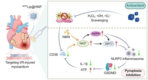
Rationale: Cardiovascular disease imposes a substantial health burden worldwide and remains the primary cause of global mortality, with acute myocardial infarction (AMI) being the most severe acute manifestation. Despite the clinical benefits of reperfusion therapy, the accompanying ischemia/reperfusion (I/R) injury often triggers irreversible myocardial damage through NLRP3 inflammasome-mediated pyroptosis and oxidative stress, for which no effective targeted therapy currently exists. Hence, novel effective therapies for AMI-related I/R injury are urgently required clinically.
Methods: We developed a novel nanoprotectant (IMTPLip@HNP) via the double-emulsification method, which exerts dual functions: NAD⁺ replenishment and ROS scavenging. Using hollow mesoporous Prussian blue (HMPB) nanozymes as core, we loaded nicotinamide mononucleotide (NMN), emulsified with perfluoropentane for ultrasound-controlled release, and encapsulated in IMTP-functionalized lipid bilayers for targeting. We validated efficacy by assessing ROS clearance, NAD⁺ restoration, NLRP3 deacetylation, and pyroptosis suppression, alongside 3D imaging and transcriptomic analysis.
Results: IMTPLip@HNP targeted and accumulated in ischemic myocardium. It exhibited potent catalase- and superoxide dismutase-like activities, eliminating ROS and restoring intracellular NAD⁺ levels. Elevated NAD⁺ further facilitated SIRT2-mediated NLRP3 deacetylation and thereafter blocked downstream pyroptotic signaling cascades. In vivo animal assays verified decreased myocardial fibrosis, improved angiogenesis and reversal of pathological cardiac remodeling. Transcriptome sequencing data identified upregulated genes related to mitochondrial oxidative phosphorylation, which confirmed multi-level cardioprotective effects of the formulation.
Conclusions: The IMTPLip@HNP nanoplatform synergistically strengthens endogenous antioxidant defense, restrains pyroptosis progression and optimizes mitochondrial homeostasis, thus effectively alleviating post-AMI myocardial I/R injury. This combinatorial nanotherapeutic strategy possesses promising translational prospect for mitigating I/R lesion and improving long-term prognosis of AMI patients.
Keywords: myocardial ischemia/reperfusion injury, NAD+ replenishment, ultrasound activation, pyroptosis, antioxidant
Introduction
Cardiovascular diseases (CVDs) remain the leading cause of death worldwide, with their incidence and prevalence continuing to increase [1]. Acute myocardial infarction (AMI), a pivotal clinical manifestation of CVDs, arises from abrupt coronary artery occlusion, resulting in regional myocardial ischemia and, if untreated, rapid progression to irreversible myocardial necrosis [2]. Existing reperfusion modalities consisting of thrombolytic therapy and percutaneous coronary intervention (PCI) effectively relieve acute myocardial injury, shrink infarct size and improve long-term clinical outcomes [3]. Nevertheless, a considerable proportion of AMI survivors suffer from early or delayed heart failure, which is predominantly induced by ischemia/reperfusion (I/R) injury, a secondary pathological lesion induced by blood reperfusion after ischemic blockade [4]. Cumulative research demonstrates that uncontrolled inflammatory cascades and out-of-balance oxidative stress serve as core pathogenic drivers of myocardial I/R injury [5-7]. Regrettably, no clinically authorized specific drugs can efficiently alleviate myocardial I/R damage so far; accordingly, patients receiving reperfusion therapy remain susceptible to fatal complications including malignant arrhythmia and cardiogenic shock [8]. Hence, rationally designed therapeutic agents that precisely block pivotal molecular targets of I/R pathogenesis are in urgent demand to avoid secondary myocardial impairment and optimize overall AMI therapeutic efficiency.
Numerous previous studies have validated NLRP3 inflammasome as a core regulatory hub of I/R-triggered myocardial damage [9-11]. Once activated, NLRP3 inflammasome initiates the cleavage and activation of caspase-1, further inducing Gasdermin D (GSDMD)-dependent pyroptosis accompanied by massive secretion of pro-inflammatory cytokines IL-1β and IL-18, which aggravates cardiomyocyte death, microvascular destruction and ventricular remodeling [12-14]. Targeted inhibition of NLRP3-mediated pyroptosis has thereby become a promising approach to limit infarct expansion and preserve cardiac function post AMI. As an essential redox coenzyme, NAD⁺ participates in the modulation of metabolic homeostasis, programmed cell death and DNA repair [15]. NAD⁺ is capable of activating sirtuin deacetylase family (represented by SIRT2) to catalyze NLRP3 protein deacetylation [16]. Deacetylation decreases the activity of the NLRP3 protein and its ability to oligomerize, preventing it from effectively assembling into an inflammasome complex, thereby blocking the maturation and release of proinflammatory factors and inhibiting the inflammatory response [17]. In addition, NAD+-dependent adenosine monophosphate-activated protein kinase (AMPK) can negatively regulate the NLRP3 inflammasome [18, 19]. However, I/R injury is often accompanied by the large consumption of NAD+ in myocardial cells. Studies have confirmed that large amounts of reactive oxygen species (ROS) are generated instantaneously during reperfusion, which leads to overactivation of the NAD+-consuming repair enzyme poly (ADP-ribose) polymerase (PARP) and rapidly depletes intracellular NAD+ stores [20, 21]. Moreover, the expression level of nicotinamide phosphoribosyltransferase (Nampt), a key rate-limiting enzyme for NAD+ synthesis through the “rescue synthesis pathway” in myocardial cells, also significantly decreases after ischemia-reperfusion and under pressure overload conditions, directly blocking NAD+ synthesis [22]. Therefore, supplementation with NAD+ and reducing its consumption are key steps in blocking NLRP3 inflammasome-mediated I/R injury. Nicotinamide mononucleotide (NMN), an endogenous precursor for NAD+ biosynthesis, can efficiently increase intracellular NAD+ levels [23]. However, free NMN molecules have inherent defects, such as a short half-life and poor stability. In addition, the dynamic contraction of the heart often leads to low bioavailability and insufficient retention of therapeutic drugs in the body, ultimately severely restricting its clinical translation and practical application [24].
Oxidative stress constitutes another core pathogenic pathway of I/R damage [25, 26]. Excessive ROS impairs tricarboxylic acid (TCA) cycle flux and fatty acid β-oxidation to disrupt mitochondrial energy metabolism, curtail ATP synthesis and upregulate pro-inflammatory gene transcription, eventually accelerating cardiomyocyte pyroptosis [27]. CD38, a membrane-located NAD⁺ glycohydrolase, is significantly upregulated in I/R microenvironment under the synergistic induction of ROS and inflammatory cytokines [28]. Elevated CD38 expression and enzymatic activity accelerate NAD⁺ hydrolysis and intracellular NAD⁺ pool depletion. Reduced NAD⁺ content further compromises the catalytic activity of sirtuin family deacetylases [29,30], which inherently exert potent antioxidant effects and protect mitochondrial electron transport chain integrity [31]. Sirtuin inactivation impairs cellular ROS clearance capacity to exacerbate oxidative stress and form a self-amplifying vicious cycle that ultimately triggers pyroptosis-related signaling cascades [32]. Accordingly, breaking such pathological cycle requires combinatorial therapies that simultaneously recover cellular NAD⁺ bioavailability and neutralize pathological ROS.
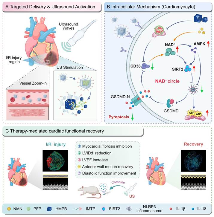
Multifunctional nanocarriers overcome inherent pharmacokinetic and pharmacodynamic drawbacks of conventional drugs via targeted lesion delivery and spatiotemporal controllable drug release while minimizing off-target toxic effects [33, 34]. In this work, we constructed a rationally optimized dual-functional nanoprotector designated IMTPLip@HMPB-NMN/PFP (abbreviated as IMTPLip@HNP). Prussian blue nanoparticles (PBNPs), which have been clinically approved for heavy metal chelation, were repurposed as nanozymes that exhibit stable, bifunctional catalase (CAT)- and superoxide dismutase (SOD)-mimetic activity, enabling efficient scavenging of H₂O₂ and •O₂⁻ [35-37]. Through controlled etching, the PB nanozymes were transformed into hollow mesoporous Prussian blue (HMPB) nanocarriers, leveraging their high surface area and tunable porosity to achieve a high loading capacity and sustained release of NMN, a potent endogenous NAD⁺ precursor. In addition, to endow the ischemic myocardium with specific targeting and ultrasound (US)-responsive release functions, HMPB was encapsulated in a lipid bilayer, which was functionalized with the ischemia-targeting peptide IMTP (CSTSMLKAC) [38, 39]; furthermore, perfluoropentane (PFP), a phase-change contrast agent, was coemulsified with NMN and stably encapsulated within the hollow mesoporous architecture of the HMPB nanocarrier. In vitro studies demonstrated that IMTPLip@HNP combined with US treatment effectively reduced intracellular ROS levels, restored NAD⁺ levels, suppressed NLRP3 inflammasome activation and downstream pyroptosis pathways, and conferred robust cytoprotection against I/R insult (Scheme 1). Ex vivo 3D whole-heart imaging demonstrated alleviated cardiac fibrotic remodeling, improved microvascular construction and intact myocardial tissue morphology. Transcriptomic profiling verified selective upregulation of mitochondrial oxidative phosphory-lation-related genes to enhance electron transport chain activity and ATP production, achieving multi-dimensional cardioprotection at metabolic, structural and genetic levels. Overall, US-assisted IMTPLip@HNP administration represents not merely a simple combination of HMPB, liposome, and targeting peptide, but rather a conceptual-level breakthrough: a mechanistic innovation of “NAD+ metabolic regulation + ROS scavenging” dual-pathway synergy to break the vicious cycle. This integrated therapeutic strategy that synergistically replenishes NAD⁺ and eliminates pathological ROS to block SIRT2-NLRP3-pyroptosis axis, restore mitochondrial energy metabolism and ameliorate post-AMI cardiac dysfunction, possessing great translational potential for clinical I/R injury intervention.
Schematic illustration showing how IMTPLip@HNP protects against myocardial ischemia/reperfusion (I/R) injury. (A) IMTPLip@HNP is delivered specifically to the I/R-injured area and activated by ultrasound. (B) The therapy works through a mechanism where NAD+-mediated positive feedback combines with antioxidant treatment to strongly inhibit NLRP3 inflammasome-induced pyroptosis. (C) When combined with ultrasound, IMTPLip@HNP treatment significantly improves cardiac function. The M-mode ultrasound and heart reconstruction images shown in this scheme were reused from Figure 7B and 7J for illustrative purposes to demonstrate the treatment outcomes.
Results and Discussion
Bioinformatic analysis of pyroptosis in cardiomyocytes
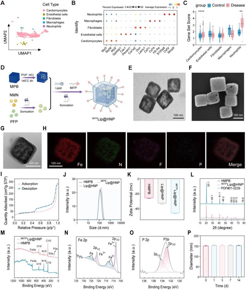
To systematically clarify the core regulatory function of pyroptosis during myocardial I/R injury, integrative bioinformatics analysis was implemented based on publicly available single-cell transcriptome datasets. Raw scRNA-seq data of dataset GSE146285 were retrieved from NCBI GEO database. After rigorous quality filtering, low-quality cells were eliminated (Figure S1). Principal component analysis (PCA) was performed for dimensionality reduction (Figure S2A), and Harmony algorithm was applied to remove inter-sample batch effects (Figure S2B). Cell subpopulations were subsequently visualized and clustered using uniform manifold approximation and projection (UMAP), revealing nine transcriptionally distinct subpopulations (Figure 1A). These clusters were annotated into five major cell types, cardiomyocytes, endothelial cells, fibroblasts, macrophages, and neutrophils, on the basis of canonical marker gene expression (Figure 1B). Functional enrichment analysis indicated that the cardiomyocyte subpopulation was significantly enriched in pathways associated with cardiac hypertrophy (Figure S3), suggesting that surviving cardiomyocytes post-I/R may contribute to pathological myocardial remodeling through compensatory hypertrophy. Subsequently, five classic algorithms, including AddModuleScore and ssGSEA, were used to quantify pyroptosis activity in each cell subpopulation on the basis of the pyroptosis feature gene set (Figure S4). Compared with cardiomyocytes in the control group, cardiomyocytes in the I/R group exhibited significantly higher pyroptosis scores (Figure 1C, S5A). The cardiomyocytes were divided into high and low pyroptosis subpopulations on the basis of the median pyroptosis score. The FindMarkers function of Seurat and the Wilcoxon rank sum test were used to screen differentially expressed genes, defined as those with LogFC > 0 and adjusted P < 0.05, that were expressed in ≥ 10% of cardiomyocytes. Finally, the 10 most significantly differentially expressed genes were identified (Figure S5B), and the expression of inflammation-related genes, such as Pkm and Cebpb, significantly increased in the I/R group. Collectively, the results of single-cell transcriptomic profiling confirm robust activation of pyroptosis in cardiomyocytes following I/R injury, suggesting that targeted inhibition of cardiomyocyte pyroptosis is a promising therapeutic strategy against I/R-induced myocardial damage.
Integrated bioinformatic analysis of cardiomyocyte pyroptosis and structural characterization of IMTPLip@HNP. (A) Identification of cell subpopulations by UMAP clustering. (B) Annotation of cell types based on canonical marker genes. (C) Pyroptosis scores of cardiomyocytes in control and I/R groups. (D) Schematic illustration of the synthesis of IMTPLip@HNP. (E) TEM images of HMPB (scale bar: 100 nm). (F) SEM images of HMPB (scale bar: 100 nm). (G) TEM image of IMTPLip@HNP (scale bar: 100 nm). (H) Elemental mapping of IMTPLip@HNP (scale bar: 100 nm). (I) Nitrogen adsorption-desorption isotherms of HMPB. (J) The hydrodynamic sizes and (K) zeta potentials of HMPB, Lip@HNP and IMTPLip@HNP (n = 3). (L) XRD patterns and (M) XPS spectrum of HMPB and IMTPLip@HNP. XPS high-resolution spectrum of (N) Fe and (O) P in IMTPLip@HNP. (P) The hydrodynamic sizes of IMTPLip@HNP during 14-day observation (n = 3). Data are mean ± SD.
Design and structural characterization of IMTPLip@HNP
To construct the multifunctional phase-change nanozyme delivery platform, IMTPLip@HNP, for the IMTP-mediated targeted delivery of NMN, we first synthesized HMPB by hydrothermal etching (Figure 1D). Transmission electron microscopy (TEM) revealed the uniform hollow cubic morphology of the IMTPLip@HNP (Figure 1E), whereas scanning electron microscopy (SEM) confirmed their well-defined, individually dispersed mesoporous cubic architecture (Figure 1F). Nitrogen adsorption-desorption isotherm analysis demonstrated a typical type IV isotherm with an H1-type hysteresis loop (Figure 1I), indicating highly ordered mesoporosity. Brunauer-Emmett-Teller (BET) measurements demonstrated the highly porous nature of HMPB, exhibiting a surface area of 122.4 ± 0.1 m²/g, a cumulative pore volume of 0.426 ± 0.001 cm³/g, and a BJH-derived pore diameter of 10.7 ± 0.1nm (Figure S6). The abundant accessible pores endow HMPB with advantageous structural characteristics for efficient drug encapsulation. IMTPLip@HNP nanoparticles were obtained using a sequential emulsification strategy, comprising initial O/W emulsification followed by re-emulsification in the lipid phase to generate a W/O/W multiple emulsion. TEM characterization confirmed that the HMPB core was surrounded by a continuous lipid bilayer (Figure 1G).
EDS elemental mapping displayed homogeneous distribution of Fe, N, F and P throughout individual nanoparticles, which validated successful co-loading of NMN and PFP inside HMPB cavities (Figure 1H). Dynamic light scattering (DLS) detection showed hydrated particle sizes of HMPB, Lip@HNP and IMTPLip@HNP were 119.98 ± 0.96 nm, 145.12 ± 2.46 nm and 151.43 ± 2.00 nm separately (Figure 1J), proving size increment after lipid coating. Corresponding zeta potentials were -25.98 ± 0.42 mV, -32.91 ± 1.09 mV and -46.06 ± 0.71 mV (Figure 1K); gradual potential decrease originated from successive modification of anionic DSPE-PEG2000-IMTP, verifying successful lipid functionalization. High-performance liquid chromatography (HPLC) revealed an NMN entrapment efficiency of 49.48 ± 0.12% and a drug loading capacity of 9.90 ± 0.01%. X-ray diffraction (XRD) analysis was conducted to probe the structural characteristics, while X-ray photoelectron spectroscopy (XPS) was used to analyze the surface chemical composition. The XRD analysis showed that the diffraction patterns of both HMPB and IMTPLip@HNP were consistent with the reference pattern of the Prussian blue analog (JCPDS no. 01-0239) (Figure 1L); however, the peak intensities were attenuated in IMTPLip@HNP because of the amorphous lipid shell, although the underlying crystalline phase was unaltered. The XPS survey spectrum of IMTPLip@HNP displayed broadened and less resolved peaks relative to those of the HMPB spectrum (Figure 1M), which was attributed to surface coverage by the organic lipid layer. The high-resolution Fe 2p spectra allowed resolution of the characteristic binding energies at 708.30 eV and 721.10 eV (Fe2⁺) and at 710.00, 713.16, 722.81, and 726.40 eV (Fe3⁺) (Figure 1N), whereas P 2p signals at 133.00 eV and 133.91 eV were assigned to the 2p3/2 and 2p1/2 spin-orbit components of phosphate (PO43⁻) (Figure 1O), confirming the integrity of the iron-phosphate framework. Besides, IMTPLip@HNP exhibited outstanding colloidal stability with unchanged hydrodynamic diameter for over 14 days and negligible size fluctuation in diverse physiological media (Figure 1P, S7).
Multiple enzyme-mimicking properties of IMTPLip@HNP
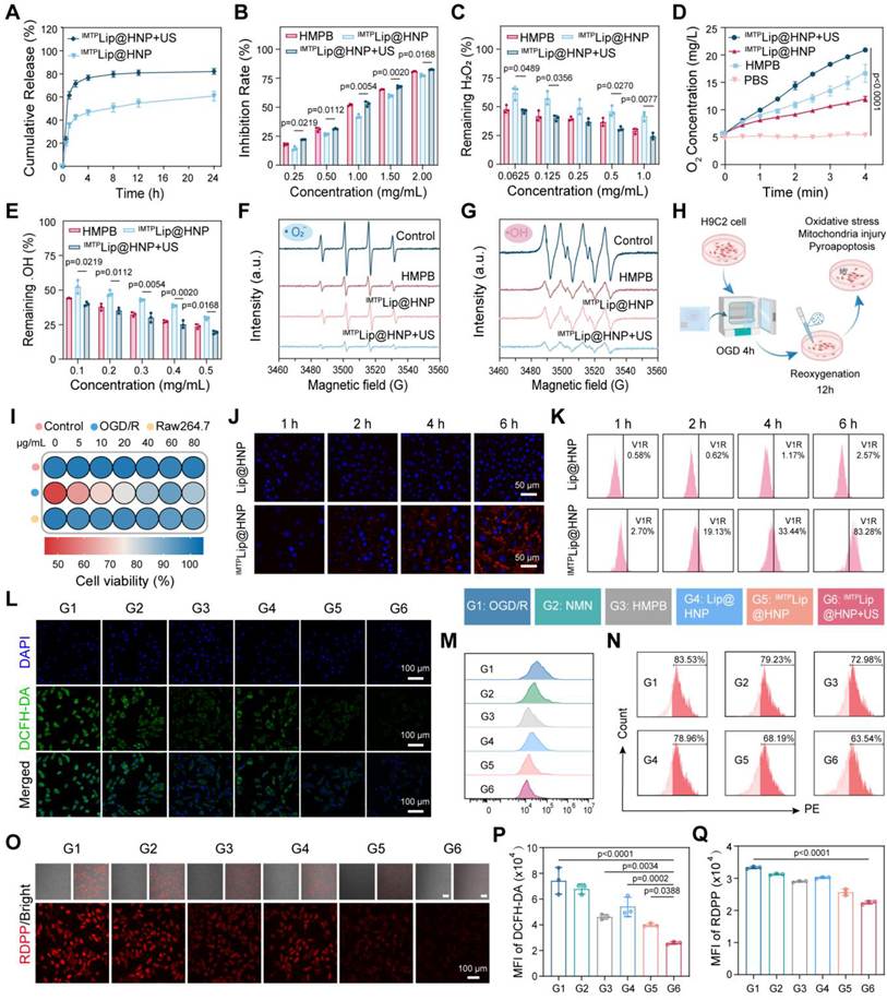
PFP, which was encapsulated within IMTPLip@HNP, is a well-established phase-change material capable of undergoing reversible liquid-to-gas transition in response to multiple external stimuli, including low-intensity pulsed ultrasound (LIPUS), mild hyperthermia, and near-infrared irradiation [39]. Critically, US-induced inertial cavitation selectively compromises the integrity of the outer lipid bilayer, thereby enabling spatiotemporally controlled release of both the HMPB nanozyme core and the coencapsulated NMN. As shown in Figure 2A, the IMTPLip@HNP + US group exhibited significantly greater cumulative NMN release than the non-irradiated IMTPLip@HNP group. Ultrasound-triggered release was sensitive to the applied intensity, duty cycle, and exposure duration, reaching a maximum at 0.8 W/cm2, 50% duty cycle, and 4 min of insonation (Figure S8). Thus, it can be concluded that US irradiation effectively induced on-demand payload release from the lipid-shielded nanocarrier. Subsequently, we systematically assessed ROS-scavenging and multi-enzyme mimetic performance of IMTPLip@HNP. SOD catalyzes •O₂⁻ disproportionation into H₂O₂ and O₂, and WST-8 colorimetric assay was adopted to quantify SOD-like activity via measuring absorbance of yellow formazan at 450 nm. Post-US IMTPLip@HNP displayed prominent dose-dependent SOD-mimetic capacity significantly superior to free HMPB (Figure 2B), which was ascribed to synergistic free radical elimination and iron redox cycle modulation induced by co-loaded NMN. By contrast, non-irradiated IMTPLip@HNP showed weakened SOD activity because lipid bilayer generated steric hindrance to block substrate access toward surface catalytic Fe sites. CAT eliminates cytotoxic H₂O₂ via disproportionation into H₂O and O₂, and TPA-based fluorescence assay was used to test CAT-mimetic activity by detecting fluorescent 2-hydroxyterephthalic acid produced from •OH-TPA reaction. US-activated IMTPLip@HNP rapidly consumed abundant H₂O₂ (Figure 2C), confirming robust CAT-mimetic function. Consistently, only IMTPLip@HNP + US group achieved sustained dissolved oxygen elevation via catalytic H₂O₂ decomposition (Figure 2D), and macroscopic bubble generation further visually verified efficient H₂O₂ breakdown. Even physiological concentration of free NMN possessed intrinsic CAT-like activity (Figure S9), contributing to composite antioxidant capacity of nanosystem. Highly toxic •OH was quantified via TMB-H₂O₂ colorimetric method based on blue complex absorbance at 652 nm, and US-treated IMTPLip@HNP exerted concentration-dependent POD-like activity (Figure 2E). EPR spectra further validated potent •O₂⁻ and •OH scavenging capacity of US-activated IMTPLip@HNP, and free NMN also displayed obvious radical quenching ability (Figure 2F-G, S10-11). In summary, comprehensive functional characterization verified SOD/CAT/POD triple enzyme-mimicking features of IMTPLip@HNP to synergistically clear diverse I/R-derived ROS, laying solid mechanistic foundation for its application as targeted antioxidant nanoprotector.
Multi-enzyme-like activities and in vitro antioxidant protection of IMTPLip@HNP. (A) Controlled release of NMN from IMTPLip@HNP triggered by US irradiation. (B) Concentration-dependent SOD-like activity of HMPB, IMTPLip@HNP, and IMTPLip@HNP + US. (C) Residual H2O2 levels after different treatments with different concentrations. (D) Time-dependent CAT-like activity of the related groups. (E) The remaining •OH after different treatments was measured by TMB colorimetric reaction. EPR spectroscopy analysis of (F) •O2⁻ and (G) •OH scavenging effects of HMPB, IMTPLip@HNP, and IMTPLip@HNP + US. (H) Schematic diagram of the experimental approach. (I) Cell viability of normal H9c2, OGD/R-injured H9c2, and RAW264.7 cells after incubation with IMTPLip@HNP. (J) CLSM images and (K) flow cytometry results of OGD/R-injured cells after incubation with Lip@HNP or IMTPLip@HNP for 1, 2, 4, and 6 h. Scale bar: 50 μm. (L) CLSM images of intracellular ROS levels stained with DCFH-DA after different treatments. Scale bar: 100 μm. (M) Flow cytometry analysis and (P) quantitative determination of intracellular ROS production under different treatments. (N) Flow cytometry analysis and (Q) quantitative determination of intracellular RDPP production under different treatments. (O) CLSM images of intracellular RDPP after different treatments. Scale bar: 100 μm. Data are mean ± SD, n = 3.
Antioxidant effect of IMTPLip@HNP after in vitro OGD/R injury
Prior to therapeutic assessment, systematic biocompatibility evaluation was performed on normal H9c2 cardiomyocytes, OGD/R-injured cardio-myocytes and RAW264.7 macrophages within 0-80 μg/mL concentration range. Cell viability of all three cell types remained above 80% below 80 μg/mL; 60 μg/mL was selected as optimal working concentration due to maximal active ingredient utilization alongside high cell survival (Figure 2I). An OGD/R cellular injury model was constructed on H9c2 to explore in vitro protective efficacy (Figure 2H). CLSM results revealed time-dependent cellular uptake of DiI-labeled IMTPLip@HNP by damaged cardiomyocytes; fluorescence intensity of IMTPLip@HNP group was persistently higher than Lip@HNP control at 1, 2, 4, 6 h, proving IMTP modification enhanced cellular internalization, which was further quantified via flow cytometry with uptake rates of 2.70%, 19.13%, 33.44%, 83.28% sequentially (Figure 2J-K). We next examined whether free IMTP could interfere with IMTPLip@HNP uptake by cardiomyocytes. The addition of free IMTP substantially diminished the intracellular red fluorescence signal from DiI-labeled IMTPLip@HNP (Figure S12A). Flow cytometry confirmed a concentration-dependent reduction in IMTPLip@HNP accumulation in H9c2 cells following free IMTP treatment (Figure S12B). These results support the involvement of an IMTP-dependent uptake mechanism. Given that cardiac troponin I (cTnI) has been proposed as a candidate binding partner for IMTP [5], the observed inhibition may reflect competition between free IMTP and the IMTP-decorated nanocarriers for cellular binding sites. Direct validation of this interaction, however, will require further binding and receptor-blocking studies. US stimulation at ≤ 1.0 W/cm2 was confirmed to be noncytotoxic to both healthy and OGD/R-injured H9c2 cells (Figure S13), supporting its safe use in acoustic-responsive applications. To exclude potential effects arising from US exposure itself, we first evaluated a US-only control in the absence of nanoparticles. Under the same exposure conditions, US alone had no appreciable effect on intracellular ROS levels (Figure S14), NAD⁺/NADH content (Figure S15), mitochondrial membrane potential (Figure S16), and GSDMD expression (Figure S17), consistent with the cell viability results (Figure S13). These results indicate that the protective effects observed following IMTPLip@HNP-mediated treatment were primarily attributable to the nanoparticle-mediated intervention rather than US exposure alone. Moreover, US-triggered cavitation and mechanical stress facilitated the controlled release of the coencapsulated therapeutics HMPB and NMN, thereby improving intracellular drug delivery and enabling synergistic, cascade-regulated cardio-protection by IMTPLip@HNP. During I/R injury, excessive ROS generation induces severe oxidative stress, thereby exacerbating myocardial injury. CLSM revealed that the ROS levels in cardiomyocytes significantly increased after OGD/R. IMTPLip@HNP has an antioxidant capacity comparable to that of free HMPB. This might be because even when the membrane structure of IMTPLip@HNP is not disrupted, some HMPB activity remains and IMTP peptide-mediated targeted recognition significantly promotes the efficient endocytosis of the nanoprotective agent by cardiomyocytes, ultimately demonstrating excellent antioxidant performance. After IMTPLip@HNP + US treatment, the intracellular ROS content was lower than that in the OGD/R group and was the lowest among all the experimental groups (Figure 2L). Flow cytometry verified the ability of this system to scavenge ROS (Figure 2M, P). Compared with that in the OGD/R group, the intracellular ROS concentration in the IMTPLip@HNP + US group was reduced by approximately 2.8-fold. These results indicate that IMTPLip@HNP can specifically target OGD/R-injured cardiomyocytes and, under US irradiation, release HMPB and NMN, thereby rapidly and efficiently eliminating most of the intracellular ROS. Next, the Tris (4,7-diphenyl-1,10-phenan-throline) ruthenium (II) dichloride (RDPP) probe was used to assess the production of O2 within cells. This probe emits characteristic red fluorescence upon excitation by light, and its fluorescence intensity is specifically quenched by O2; thus, its fluorescence intensity is inversely linearly related to the intracellular O2 concentration. The red fluorescence signal in the IMTPLip@HNP + US group was significantly weaker than that in the OGD/R group (Figure 2O). Flow cytometry detection and quantitative analysis confirmed this trend (Figure 2N, Q), demonstrating that IMTPLip@HNP can also catalyze the decomposition of H2O2 and generate O2 within cells, alleviating intracellular hypoxia.
IMTPLip@HNP maintains mitochondrial function after OGD/R injury by increasing the level of NAD+ in vitro
NAD⁺ functions as a central redox cofactor (coenzyme I) essential for cellular energy metabolism, DNA repair, and mitochondrial homeostasis [40]. Stress-triggered CD38 overactivation accelerates intracellular NAD⁺ consumption [28], while NMN as direct NAD⁺ precursor efficiently boosts cardiomyocyte NAD⁺ concentration to relieve oxidative damage and preserve mitochondrial integrity (Figure 3A). I/R-induced ROS accumulation triggers oxidative damage against intracellular biomacromolecules and organelles [41-43]. MitoSOX Red fluorescent probe was utilized to quantify mitochondrial •O₂⁻ level, and OGD/R insult induced dramatic mitochondrial superoxide overproduction, which was reduced dose-dependently by free HMPB or IMTPLip@HNP.
IMTPLip@HNP preserves mitochondrial function via NAD+ promotion after OGD/R injury in vitro. (A) Schematic diagram of intracellular NAD+ synthesis. CLSM images (B) and quantitative determination (D) of intracellular MitoSOX after different treatments. Scale bar: 100 μm. (C) Representative confocal images of JC-1 staining in H9c2 cells after OGD/R. Scale bar: 100 μm. Cellular levels of (E) MDA, (F) GSH and (G) ATP concentration after different treatments. (H) Mitochondrial membrane potential changes and elevation ratios (I) after different treatments determined by flow cytometry. CLSM images (J) and quantification (K) of CD38 IF staining after different treatments. Scale bar: 100 μm. (L) The levels of intracellular NAD+/NADH after different treatments. (M) Mitochondrial ultrastructure in cardiomyocytes (red arrow representing mitochondrial cristae). Scale bar: 1 μm. Data are mean ± SD, n = 3.
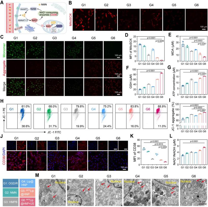
Notably, US-assisted IMTPLip@HNP completely eliminated excess mitochondrial •O₂⁻ with 95.30% reduction of MitoSOX fluorescence versus OGD/R model group (Figure 3B, D), which was attributed to US-triggered synchronous release of HMPB and NMN. Mitochondrial membrane potential (ΔΨm) is a key marker of mitochondrial integrity and drops rapidly under oxidative stress. JC-1 staining demonstrated that IMTPLip@HNP + US not only prevented ΔΨm dissipation but also restored it to a normal physiological level, with the percentage of JC-1 red fluorescence increasing by 23.57% compared with the OGD/R group, as determined by flow cytometry (Figure 3C, H, and I). Moreover, combined treatment markedly lowered intracellular MDA level and recovered depleted GSH and ATP contents simultaneously (Figure 3E-G). Collectively, these findings indicate that IMTPLip@HNP + US intervention alleviates lipid peroxidation of the mitochondrial membrane and restores the mitochondrial redox balance and oxidative phosphorylation capacity, thereby preserving both structural architecture and bioenergetic function. Next, we investigated the expression of CD38 protein in major cell populations involved in myocardial I/R injury using the OGD/R model. Immunofluorescence staining revealed that, compared to normoxic controls, CD38 fluorescence expression was increased not only in endothelial cells but also in cardiomyocytes and macrophages following OGD/R treatment, indicating that this stress condition leads to a general upregulation of CD38 expression across major cell populations in myocardial tissue (Figure S18). Notably, nanoparticle treatment significantly reduced CD38 expression in cardiomyocytes. This effect may be attributed to the multienzyme mimetic activities of the HMPB component, such as CAT-like and SOD-like activities, which effectively alleviate mitochondrial oxidative stress and subsequently inhibit the pathological upregulation of CD38 (Figure 3J-K). Notably, IMTPLip@HNP + US treatment significantly restored the intracellular NAD⁺/NADH levels in OGD/R-treated cells compared with all other groups (Figure 3L). To further examine the contribution of CD38 inhibition to NAD⁺ restoration, OGD/R-treated H9c2 cells were subjected to treatment with 78c + NMN, 78c + IMTPLip@HNP + US, or IMTPLip@HNP + US. Although 78c + NMN effectively increased the NAD⁺/NADH levels, the addition of 78c to IMTPLip@HNP + US did not further enhance NAD⁺/NADH levels compared with IMTPLip@HNP + US alone (Figure S19). This finding suggests that the NAD⁺-restoring effect of IMTPLip@HNP + US may be associated with modulation of the CD38-dependent NAD⁺ metabolic pathway. We next investigated whether NAD+ restoration was functionally required for the downstream protective effects of IMTPLip@HNP + US. Pharmacological inhibition of NAD+ biosynthesis with FK866 largely abolished the reduction in mitochondrial •O₂⁻ levels induced by IMTPLip@HNP + US (Figure S20). Consistently, FK866 also reversed the suppressive effect of IMTPLip@HNP + US on total GSDMD expression (Figure S21). Collectively, these findings suggest a potential mechanistic link whereby IMTPLip@HNP + US preserves intracellular NAD+ by suppressing CD38-mediated NAD+ consumption and enhancing NMN delivery, thereby attenuating I/R-induced mitochondrial oxidative stress, preserving oxidative phosphorylation, and ultimately limiting GSDMD-mediated pyroptotic injury. The bio-TEM morphological observations revealed that OGD/R injury led to damage, breakage, and even complete disappearance of mitochondrial cristae in cardiomyocytes, suggesting significant damage to the mitochondrial inner membrane structure; however, in the IMTPLip@HNP + US intervention group, the mitochondrial cristae were densely arranged, their morphology was clear, and their inner membrane structure was intact (Figure 3M).
IMTPLip@HNP exhibits antipyroptotic activity and regulates macrophage polarization after OGD/R injury in vitro
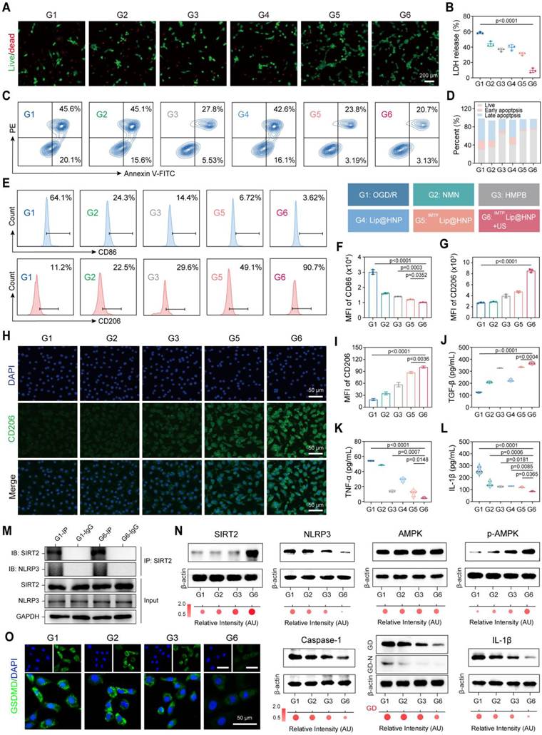
Given that NAD+ depletion in cardiomyocytes induced by I/R injury is a key pathological mechanism that mediates NLRP3 inflammasome activation and pyroptosis, we further investigated the effect of IMTPLip@HNP on pyroptosis in OGD/R-injured cardiomyocytes. The live/dead fluorescence staining results revealed that OGD/R treatment significantly reduced cell viability and that IMTPLip@HNP + US treatment significantly reduced cell death (Figure 4A). A CCK-8 assay quantitatively corroborated this protective effect, demonstrating robust recovery of metabolic activity following IMTPLip@HNP + US treatment (Figure S22). Consistent with these findings, lactate dehydrogenase (LDH) release, a direct indicator of plasma membrane rupture, increased to 58.69 ± 1.38% in the OGD/R group, whereas IMTPLip@HNP + US treatment reduced LDH leakage by 83.3% compared with that in the OGD/R control group (Figure 4B), confirming the strong preservation of membrane integrity. Annexin V-FITC/PI double staining indicated total apoptotic ratio of 65.70% after OGD/R modeling; monotherapy of free HMPB or IMTPLip@HNP achieved partial protection, while US-combined treatment synergistically restrained early and late apoptosis to decrease total death rate down to 23.83% (Figure 4C-D). In addition to direct lytic cell death, pyroptosis initiates self-amplifying inflammatory cascade through massive IL-1β and IL-18 secretion to aggravate myocardial pathological injury. Reprogramming cardiac-resident or infiltrating macrophages from the proinflammatory M1 phenotype toward the reparative M2 phenotype represents a promising strategy to break this cycle. To assess the immunomodulatory potential of IMTPLip@HNP, we established an LPS-induced M1-polarized macrophage model (via 12 h of stimulation) and then treated the cells with the experimental nanotherapeutics. Flow cytometry demonstrated that IMTPLip@HNP + US significantly downregulated the M1 marker CD86 while concurrently upregulating the M2 marker CD206 (Figure 4E-G). Immuno-fluorescence imaging confirmed this phenotypic shift at the single-cell level (Figure 4H), and quantitative analysis revealed that the CD206 fluorescence intensity in the IMTPLip@HNP + US group was 5.2-fold greater than that in the untreated control group (Figure 4I), providing strong evidence for functional M1-to-M2 macrophage reprogramming. To clarify whether the immune-regulatory effects of the nanozyme were attributable to restored cardiomyocyte homeostasis, macrophages were exposed to conditioned media (CM) from OGD/R-stressed H9c2 cardiomyocytes treated with IMTPLip@HNP + US or the caspase-1 inhibitor VX-765. CM from untreated injured cardiomyocytes promoted a pro-inflammatory M1 phenotype, whereas media from IMTPLip@HNP + US- or VX-765-treated cells shifted macrophages toward an M2 reparative phenotype (Figure S23). Because pharmacological inhibition of pyroptosis by VX-765 phenocopied the effects of the nanozyme, the macrophage polarization response is likely driven by reduced cardiomyocyte pyroptotic signaling and altered paracrine communication, rather than a direct action of IMTPLip@HNP on macrophages. ELISAs confirmed that IMTPLip@HNP + US intervention significantly increased the level of the anti-inflammatory factor TGF-β in the cardiomyocyte culture supernatant, while the levels of the proinflammatory factors TNF-α and IL-1β were significantly reduced (Figure 4J-L); this immunomodulatory effect was speculated to be mediated by the synergistic anti-inflammatory effect of NMN and HMPB. To determine whether this anti-inflammatory phenotype stems from upstream inhibition of cardiomyocyte pyroptosis, we profiled the canonical AMPK-SIRT2-NLRP3-caspase-1-GSDMD signaling axis. The molecular association between SIRT2 and NLRP3 was subsequently examined by co-immunoprecipitation (Co-IP) analysis. NLRP3 was enriched in SIRT2 pull-down complexes, and the interaction was moderately enhanced following IMTPLip@HNP + US treatment relative to OGD/R stimulation (Figure 4M). These results demonstrate a physical association between SIRT2 and NLRP3 in cardiomyocytes and reinforce the proposed role of the NAD⁺-SIRT2-NLRP3 pathway in nanozyme-mediated cardioprotection. Western blotting indicated that OGD/R stimulation promoted the accumulation of several pyroptosis-associated proteins, including NLRP3, caspase-1, GSDMD, GSDMD-N, and IL-1β. Although the total expression of AMPK was not altered, its phosphorylation level, together with SIRT2 phosphorylation, was significantly suppressed after OGD/R exposure. These observations suggest that OGD/R triggers pyroptotic cell death through activation of the related signaling cascade. In contrast, in the NMN group, the expression of pyroptosis proteins was slightly downregulated by supplementation with NAD+ precursors, and the HMPB group showed a moderate inhibitory effect owing to its multienzyme mimetic antioxidant activity; however, the combined intervention of IMTPLip@HNP + US reversed the above abnormalities to the greatest extent (Figure 4N, S24). Furthermore, inhibition of AMPK with Compound C reversed the nanozyme-mediated suppression of NLRP3 expression (Figure S25), indicating that AMPK activation is required for NLRP3 inhibition. Overall, these protein-level results suggest that IMTPLip@HNP + US may suppress the NLRP3 inflammasome-mediated caspase-1/GSDMD pyroptosis pathway, potentially by regulating CD38 to restore NAD+ homeostasis, enhancing the SIRT2-NLRP3 interaction, and restoring AMPK/SIRT2 signaling, thereby limiting NLRP3 activation. Immunofluorescence staining further showed that OGD/R markedly increased total GSDMD immunostaining, whereas treatment with IMTPLip@HNP + US substantially reduced it (Figure 4O), providing additional evidence that the nanozyme suppresses pyroptosis. Collectively, these data demonstrate that IMTPLip@HNP + US exerts cardioprotection on multiple levels: (i) intracellularly, by restoring NAD⁺ homeostasis and scavenging mitochondrial ROS to suppress cardiomyocyte pyroptosis; and (ii) extracellularly, by polarizing macrophages toward an anti-inflammatory, tissue-reparative M2 phenotype. This coordinated action disrupts the vicious “pyroptosis-inflammation-secondary injury” cycle, positioning IMTPLip@HNP + US as a rationally designed, multimodal therapeutic platform for I/R injury.
IMTPLip@HNP exerts antipyroptotic effects and regulates macrophage polarization after OGD/R injury in vitro. (A) Live/dead fluorescence microscopy imaging after different treatments. Scale bar: 200 μm. (B) The released content of LDH after different treatments. (C) Flow cytometric analysis of apoptosis and (D) quantification after different treatments. (E) Flow cytometric results of RAW264.7 macrophage polarization and corresponding (F-G) quantitative analysis after different treatments. (H) CLSM images and (I) quantification of CD206 IF staining after different treatments. Scale bar: 50 μm. Inflammatory factors secretion of (J) TGF-β, (K) TNF-α and (L) IL-1β after corresponding treatments. (M) SIRT2 co-immunoprecipitates with NLRP3 in H9c2 cells (n = 1). (N) Western blot of SIRT2, NLRP3, AMPK, p-AMPK, Caspase-1, GSDMD, GSDMD-N, and IL-1β expression at different groups. (O) Immunofluorescence staining of total GSDMD in OGD/R-injured H9c2 cells after different treatments. Scale bar: 50 μm. Data are mean ± SD, n = 3, except for the Co-IP assay (M).
Targeting ability and ultrasound imaging performance of IMTPLip@HNP in I/R rats
Ultrasound imaging, as a noninvasive, real-time and dynamically repeatable imaging modality, holds significant value in the study of myocardial I/R injury [44]. The PFP loaded in core of the IMTPLip@HNP NPs has a low boiling point and a high latent heat of vaporization, enabling a controllable liquid-gas phase transition under ultrasound irradiation and generating strong microbubble echo signals, which endows it with the potential to serve as a targeted phase-change ultrasound contrast agent [45]. This study first verified the feasibility of using IMTPLip@HNP NPs for in vitro contrast-enhanced ultrasound (CEUS) imaging and systematically determined the optimal imaging parameters under multiple gradient ultrasound powers and irradiation times (Figure 5A). After US irradiation, the echo signal intensities of the IMTPLip@HNP NPs in both B-mode and CEUS mode gradually increased, with a maximum echo intensity at 0.8 W/cm² ultrasound power and an irradiation time of 4 min (Figure 5B-C).
Targeting ability and ultrasound imaging performance of IMTPLip@HNP in I/R rats. (A) In vitro B-mode and CEUS imaging of IMTPLip@HNP NPs under US irradiation at different powers and durations. (B) Gray value analysis of B-mode imaging and (C) CEUS imaging. (D) In vivo US imaging procedure in I/R rats injected with IMTPLip@HNP or Lip@HNP. (E) Representative ex vivo fluorescence images of I/R hearts obtained at different time points after intravenous delivery of DiR-labeled IMTPLip@HNP or Lip@HNP NPs, with (G) quantitative evaluation of the fluorescence intensity. (F) US images and (H) quantitative analysis reveal enhanced signal intensity in I/R rats administered IMTPLip@HNP NPs after US irradiation (0.8 W/cm², 4 min). Red arrows indicate the myocardial I/R injury region. Data are mean ± SD, n = 3.
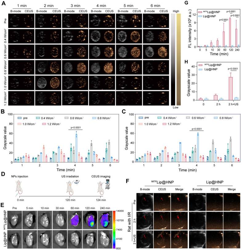
Optical microscopy provided direct visualization of PFP vaporization under the corresponding US conditions. US irradiation induced extensive microbubble formation in the IMTPLip@HNP NP suspension, and the response was most prominent at 0.8 W/cm2 with 4 min of exposure (Figure S26). Together, these findings indicate that IMTPLip@HNP NPs encapsulating PFP respond well to US. To explore the therapeutic effect of IMTPLip@HNP in vivo, the targeted accumulation characteristics of DiR-labeled IMTPLip@HNP and Lip@HNP NPs at various time points in I/R model rats were first investigated. Ex vivo fluorescence imaging revealed that after tail vein administration to I/R rats, the fluorescence intensity of the heart tissue in the IMTPLip@HNP group reached a stable peak at 2 h, whereas no obvious drug accumulation was observed in the heart tissue in the Lip@HNP group (Figure 5E, G). We also collected the main organs of both groups of rats. Affected by reticuloendothelial system (RES) phagocytosis, large amounts of NPs accumulated in liver and spleen tissues in both groups (Figure S27-28). On this basis, in this study, we chose to perform US irradiation 2 h after tail vein injection of the NPs and to conduct CEUS imaging (Figure 5D). IMTPLip@HNP and Lip@HNP NPs were injected into the tail vein of I/R model rats. Two hours after administration, only weak contrast signals were detected in the IMTPLip@HNP group by CEUS; however, after treatment at an ultrasonic power of 0.8 W/cm² for 4 min, the CEUS grayscale value of this group significantly increased and was significantly greater than that of the Lip@HNP group (Figure 5F, H). These results indicate that IMTP exhibits good biological targeting in I/R model rats and provide strong evidence for the use of IMTPLip@HNP NPs as ultrasound contrast agents.
In vivo therapeutic effects and underlying mechanisms of IMTPLip@HNP NPs on acute myocardial I/R injury
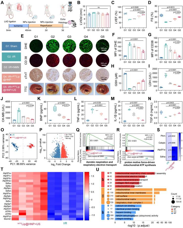
Given the significant ROS scavenging and antipyroptotic effects of IMTPLip@HNP in vitro, we further investigated their antioxidant, anti-inflammatory and cardioprotective effects on acute myocardial I/R injury in vivo. Given the limited stability of free NMN in the circulation, we next examined whether encapsulation within IMTPLip@HNP could improve its blood retention [46]. Free NMN and IMTPLip@HNP were incubated with whole blood ex vivo, and the residual NMN levels were monitored over time. Free NMN declined rapidly, whereas NMN associated with IMTPLip@HNP remained substantially higher throughout the observation period (Figure S29). These results suggest that incorporation into the nanocarrier markedly improves the blood stability of NMN and prolongs its retention, potentially addressing its otherwise short circulation lifetime. We then established an in vivo I/R model via minimally invasive thoracotomy with transient ligation of the left anterior descending artery (LAD) for 40 minutes, followed by reperfusion. Endpoint analyses were performed on day 3 postreperfusion (Figure 6A). Representative electrocardiographic tracings before and after LAD ligation are presented in Figure S30. M-mode echocardiography revealed that after different treatments, the cardiac function of each group of rat improved to varying degrees compared with that of the model group. Among them, the protective effect of IMTPLip@HNP + US treatment on cardiac function was the most significant, as evidenced by increases of 58.0% and 85.2% in the left ventricular ejection fraction (LVEF) and fractional shortening (FS), respectively, compared with those in the I/R group. Unchanged left ventricular end-diastolic diameter (LVIDd) among all experimental groups demonstrated early-stage I/R injury mainly manifested as reversible cellular dysfunction and acute inflammatory activation rather than irreversible ventricular structural remodeling (Figure 6B-D). Quantification of dihydroethidium (DHE) fluorescence staining and CD38 immunofluorescence in myocardial tissue sections confirmed profound oxidative stress and NAD⁺ catabolic dysregulation in the I/R group: the DHE signal intensity in the ischemic zone was elevated > 3-fold compared with that in the sham group, and CD38 expression was significantly upregulated (Figure 6E). In contrast, compared with I/R model rats, those in the IMTPLip@HNP + US group exhibited marked attenuation of both parameters, which corresponded to a 47.62% reduction in DHE fluorescence intensity and 77.86% decrease in CD38 fluorescence intensity (Figure 6F-G). Consistent with these findings, systemic redox imbalance was evident in I/R rats: reduced GSH in the serum was significantly depleted (Figure 6H), while the MDA concentration was elevated (Figure S31). IMTPLip@HNP + US restored the level of GSH to near-basal levels and suppressed the accumulation of MDA, demonstrating the systemic improvement in antioxidant capacity and the mitigation of membrane oxidative damage. Cardiac troponin T (cTnT), the specific biomarker reflecting cardiomyocyte structural integrity, almost vanished at infarct border zone of I/R hearts accompanied by sarcomere breakdown and massive necrosis; conversely, combinatorial therapy retained regular striated cTnT distribution to preserve intact myofibrillar construction (Figure 6E). Hematoxylin-eosin (H&E) staining corroborated this protection, revealing reduced neutrophil infiltration and interstitial edema and preserved cardiomyocyte morphology in the treated group (Figure S32). 2,3,5-Triphenyltetrazolium chloride (TTC) staining demonstrated that the infarct size in I/R rats treated with IMTPLip@HNP combined with US was markedly reduced compared with that in the untreated I/R group (Figure 6E). Furthermore, IMTPLip@HNP + US treatment significantly reduced the serum levels of LDH, creatine kinase-MB (CK-MB), and B-type natriuretic peptide (BNP) relative to the I/R group (Figure 6I-K). Analysis of plasma inflammatory mediators further showed decreased TNF-α and IL-1β levels, together with an elevated TGF-β level, after IMTPLip@HNP + US intervention (Figure 6L-N). These results indirectly confirm that IMTPLip@HNP + US can synergistically improve oxidative stress through the positive feedback effect of NAD+, ultimately achieving a potent anti-inflammatory effect. To further elucidate the cardioprotective mechanism of IMTPLip@HNP + US against I/R injury, transcriptomic analysis was performed on myocardial tissues after treatment. Three days post-reperfusion, ischemic myocardial tissues were collected from rats in the I/R model and IMTPLip@HNP + US intervention groups. Total RNA was extracted for high-throughput mRNA sequencing, and PCA was used to assess overall differences in gene expression profiles and sample reproducibility. PCA results showed clear separation of samples between the I/R and IMTPLip@HNP + US groups, indicating that IMTPLip@HNP + US systematically reshapes the transcriptomic landscape of I/R-injured myocardium (Figure 6O). Gene expression profiling identified 3742 significantly upregulated and 3781 significantly downregulated differentially expressed genes (DEGs) between the two groups (Figure 6P). GSEA demonstrated that mitochondrial energy metabolism-related pathways-including aerobic respiration, electron transport chain, and proton-driven ATP synthesis-were markedly activated in the IMTPLip@HNP + US group compared with the I/R group (Figure 6Q-R). Heatmap verification confirmed that core genes of these pathways (e.g., Atp5f1a, Sdha, ATP6) were significantly upregulated in the IMTPLip@HNP + US group (Figure 6T). The sequencing findings were further validated by qPCR in an independent cohort of heart samples. Atp5f1a and Sdha expression was elevated in the IMTPLip@HNP + US group relative to the I/R group, whereas Pkm and Cebpb expression was decreased (Figure 6S). Functional assessment in OGD/R cells revealed significantly higher activities of mitochondrial complex I and complex IV following IMTPLip@HNP + US treatment than in the OGD/R group (Figure S33), indicating improved mitochondrial respiratory capacity. Gene Ontology (GO) enrichment analysis (biological process [BP], cellular component [CC], molecular function [MF]) further confirmed that DEGs were primarily enriched in mitochondrial respiratory chain complex I assembly, mitochondrial electron transport, and NADH dehydrogenase activity, and localized to key energy metabolism structures (mitochondrial inner membrane, mitochondrial matrix; Figure 6U). Kyoto Encyclopedia of Genes and Genomes (KEGG) pathway enrichment analysis showed that oxidative phosphorylation and propanoate metabolism-core energy metabolism pathways-exhibited the highest enrichment and significance (Figure S34). Collectively, these multi-omics findings confirm that IMTPLip@HNP + US restores energy metabolic homeostasis in I/R-injured cardiomyocytes by upregulating core mitochondrial energy metabolism genes and activating respiratory chain electron transport and ATP synthesis pathways.
In vivo therapeutic effects and underlying mechanisms of IMTPLip@HNP NPs on acute myocardial I/R injury. (A) Schematic illustration of animal intervention. (B-D) Cardiac function assessed by echocardiography. (E) Representative images of myocardial tissues by DHE staining, CD38, cTnT, and TTC staining. DHE and cTnT staining, scale bar: 200 μm; CD38 staining, scale bar: 100 μm. Quantitative analysis of (F) DHE and (G) CD38 IF staining. The levels of (H) GSH, (I) LDH, (J) CK-MB, and (K) BNP in serum. (L-N) Levels of inflammatory cytokines (TGF-β, TNF-α, and IL-1β) in rats with I/R injury. (O) PCA of myocardial transcriptomes in I/R rats treated with PBS or IMTPLip@HNP + US. (P) Volcano plot of differently expressed genes of myocardial tissue after treated with saline or IMTPLip@HNP + US. (Q-R) GSEA enrichment plots of IMTPLip@HNP + US versus the I/R control group. (S) qPCR validation of Atp5f1a, Sdha, Pkm, and Cebpb in heart tissue (n = 3). (T) Heatmap of mitochondrial respiratory chain and ATP synthase-related genes. (U) GO enrichment analysis of genes differentially expressed between groups. Data are mean ± SD, n = 5.
In vivo therapeutic effects of IMTPLip@HNP NPs on chronic myocardial I/R injury
Myocardial repair following ischemia is a highly orchestrated pathophysiological process in which cardiac fibrosis emerges as a defining pathological hallmark of the late phase and persists throughout the progressive course of adverse myocardial remodeling [47,48]. To assess the long-term therapeutic effect of IMTPLip@HNP on cardiac recovery following I/R injury, IMTPLip@HNP was administered on the day of surgery and on postoperative days 4, 8, 13, and 27, followed by echocardiographic monitoring of cardiac structure and function at multiple time points (Figure 7A). From day 1 postoperative through the endpoint of the study, the systolic performance of the IMTPLip@HNP + US group was significantly preserved relative to that of the I/R control group, as evidenced by sustained increases in the LVEF and FS and the significant reduction in the LVIDd (Figure 7B-E). These findings indicate that IMTPLip@HNP + US not only promotes contractile recovery but also actively suppresses maladaptive ventricular dilation. In the I/R group, the mitral valve E/A ratio first decreased, then rose, and subsequently fell slightly but remained elevated. This indicates that left ventricular diastolic function progressed from early relaxation disorder to pseudonormalization, with continuous deterioration of diastolic function (Figure S35). Additionally, this loss of diastolic function was accompanied by a significant reduction in the amplitude of motion of the anterior and posterior walls of the left ventricle (Figure S36-37), whereas IMTPLip@HNP + US treatment significantly reversed these pathological changes and effectively protected left ventricular diastolic function. Gross anatomical examination of the excised hearts confirmed that the I/R group displayed the largest pale infarct zone, whereas the IMTPLip@HNP + US group exhibited the smallest infarct area (Figure S38). H&E and Masson’s trichrome staining results also revealed that after IMTPLip@HNP + US treatment, the degree of myocardial injury and fibrosis caused by I/R was significantly reduced (Figure 7F-G). Myocardial cell loss and fibrosis aggravation can induce compensatory hypertrophy in the remaining myocardial cells. Wheat germ agglutinin (WGA) staining revealed that the degree of myocardial cell hypertrophy in the periphery of the I/R area in the IMTPLip@HNP + US group was significantly reduced, with the relative cell area being 48.0% lower than that in the I/R group, confirming that the nanoprotective agent can effectively reverse myocardial remodeling. Free NMN and IMTPLip@HNP, which lack cardiac tissue targeting and ultrasound-triggered drug release functions, had significantly weaker myocardial protective effects than IMTPLip@HNP + US did (Figure 7H-I).
In vivo therapeutic effects of IMTPLip@HNP NPs on chronic myocardial I/R injury. (A) Schematic illustration of in vivo therapeutic strategy. (B) M-mode echocardiographic images from different groups on day 28 post-treatment. (C-E) Quantitative analysis of left ventricular cardiac function parameters, including LVEF, FS, and LVIDd on day 28 post-treatment. (F) Representative images of H&E and Masson’s trichrome staining of hearts on day 14 post-treatment. Scale bar: 200 μm. (G) Quantification of the myocardial fibrosis area. (H) WGA staining and (I) Quantitative analysis of myocardial tissues at 14 days post-treatment. Scale bar: 100 μm. (J) 3D LSFM imaging and 3D vascular reconstruction of hearts treated with PBS or IMTPLip@HNP + US. Scale bar: 2000 μm. (K) Ventricular wall thickness measurement and (M) quantitative analysis in I/R and IMTPLip@HNP + US groups. Scale bar: 1000 μm. (L) Local high-magnification vascular imaging of hearts. Scale bar: 200 μm. (N) Ultrastructure of myocardial mitochondria (red arrow representing mitochondrial cristae). Scale bar: 1 μm. Data are mean ± SD, n = 5.
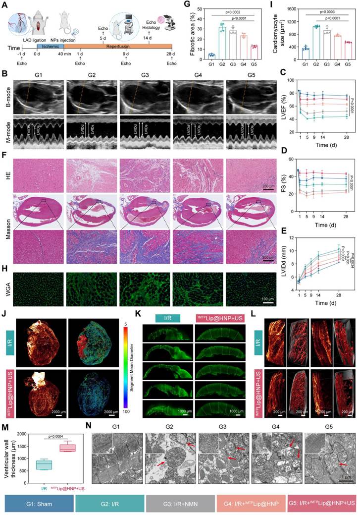
To decipher the cellular mechanisms responsible for the antifibrotic effects, we integrated whole-heart tissue clearing, α-smooth muscle actin (α-SMA) immunostaining, and light-sheet fluorescence microscopy (LSFM) to construct a comprehensive high-resolution three-dimensional map of myofibroblast activation across the cardiac tissue [49]. LSFM imaging unambiguously demonstrated extensive, spatially heterogeneous α-SMA⁺ fibrotic foci throughout the myocardium in the I/R group, whereas the IMTPLip@HNP + US group exhibited markedly diminished lesion burden and the absence of diffuse fibrotic networks (Figure 7J, Video S1-2). Three-dimensional vascular reconstruction revealed pathological arterial/arteriolar dilation in the I/R group, which was consistent with fibrosis-induced vascular stiffening and impaired vasoreactivity, while vessel caliber in the IMTPLip@HNP + US group remained within physiological limits (Figure 7J). Moreover, local high-magnification vascular imaging revealed that the microvessel density was significantly greater and the vessel diameter within the injury border zone was normalized in the IMTPLip@HNP + US group (Figure 7L, Table S1). Moreover, quantitative analysis of multilayered ventricular wall thickness in the area of I/R injury confirmed that the ventricular wall thickness in the I/R group decreased by 47.2% compared with that in the IMTPLip@HNP + US group (Figure 7K, M), indicating that extensive infarction and myocardial fibrosis led to significant thinning of the ventricular wall in the injured area. These results confirmed that IMTPLip@HNP + US intervention significantly inhibited myocardial fibrosis induced by I/R, effectively alleviating myocardial cell hypertrophy and abnormal angiogenesis and reducing myocardial remodeling, thereby exerting a significant protective effect on the myocardium. Bio-TEM revealed profound ultrastructural damage in I/R cardiomyocytes, including mitochondrial swelling, cristae disintegration, matrix vacuolization, and outer membrane rupture. IMTPLip@HNP + US treatment robustly preserved mitochondrial architecture and restored cristae density and membrane integrity (Figure 7N), indicating that mitochondrial protection is a key subcellular mechanism underlying functional recovery. Furthermore, H&E staining of major organs showed no abnormal inflammatory response or histomorphological damage in vivo (Figure S39).
In vivo biosafety of IMTPLip@HNP
To evaluate the biocompatibility and clinical translation potential of this nanoprotective agent, a systematic evaluation of the biological safety of IMTPLip@HNP was conducted in this study. Serum iron levels and SOD inhibition rates were monitored over 84 days following administration. Neither parameter differed significantly from the saline control at any measured time point (0, 1, 5, 14, 28, 56, and 84 days). Iron accumulation in major organs was subsequently quantified by inductively coupled plasma mass spectrometry (ICP-MS) on days 0 and 84. No significant differences in iron levels were detected in the heart, liver, spleen, lung, or kidney between the treatment groups, and no time-dependent increase was observed (Figure S40). Consistent with these quantitative measurements, enhanced Prussian blue staining of cardiac sections revealed no appreciable differences in iron deposition among the groups (Figure S41). Together, these results provide no evidence of systemic iron overload or progressive iron accumulation in cardiac tissue or major reticuloendothelial organs. Blood compatibility was then evaluated by a hemolysis assay. Hemocompatibility evaluation demonstrated that IMTPLip@HNP caused negligible hemolysis even at 3 mg/mL, satisfying the established biosafety requirement of a hemolysis rate below 5% (Figure S42). In addition, blood samples were collected from rats that received IMTPLip@HNP via tail vein injection at different time points for complete blood cell count and key blood biochemical index tests, such as liver and kidney function, and the results indicated that there were no statistically significant differences in any of the parameters compared with those of the normal saline control group (Figure S43). Histopathological analysis of major organs (heart, liver, spleen, lung, and kidney) after H&E staining revealed no pathological changes, such as necrosis, inflammatory infiltration, hemorrhage, or fibrosis (Figure S44). Collectively, these findings support the favorable biocompatibility and safety profile of IMTPLip@HNP and provide a foundation for its further preclinical development.
Although encouraging results have been obtained, several limitations of the current study warrant further investigation. First, the US parameters used in this study were primarily determined based on in vitro optimization and previous reports, rather than systematic in vivo optimization. Future studies should optimize acoustic power, sonication duration, and treatment timing in vivo to define an effective therapeutic window. Second, Co-IP provided supportive evidence for the interaction between SIRT2 and NLRP3, further validation using additional independent biological replicates would strengthen the robustness of this mechanistic finding. Third, although cardiac function was evaluated by echocardiography for up to 28 days and in vivo biosafety was monitored for 84 days, the histological assessment was limited to the first 14 days. Therefore, the present study may not fully capture the longer-term histological responses and structural remodeling of the treated tissue. Future studies with extended histological follow-up beyond 14 days are warranted to further characterize the long-term tissue response and structural remodeling. Fourth, although improved ex vivo blood stability suggests enhanced stability under blood-mimicking conditions, it cannot substitute for a comprehensive in vivo PK and clearance study or establish a prolonged circulation half-life. Future studies should therefore incorporate systematic in vivo PK, biodistribution, and clearance analyses.
Conclusion
In conclusion, we successfully constructed an ultrasound-responsive targeted nanoprotector to realize multi-pathway cardioprotection against myocardial I/R injury. By integrating HMPB’s inherent CAT/SOD-mimetic antioxidant activity and NMN’s NAD⁺ replenishing function within functionalized lipid coating, the nanoplatform effectively recovered intracellular NAD⁺ homeostasis, eliminated pathological ROS and blocked NLRP3 inflammasome-mediated pyroptotic cascade. Such synergistic intervention optimized mitochondrial energy metabolism, restrained pathological fibrotic remodeling and facilitated reparative cardiac microenvironment formation, thereby preserving left ventricular systolic function and mitigating systemic inflammatory response without obvious in vivo toxicity. By coupling precise lesion targeting with effects via a dual mechanism, IMTPLip@HNP offers a robust strategy to halt the self-amplifying cycle of NAD⁺ depletion and oxidative damage, presenting a promising and clinically translatable paradigm for improving patient outcomes after AMI.
Materials and Methods
Materials
All experimental consumables and chemical agents were acquired from certified commercial vendors. Basal cell culture products including high-glucose and glucose-free DMEM formulations were separately sourced from Gibco and Procell, alongside penicillin-streptomycin mixture and fetal bovine serum (FBS, catalog No.164210) supplied by Procell Life Science & Technology (Wuhan, China). Lipid raw materials (DPPC, cholesterol) plus LPS originated from Sigma-Aldrich, while NMN (HY-F0004) and RDPP (HY-W074143) were procured via MedChemExpress. Custom IMTP polypeptide was obtained from China Peptide, and synthetic additives including PVP-30 and glutaraldehyde as well as inorganic salt K₃[Fe(CN)₆] were purchased from Macklin. Mitochondrial complex I and complex IV activity assay kits were purchased from Abbkine (Wuhan, China). Fluorescent markers (DAPI, DiI) and assay kits targeting ROS, mitochondrial superoxide, NAD+/NADH, SOD and H₂O₂ were split between Beyotime and Boxbio suppliers. Beyotime provided the CCK-8 cell viability kit (C0040), and Boster Biological Technology (Wuhan, China) supplied ELISA test boxes for quantitative measurement of TNF-α, IL-1β and TGF-β levels. Antibody resources covered multiple brands: p-AMPK (CST:2531), CD38 (Abmart: TD6551), GSDMD (N terminal) (Huabio: ER1901-37); AMPK, NLRP3 and caspase-1 were purchased from Abcam under respective item codes ab32047, ab263899, ab179515. Proteintech delivered primary antibodies against SIRT2 (19655-1-AP), CD206 (18704-1-AP), cTnT (15513-1-AP), IL-1β (26048-1-AP) and β-actin (20536-1-AP), and Biolegend offered fluorophore-conjugated CD86-FITC and CD206-APC antibodies required for immunofluorescence and western blotting work. Conventional organic solvents like chloroform and hydrochloric acid together with H₂O₂ were obtained from Chuandong Chemical Co., Ltd., Chongqing.
Animals
All in vivo animal manipulations gained formal approval from the Institutional Animal Care and Use Committee at Chongqing Medical University (authorization number Kelun 2023-010), with every operation implemented in compliance with standardized ethical protocols laid down for laboratory animal research. Eight-week-old male Sprague-Dawley rats served as experimental subjects throughout this study. Rodents were housed inside SPF-grade breeding facilities featuring alternating 12-hour light-dark cycles; ambient temperature was stabilized between 18 and 25 °C with relative humidity fixed at 40%-60%, and unlimited access to standard feed and purified drinking water was guaranteed for all individuals.
Preparation of IMTPLip@HNP
The W/O/W double-emulsification tactic was adopted to synthesize target IMTPLip@HNP nanocarriers. For mesoporous Prussian blue (MPB) precursor preparation, 132 mg K₃[Fe(CN)₆] mixed with 3 g PVP was fully dissolved within 40 mL 0.01 M HCl solution under stirring for 30 min, followed by continuous heating at 80 °C over a 24-hour period; harvested sediments underwent centrifugal separation and freeze-drying procedures afterwards. Subsequent hollow-structured HMPB production started by dissolving 100 mg PVP plus 20 mg prefabricated MPB in 20 mL 1 M HCl, with four hours’ stirring before additional 4-hour thermal incubation at 140 °C. After natural cooling down to room environment, final HMPB powders were collected via centrifugation and lyophilization treatment.
To prepare DSPE-PEG2000-IMTP conjugate, 100 mg DSPE-PEG2000-NHS was first dissolved in 3 mL DMF solvent, with sequential addition of CSTSMLKAC peptide and triethylamine catalyst; ambient-temperature stirring lasting 12 h facilitated complete amidation reaction. Post-reaction raw liquor was dialyzed against deionized water over 24 h using 2000 Da cutoff dialysis bags, and lyophilization yielded purified conjugated products. Lipid membrane precursor was generated by dissolving 4 mg cholesterol, 4 mg DSPE-PEG2000-IMTP and 12 mg DPPC within 10 mL chloroform; solvent evaporation via 45 °C rotary evaporator formed uniform lipid thin film, which received 5 mL PBS supplementation and 5 min atmospheric hydration before ultrasonic homogenization to get homogeneous IMTP-modified lipid stock solution.
Primary emulsion construction: 2 mg NMN solid dissolved in 200 μL ultrapure water blended with 100 μL liquid PFP, followed by 6 min ice-bath sonication at 100 W power setting. Secondary emulsification combined pre-made primary emulsion, 1 mg HMPB powder and 1 mL 4 mg/mL IMTP-containing lipid liquid, adopting identical 100 W, six-minute ultrasonic treatment under ice surroundings; finished IMTPLip@HNP was separated through centrifugal precipitation.
Fluorescently tagged nanoparticles were manufactured via slight formulation adjustment: DiI-incorporated IMTPLip@HNP was prepared by mixing DiI substance during lipid film formation stage, while Lip@HNP without targeting segments replaced DSPE-PEG2000-IMTP with plain DSPE-PEG2000 under otherwise identical operating conditions. DiR-labeled nanoparticle variants were produced in parallel by swapping DiI for DiR with all remaining synthesis parameters unchanged.
Characterization of IMTPLip@HNP
Multiple instrumental approaches characterized particle morphological features and chemical compositions. TEM (FEI Tecnai G2) captured structural information for HMPB and finished IMTPLip@HNP, whereas Hitachi SU8020 field-emission SEM visualized HMPB surface topography. Talos F200 X high-resolution TEM assisted crystalline feature and elemental distribution analysis. Bulk crystalline phase and chemical makeup were separately identified through Bruker D8 ADVANCE XRD and Thermo Scientific KAlpha XPS equipment. HMPB’s specific surface area and pore dimension distribution data were quantified using Micromeritics ASAP 2460 BET testing equipment.
HPLC quantification established NMN standard calibration lines based on chromatographic peak magnitudes to compute free drug quantities inside nanoparticle supernatants; encapsulation efficiency (EE) and drug loading content (DL) were calculated using preset mathematic formulas kept consistent with original experimental design. IMTP peptide and DSPE-PEG2000-IMTP conjugate structural confirmation relied on AB5800 TOF-MS alongside Bruker AVANCE III NMR spectroscopy. Malvern NanoZS dynamic light scattering equipment recorded hydrodynamic particle diameters and corresponding zeta potential readings for all prepared nanosamples.
Drug release
In vitro drug release was evaluated under two conditions: (i) Two equal-volume IMTPLip@HNP suspensions dispersed in 25 mL PBS were incubated at 37 °C under 100 r/min continuous shaking to mimic physiological in-vivo drug liberation environments. Half an hour post-incubation initiation, one experimental subset received targeted LIPUS exposure (1.0 MHz, 0.8 W/cm², 50% duty cycle for 4 min), whereas the counterpart group stayed free from ultrasonic stimulation with all surrounding conditions matched entirely. At predetermined time nodes (0, 0.5, 1, 2, 4, 8, 12, 24 h), 0.5 mL supernatant was withdrawn after centrifugal separation, and equivalent volume of fresh PBS was supplemented to sustain fixed total liquid volume. (ii) Six equal-volume aliquots of IMTPLip@HNP suspension were separately irradiated with US at three acoustic power densities (0.4, 0.6, and 0.8 W/cm²) and two duty cycles (20% and 50%) for 4 min. The samples were subsequently dispersed in 25 mL of PBS and incubated at 37 ℃ with continuous shaking at 100 rpm for 24 h. HPLC detection quantified residual NMN concentration in collected supernatant specimens to compute cumulative drug release percentages.
SOD-mimicking capacity of IMTPLip@HNP
Commercial Boxbio SOD detection kits were utilized following manufacturer’s official specifications to quantify the superoxide dismutase-like catalytic performance of prepared IMTPLip@HNP.
CAT-mimicking property assessment
Two independent test schemes validated catalase mimic effects. First experimental route mixed gradient-concentration specimens (0.0625-1.0 mg/mL calculated on HMPB weight: raw HMPB, untreated IMTPLip@HNP, US-pretreated IMTPLip@HNP) into 200 μM H₂O₂ working solution with three-hour ambient incubation; residual hydrogen peroxide content was assayed with dedicated test kits to reflect decomposition efficiency. Second detection tracked real-time dissolved oxygen variation: equal concentration gradient samples were added into 2.5 mL 30% concentrated H₂O₂ solution with blank PBS control group, and continuous four-minute oxygen level recording tracked catalytic oxygen generation kinetics.
POD and free radical scavenging tests
TMB colorimetric quantification tracked •OH-associated peroxidase activity, relying on absorbance readings at 652 nm captured by SpectraMax M5 microplate reader after blue complex formation triggered by hydroxyl radical oxidation of TMB substrates. TiO2-UV irradiation (340 nm wavelength) generated free •OH radicals for EPR-based radical elimination measurement; three sample cohorts (bare HMPB, regular IMTPLip@HNP, US-pretreated IMTPLip@HNP) were incubated for 10 min before final EPR spectrum collection. Xanthine plus xanthine oxidase reaction generated •O2⁻ species for superoxide scavenging evaluation under identical sample grouping and incubation timing, with residual radical amounts ultimately detected via EPR spectral analysis.
Cell culture
H9c2 cardiomyocyte and RAW 264.7 macrophage cell strains were obtained from ATCC repository under catalog numbers CL-0089 and CL-0190 respectively. Complete growth medium consisted of high-glucose DMEM supplemented with 10% FBS plus 1% penicillin-streptomycin mixture; incubation settings were fixed at 37 °C within humidified incubators filled with 5% carbon dioxide atmosphere.
OGD/R injury cell model construction
H9c2 cultures reaching 70%-80% confluency underwent glucose/serum deprivation before being sealed inside anaerobic containers equipped with hypoxic gas packs for four-hour 37 °C hypoxic cultivation. After hypoxic incubation finished, original culture medium was substituted with complete high-glucose DMEM containing designated therapeutic formulations, and cells received 12-hour normoxic reoxygenation inside standard incubator environments (37 °C, 95% air +5% CO2).
Cell viability assay
H9c2 and RAW264.7 cells were plated onto 96-well culture plates and cultured for a full day prior to medium replacement with 100 μL serially diluted IMTPLip@HNP working solutions for another 24-hour normoxic cultivation. Separate H9c2 subgroups received exclusive OGD/R damage or single ultrasonic intervention according to preset grouping layout. Double PBS rinsing preceded CCK-8 color development, with absorbance figures collected at 450 nm wavelength for cellular viability statistics following kit guidelines.
Cellular uptake test
Confocal culture dish-grown H9c2 cells reaching a suitable density were subjected to OGD/R injury and subsequently incubated with 60 μg/mL DiI-labeled IMTPLip@HNP or nontargeted Lip@HNP for 1, 2, 4, or 6 h to characterize the time-dependent cellular uptake. For the competitive inhibition assay, cells were separately pretreated with IMTP at three graded concentrations (0.1, 0.4 and 0.8 mg/mL) for 1 h, followed by the addition of 60 μg/mL DiI-labeled IMTPLip@HNP and further incubation for 6 h. Post-PBS cleaning and DAPI nuclear counterstaining, specimens were fixed with 4% paraformaldehyde for Nikon A1 CLSM imaging; ImageJ computed average fluorescence intensity values, and BD FACS Vantage flow cytometry further numerically quantified internalized particle proportions at scheduled time points.
In vitro oxidative stress assessment
Post-intervention H9c2 cells received diverse fluorescent probe staining for distinct ROS subtypes: DCFH-DA incubation (37 °C, 20 min) traced total reactive oxygen species via CLSM (488 nm excitation) and flow cytometry quantification; 1-hour 10 μM RDPP labeling monitored cytosolic superoxide at 615 nm detection wavelength, while dark-condition 20 min MitoSOX Red incubation pinpointed mitochondrial •O2⁻ with 510 nm CLSM observation and subsequent MFI calculation.
Mitochondrial membrane potential (ΔΨm) test
Discarded culture medium from treated H9c2 cells was replaced by commercial JC-1 working liquid, with 30-minute 37 °C incubation enabling potential-dependent fluorescent shift observation under fluorescence microscopy; flow cytometry was used to determine the proportion of cells exhibiting high JC-1 red fluorescence, which was used as a relative measure of ΔΨm.
Biochemical index detection (MDA/GSH/ATP/NAD⁺/NADH)
Cell lysate samples or in-vivo rat serum harvested after different therapeutic interventions were processed using matched commercial assay kits, with all operating steps strictly implemented in accordance with enclosed manufacturer operating manuals for target biomarker concentration detection.
Cellular ultrastructural TEM observation
Post-OGD/R treated cardiomyocytes were collected and fixed overnight using 2.5% glutaraldehyde solution at 4 °C. After PBS washing and centrifugal enrichment, cell pellets received one-hour post-fixation via 1% osmium tetroxide. Specimens went through gradient acetone dehydration and propylene oxide infiltration before resin embedding and ultrathin slice preparation; copper-grid-mounted ultrathin slices underwent uranyl acetate and lead citrate double staining for Hitachi HT7700 TEM ultrastructure inspection.
Live/dead & apoptosis, LDH assays
Calcein AM/PI mixed staining liquor was added onto PBS-washed adherent H9c2 cells after OGD/R treatments, followed by half-hour dark incubation and microscopic observation using Olympus IX73 inverted fluorescence equipment. Annexin V-FITC/PI dual staining from Boster (MK1028) generated single-cell suspensions for flow cytometric apoptosis detection, with FlowJo v10.8.1 used for final numerical analysis. LDH leakage levels reflected cellular membrane damage degrees measured by matching cytotoxicity detection kits per official instructions.
In vitro macrophage polarization assay
RAW264.7 macrophages were exposed to 200 ng/mL LPS over 12 h to build inflammatory cell microenvironment before co-cultivation with distinct treatment formulations for another 12 h. Fixed cells underwent sequential permeabilization (0.5% Triton X-100), 5% BSA blocking, overnight 4 °C anti-CD206 primary antibody incubation and room-temperature secondary antibody labeling; DAPI counterstaining preceded CLSM imaging and MFI computation. Suspended macrophage specimens were stained with CD86-FITC/CD206-APC for flow cytometric phenotypic proportion calculation.
Conditioned medium collection and macrophage polarization assay
To assess the effect of cardiomyocyte-derived conditioned medium (CM) on macrophage polarization, H9c2 cells were subjected to OGD/R and then divided into three groups: OGD/R group, IMTPLip@HNP + US group, and VX-765 (20 μM) group. After treatment for 24 h, culture supernatants were collected as CM, centrifuged to remove cellular debris, and added to RAW264.7 macrophages for 24 h. Macrophage polarization was then analyzed by flow cytometry using CD86 (M1 marker) and CD206 (M2 marker).
Immunofluorescence staining for total GSDMD
For immunofluorescence analysis, cells cultured in confocal dishes were fixed with 4% paraformaldehyde and permeabilized with 0.1% Triton X-100 following the indicated treatments. After blocking with 5% bovine serum albumin (BSA), the cells were incubated overnight at 4 °C with an anti-GSDMD antibody recognizing total GSDMD (1:200). The cells were then incubated with a fluorescein isothiocyanate (FITC)-conjugated secondary antibody (1:500) for 1 h at room temperature in the dark. Nuclei were counterstained with DAPI, and fluorescence images were acquired using a confocal laser scanning microscope under identical acquisition settings for all experimental groups.
CD38 immunofluorescence staining
Cultured cells or 7-μm-thick frozen cardiac tissue sections were fixed, permeabilized, and blocked using standard immunofluorescence procedures. The samples were then incubated overnight at 4 °C with an anti-CD38 antibody (1:200; Abmart), followed by incubation with a fluorescently labeled secondary antibody. After washing with PBS, the samples were mounted with a DAPI-containing mounting medium and imaged using fluorescence microscopy.
Western blot
Total intracellular protein was extracted with protease-supplemented RIPA lysis buffer, with BCA assay determining total protein concentrations. Equal-quantity protein specimens were separated by SDS-PAGE electrophoresis then transferred onto PVDF blotting membranes; 5% skimmed milk/TBST blocking preceded overnight primary antibody incubation (4 °C) and two-hour HRP-secondary antibody binding at ambient temperature, with target protein bands visualized via chemiluminescent substrate development.
Ischemia/reperfusion model
Experimental SD rats received intraperitoneal anesthesia with 50 mg/kg 3% pentobarbital sodium before tracheotomy and mechanical ventilation (70-80 breaths/min respiratory frequency, 6-8 mL tidal volume). Thoracotomy exposed intact cardiac tissue to realize LAD coronary ligation via 6-0 surgical thread, with successful ischemic status verified through ECG waveform alteration. Forty-minute ischemia was followed by suture removal to initiate blood reperfusion, at which point experimental animals got tail-vein injection of 1 mL 2 mg/mL IMTPLip@HNP solution using 31-G fine needles. Sham-operated counterparts received identical thoracotomy and anesthesia without coronary ligation, with equivalent-volume saline administered after 40 min waiting period; thoracic cavities were sutured layer by layer using 4-0 sterile sutures upon experiment completion.
Echocardiography
I/R-modeled rats received isoflurane inhalation anesthesia at predetermined postoperative time points (1, 3, 5, 9, 14, and 28 days) for Philips EPIC 7C transthoracic ultrasound scanning. Left ventricular long-axis images (B-mode & M-mode) were captured to compute LVIDd, LVEF and FS values; every numerical result originated from average data across no less than three continuous cardiac cycles.
Optical microscopy analysis of PFP morphological changes
IMTPLip@HNP nanoparticles were subjected to ultrasound irradiation at five different acoustic intensities (0.4, 0.6, 0.8, 1.0, and 1.2 W/cm2) with a constant duty cycle of 50%. For each ultrasound condition, samples were irradiated continuously for 6 min, and morphological alterations of PFPs were recorded in real time using optical microscopy.
Quantitative real-time PCR (qRT-PCR)
Total RNA was extracted using an Animal Total RNA Isolation Kit (Foregene) according to the manufacturer’s instructions. RNA concentration and purity were determined using a nucleic acid analyzer, and equal amounts of RNA were reverse-transcribed into cDNA using a SweScript RT I First Strand cDNA Synthesis Kit (Servicebio). Quantitative real-time PCR was performed using SYBR Green qPCR Master Mix (Servicebio) on a Bio-Rad CFX Connect Real-Time PCR Detection System. The amplification specificity was confirmed by melting curve analysis. Relative gene expression levels were calculated using the 2-ΔΔCt method and normalized to the corresponding reference gene. All reactions were performed in technical triplicates.
Co-immunoprecipitation (Co-IP)
For co-immunoprecipitation, cells were collected and lysed on ice in IP lysis buffer containing protease and phosphatase inhibitor cocktails. Following centrifugation to remove insoluble material, the protein concentration of each lysate was measured using a BCA protein assay kit. Equivalent amounts of total protein were subsequently incubated with the indicated antibodies or nonspecific IgG as a negative control. Protein-antibody complexes were then isolated using Protein A/G magnetic beads. After extensive washing, the bound proteins were eluted from the beads and resolved by SDS-PAGE, followed by Western blot analysis with the indicated antibodies. Corresponding input lysates and IgG immunoprecipitation controls were analyzed in parallel to assess protein recovery and confirm the specificity of the detected interactions.
Inductively coupled plasma mass spectrometry (ICP-MS)
At the designated sampling time points, rats were euthanized, and the major organs were harvested following perfusion with ice-cold PBS. Tissue specimens were mineralized by microwave-assisted digestion in concentrated nitric acid and hydrogen peroxide. After digestion, the resulting solutions were brought to volume with 2% nitric acid and subjected to ICP-MS analysis. Elemental calibration was performed using certified multi-element standard solutions, while internal standards were included to monitor analytical performance and correct for potential signal variation. The measured elemental content was normalized to the wet weight of each tissue sample and reported as micrograms per gram (µg/g).
Pharmacological inhibitor treatment
The CD38 inhibitor 78c, NAD+ biosynthesis inhibitor FK866, and AMPK inhibitor Compound C were dissolved in DMSO to generate 10 mM stock solutions and stored at -80 ℃ until use. H9c2 cells subjected to OGD/R were pretreated with 78c, FK866, or Compound C (10 µM each) for 1 h before subsequent experimental treatments. Following inhibitor treatment, cells were cultured for an additional 12 or 24 h, after which samples were collected for NAD+/NADH quantification, Western blot analysis, immunofluorescence staining, and flow cytometric assays.
Serum biochemistry and multiplex histopathological staining
Centrifugation (2500 × g, 10 min, 4 °C) of harvested rat whole blood separated serum specimens for automatic biochemical analyzer testing covering hepatic, renal and myocardial enzyme indexes. Excised cardiac tissues were perfused with PBS before 1 mm-thick cross slicing: slices received 1% TTC dark incubation (37 °C, 15 min) then paraformaldehyde fixation for infarct-area imaging. Specimens pre-soaked overnight in 15% sucrose solution were embedded within OCT compound for frozen section preparation, undergoing routine H&E, Masson’s trichrome and WGA fluorescent staining respectively; ImageJ software quantified relative infarct percentage and average cardiomyocyte cross-sectional dimensions from acquired microscopic pictures.
Transcriptomic sequencing and whole-heart 3D light-sheet imaging
Left ventricular infarct-region myocardium from I/R and IMTPLip@HNP + US treated rat cohorts (n = 5 each group) was collected for total RNA extraction; commercial OE Biotech finished full transcriptome sequencing and subsequent bioinformatic data mining via their exclusive OECloud online analysis platform. Genes with p < 0.05 plus |fold change| > 2 were defined as statistically differentially expressed targets.
Complete rat heart specimens underwent transcardial PBS perfusion followed by 4% paraformaldehyde fixation before 5-day methanol penetration and overnight 5% H₂O₂ decolorization at 4 °C. After multi-day detergent permeation and serum blocking steps, α-SMA primary and corresponding secondary antibodies were sequentially incubated over seven days apiece, with thorough Tween-20 rinsing between different labeling stages. Heart transparency processing adopted Jarvis Biopharma FDISCO clearing kit (reagent A-F sequential incubation), and cleared samples were mounted inside customized 3D-print chambers for LiToneXL light-sheet microscopic scanning; LitScan and Imaris v7.2.3 software accomplished raw image stitching and three-dimensional volumetric reconstruction respectively.
In vivo biosafety evaluation
Thirty-five experimental SD rats were randomly split into seven separate cohorts to conduct systemic biosafety assessment. Single-dose 1 mL (2 mg/mL) IMTPLip@HNP intravenous injection was administered on days 0, 1, 5, 14, 28, 56 and 84 individually per group. Terminal blood collection and major visceral organ (heart/liver/spleen/lung/kidney) dissection were scheduled at corresponding time points; serum biochemical detection and organ H&E pathological staining were performed to judge potential systemic toxicity induced by nanomaterials.
Statistical analysis
All measured numerical outcomes were expressed in mean ± standard deviation format. Shapiro-Wilk normality testing guided the selection of appropriate parametric or nonparametric statistical algorithms. Intergroup comparisons between two datasets utilized independent Student’s t-test, whereas multi-group statistical discrepancies were verified via one-way/two-way ANOVA followed by Tukey’s post-hoc multiple comparison testing. Every biological experiment was reproduced no fewer than three independent times, and p < 0.05 was set as the statistical significance cutoff. SPSS 20.0 and GraphPad Prism 10.0 served as dual software tools for all quantitative statistical computations.
Abbreviations
AMI: acute myocardial infarction; I/R: ischemia/reperfusion; ROS: reactive oxygen species; HMPB: hollow mesoporous Prussian blue; NMN: nicotinamide mononucleotide; US: ultrasound; CVDs: cardiovascular diseases; PCI: percutaneous coronary intervention; GSDMD: Gasdermin D; AMPK: adenosine monophosphate-activated protein kinase; TCA: tricarboxylic acid; CAT: catalase; SOD: superoxide dismutase; PFP: perfluoropentane; PCA: principal component analysis; UMAP: uniform manifold approximation and projection; TEM: transmission electron microscopy; SEM: scanning electron microscopy; HRTEM: high-resolution TEM; BET: Brunauer-Emmett-Teller; EDS: energy-dispersive X-ray spectroscopy; DLS: dynamic light scattering; HPLC: high-performance liquid chromatography; XRD: X-ray diffraction; XPS: X-ray photoelectron spectroscopy; LIPUS: low-intensity pulsed ultrasound; •O2⁻: superoxide anion; H2O2: hydrogen peroxide; O2: molecular oxygen; TPA: terephthalic acid; •OH: hydroxyl radicals; TMB: 3,3',5,5'-Tetramethylbenzidine; POD: peroxidase; EPR: electron paramagnetic resonance; OGD/R: oxygen-glucose deprivation/reperfusion; CLSM: confocal laser scanning microscopy; MDA: malondialdehyde; GSH: glutathione; LDH: lactate dehydrogenase; CEUS: contrast-enhanced ultrasound; RES: reticuloendothelial system; LAD: left anterior descending artery; LVEF: left ventricular ejection fraction; FS: fractional shortening; LVIDd: left ventricular end-diastolic diameter; DHE: dihydroethidium; cTnT: cardiac troponin T; H&E: hematoxylin and eosin; TTC: 2,3,5-Triphenyltetrazolium chloride; CK-MB: creatine kinase isoenzyme MB; DEGs: differentially expressed genes; WGA: wheat germ agglutinin; α-SMA: α-smooth muscle actin; LSFM: light-sheet fluorescence microscopy; W/O/W: water-in-oil-in-water; MPB: mesoporous Prussian blue; IMTP: CSTSMLKAC; PBS: phosphate-buffered saline; EE: encapsulation efficiency; DL: drug loading; DMEM: Dulbecco’s modified Eagle medium; FBS: fetal bovine serum; PS: penicillin-streptomycin; MFI: mean fluorescence intensity; RDPP: Tris (4,7-diphenyl-1,10-phenanthroline) ruthenium (II) dichloride; LPS: lipopolysaccharide; ATP: adenosine triphosphate; BSA: bovine serum albumin; PFA: paraformaldehyde; ANOVA: analysis of variance; ICP-MS: inductively coupled plasma mass spectrometry; CO-IP: co-immunoprecipitation; qRT-PCR: quantitative real-time PCR; CM: conditioned medium.
Supplementary Material
Supplementary figures and table, video legends.

Supplementary video 1.
Supplementary video 2.
Acknowledgements
This work was supported by National Natural Science Foundation of China (Grant No. 82472011, 82302218, 82472006, 82171946, 82270486), Sichuan Provincial Natural Science Foundation (2025ZNSFSC1705), Chongqing Science and Technology & Health Commission Joint Research General Program (2025MSXM048), Joint Project of Pinnacle Disciplinary Group, the Second Affiliated Hospital of Chongqing Medical University (2024205, 2024210), Kuanren Talents Program of the Second Affiliated Hospital of Chongqing Medical University (KR2024G006, KR2024Y044), Natural Science Foundation of Chongqing (Grant No. CSTB2024NSCQ-MSX1151). Special thanks go to BioRender for providing Graphic abstract, Scheme 1 and Figure 1D, 2H, 3A, 5D, 6A, and 7A.
Author contributions
Min Xu: Conceptualization, Methodology, Visualization, Validation, Investigation, Resources, Data curation, Writing original draft, Funding acquisition. Xuemei Tang: Methodology, Visualization, Validation, Investigation, Data curation. Fang Li: Methodology, Visualization, Validation, Resources, Data curation. Leilei Zhu: Methodology, Visualization. Hongjin An: Investigation. Shengzhe Hou: Investigation. Yunfan Liu: Investigation. Haitao Ran: Supervision. Zhigang Wang: Supervision. Zhiyi Zhou: Project administration, Funding acquisition. Weixi Jiang: Writing-review & editing, Supervision, Project administration, Funding acquisition, Conceptualization. Jianli Ren: Writing-review & editing, Supervision, Funding acquisition.
Data availability statement
The data that support the findings of this study are available from the corresponding author upon reasonable request.
Ethics committee approval
All the animal experiments were approved by Institutional Animal Care and Use Committee of Chongqing Medical University and all procedures were in accordance with the ethical standards Use Committee of Chongqing Medical University: Kelun prequalification no. (2023) 010.
Competing Interests
The authors have declared that no competing interest exists.
References
1. Wang L, Li D, Yao F, Feng S, Tong C, Rao R. et al. Serpina3k lactylation protects from cardiac ischemia reperfusion injury. Nat Commun. 2025;16:1012
2. Luo Q, Li Z, Sun W, Wang G, Yao H, Wang G. et al. Myocardia-Injected Synergistically Anti-Apoptotic and Anti-Inflammatory Poly(amino acid) Hydrogel Relieves Ischemia-Reperfusion Injury. Adv Mater. 2025;37:2420171
3. Cheng N, Luo Q, Yang Y, Shao N, Nie T, Deng X. et al. Injectable pH Responsive Conductive Hydrogel for Intelligent Delivery of Metformin and Exosomes to Enhance Cardiac Repair after Myocardial Ischemia-Reperfusion Injury. Adv Sci. 2025;12:2410590
4. Yang J, Wang J, Zeng Z, Chen Z, Wang D, Wu Y. Injectable sustained-release danshensu sodium-loaded nanoparticle hydrogel targets macrophages to improve myocardial microenvironment for myocardial infarction treatment. Bioact Mater. 2025;54:159-78
5. Ma X, Fan Z, Peng J, Nie L. Ischemic Area-Targeting and Self-Monitoring Nanoprobes Ameliorate Myocardial Ischemia/Reperfusion Injury by Scavenging ROS and Counteracting Cardiac Inflammation. Adv Sci. 2025;12:2414518
6. Wang H, Li L, Luo L, Cheng Y, Zhou F, Zhao S. et al. Advanced Nanomaterial Platforms for Targeted Therapy of Myocardial Ischemia-Reperfusion Injury. Research. 2025;8:822
7. Zheng H, Feng L, Xin H, Du F, Wang Y, Wang W. et al. A microenvironment-responsive, injectable, and conductive hydrogel integrated with a catalytic nanozyme with ROS scavenging, anti-inflammation, and pro-angiogenic capabilities for synergistic myocardial infarction therapy. Acta Biomater. 2026;217:618-36
8. Ferdinandy P, Andreadou I, Baxter GF, Bøtker HE, Davidson SM, Dobrev D. et al. Interaction of Cardiovascular Nonmodifiable Risk Factors, Comorbidities and Comedications With Ischemia/Reperfusion Injury and Cardioprotection by Pharmacological Treatments and Ischemic Conditioning. Pharmacol Rev. 2023;75:159-216
9. Liu S, Bi Y, Han T, Li YE, Wang Q, Wu NN. et al. The E3 ubiquitin ligase MARCH2 protects against myocardial ischemia-reperfusion injury through inhibiting pyroptosis via negative regulation of PGAM5/MAVS/NLRP3 axis. Cell Discov. 2024;10:24
10. Zhu L, Liu Y, Wang K, Wang N. Regulated cell death in acute myocardial infarction: Molecular mechanisms and therapeutic implications. Ageing Res Rev. 2025;104:102629
11. Xiang Q, Yi X, Zhu X, Wei X, Jiang D. Regulated cell death in myocardial ischemia-reperfusion injury. Trends Endocrinol Metab. 2024;35:219-34
12. Toldo S, Abbate A. The NLRP3 inflammasome in acute myocardial infarction. Nat Rev Cardiol. 2018;15:203-14
13. Liu Y, Zhang J, Zhang D, Yu P, Zhang J, Yu S. Research Progress on the Role of Pyroptosis in Myocardial Ischemia-Reperfusion Injury. Cells. 2022;11:3271
14. Welt FGP, Batchelor W, Spears JR, Penna C, Pagliaro P, Ibanez B. et al. Reperfusion Injury in Patients with Acute Myocardial Infarction: JACC Scientific Statement. J Am Coll Cardiol. 2024;83:2196-2213
15. Yoshino J, Baur JA, Imai S. NAD+ Intermediates: The Biology and Therapeutic Potential of NMN and NR. Cell Metab. 2018;27:513-28
16. Covarrubias AJ, Perrone R, Grozio A, Verdin E. NAD+ metabolism and its roles in cellular processes during ageing. Nat Rev Mol Cell Biol. 2021;22:119-41
17. He M, Chiang H, Luo H, Zheng Z, Qiao Q, Wang L. et al. An Acetylation Switch of the NLRP3 Inflammasome Regulates Aging-Associated Chronic Inflammation and Insulin Resistance. Cell Metab. 2020;31:580-91
18. Zhang J, Huang L, Shi X, Yang L, Hua F, Ma J. et al. Metformin protects against myocardial ischemia-reperfusion injury and cell pyroptosis via AMPK/NLRP3 inflammasome pathway. Aging. 2020;12:24270-87
19. Yao M, Wang Z, Jiang L, Wang L, Yang Y, Wang Q. et al. Oxytocin ameliorates high glucose- and ischemia/reperfusion-induced myocardial injury by suppressing pyroptosis via AMPK signaling pathway. Biomed Pharmacother. 2022;153:113498
20. Lv S, Ju C, Peng J, Liang M, Zhu F, Wang C. et al. 25-Hydroxycholesterol protects against myocardial ischemia-reperfusion injury via inhibiting PARP activity. Int J Biol Sci. 2020;16:298-308
21. Abdellatif M, Sedej S, Kroemer G. NAD+ Metabolism in Cardiac Health, Aging, and Disease. Circulation. 2021;144:1795-817
22. Doan KV, Luongo TS, Ts’olo TT, Lee WD, Frederick DW, Mukherjee S. et al. Cardiac NAD+ depletion in mice promotes hypertrophic cardiomyopathy and arrhythmias prior to impaired bioenergetics. Nat Cardiovasc Res. 2024;3:1236-48
23. Wang S, Han Y, Liu R, Hou M, Neumann D, Zhang J. et al. Glycolysis-Mediated Activation of v-ATPase by Nicotinamide Mononucleotide Ameliorates Lipid-Induced Cardiomyopathy by Repressing the CD36-TLR4 Axis. Circ Res. 2024;134:505-25
24. Liang Y, Li M, Tang Y, Yang J, Wang J, Zhu Y. et al. Temperature-sensitive hydrogel dressing loaded with nicotinamide mononucleotide accelerating wound healing in diabetic mice. Biomed Pharmacother. 2023;167:115431
25. Ye T, Chen C, Wang D, Huang C, Yan Z, Chen Y. et al. Protective effects of Pt-N-C single-atom nanozymes against myocardial ischemia-reperfusion injury. Nat Commun. 2024;15:1682
26. Zhang X, Sun Y, Yang R, Liu B, Liu Y, Yang J. et al. An injectable mitochondria-targeted nanodrug loaded-hydrogel for restoring mitochondrial function and hierarchically attenuating oxidative stress to reduce myocardial ischemia-reperfusion injury. Biomaterials. 2022;287:121656
27. Liu Q, Gao Z, Zhang X, Duan Q, Zhang Y, Midgley AC. et al. Assembly of Genetically Engineered Ionizable Protein Nanocage-based Nanozymes for Intracellular Superoxide Scavenging. Nat Commun. 2025;16:1123
28. Qiu Y, Xu S, Chen X, Wu X, Zhou Z, Zhang J. et al. NAD+ exhaustion by CD38 upregulation contributes to blood pressure elevation and vascular damage in hypertension. Signal Transduct Target Ther. 2023;8:353
29. Schmauck-Medina T, Lautrup S, Di Francesco A, Mitchell SJ, Clemmensen C, Partridge L. et al. Dietary restriction in aging and longevity. Nat Aging. 2026;6:485-505
30. DiGiovanni LF, Khroud PK, Carmichael RE, Schrader TA, Gill SK, Germain K. et al. ROS transfer at peroxisome-mitochondria contact regulates mitochondrial redox. Science. 2025;389:157-62
31. Małysiak ES, Schiavoni V, Rotondo R, Zeppa SD, Vignini A, Moriconi E. et al. The Sirtuin Network: Linking NAD+ Metabolism, Mitochondrial Function, and Metabolic Homoeostasis. Diabetes Metab Res Rev. 2026;42:e70201
32. Luo Q, Sun W, Li Z, Sun J, Xiao Y, Zhang J. et al. Biomaterials-mediated targeted therapeutics of myocardial ischemia-reperfusion injury. Biomaterials. 2023;303:122368
33. Zheng Z, Tan Y, Li Y, Liu Y, Yi G, Yu C. et al. Biotherapeutic-loaded injectable hydrogels as a synergistic strategy to support myocardial repair after myocardial infarction. J Control Release. 2021;335:216-36
34. Long M, Wang L, Kang L, Liu D, Long T, Ding H. et al. Prussian Blue Nanozyme Featuring Enhanced Superoxide Dismutase-like Activity for Myocardial Ischemia Reperfusion Injury Treatment. ACS Nano. 2025;19:4561-81
35. Ding X, Hu Y, Feng X, Wang Z, Song Q, Dai C. et al. Enhanced Blood-Brain Barrier Penetrability of BACE1 SiRNA-Loaded Prussian Blue Nanocomplexes for Alzheimer’s Disease Synergy Therapy. Exploration. 2025;5:e20230178
36. Shen K, Li X, Huang G, Yuan Z, Xie B, Chen T. et al. High rapamycin-loaded hollow mesoporous Prussian blue nanozyme targets lesion area of spinal cord injury to recover locomotor function. Biomaterials. 2023;303:122358
37. Sun X, Chen H, Gao R, Qu Y, Huang Y, Zhang N. et al. Intravenous Transplantation of an Ischemic-specific Peptide-TPP-mitochondrial Compound Alleviates Myocardial Ischemic Reperfusion Injury. ACS Nano. 2023;17:896-909
38. Sun Y, Zhang P, Li Y, Hou Y, Yin C, Wang Z. et al. Light-Activated Gold-Selenium Core-Shell Nanocomposites with NIR-II Photoacoustic Imaging Performances for Heart-Targeted Repair. ACS Nano. 2022;16:18667-81
39. Zhang W, Wang J, Xie Z, Zou H, Chen Q, Xu L. et al. Antithrombotic Therapy by Regulating the ROS-Mediated Thrombosis Microenvironment and Specific Nonpharmaceutical Thrombolysis Using Prussian Blue Nanodroplets. Small. 2022;18:2106252
40. Hernandez-Resendiz S, Prakash A, Loo SJ, Semenzato M, Chinda K, Crespo-Avilan GE. et al. Targeting mitochondrial shape: at the heart of cardioprotection. Basic Res Cardiol. 2023;118:49
41. Wu Z, Chen L, Guo W, Wang J, Ni H, Liu J. et al. Oral mitochondrial transplantation using nanomotors to treat ischaemic heart disease. Nat Nanotechnol. 2024;19:1375-85
42. Zhang K, Wang Z, Zhang L, Wu H, Liu J, Zhang M. et al. Pharmacologically inherited carbon dots from Salvia miltiorrhiza with potent antioxidant activity and multi-pathway modulation for myocardial ischemia-reperfusion injury therapy. Theranostics. 2026;16:1855-76
43. Zhao T, Wu W, Sui L, Huang Q, Nan Y, Liu J. et al. Reactive oxygen species-based nanomaterials for the treatment of myocardial ischemia reperfusion injuries. Bioact Mater. 2022;7:47-72
44. Wang S, Li C, Wei Z, Tang C, Wang Y, Yan F. et al. Acoustic hydrogen delivery to treat PANoptosis induced by myocardial ischemia/reperfusion injury in rats. Biomater Adv. 2026;183:214770
45. Li F, Chen L, Zhong S, Chen J, Cao Y, Yu H. et al. Collagen-Targeting Self-Assembled Nanoprobes for Multimodal Molecular Imaging and Quantification of Myocardial Fibrosis in a Rat Model of Myocardial Infarction. ACS Nano. 2024;18:4886-902
46. Unno J, Mills KF, Ogura T, Nishimura M, Imai S. Absolute quantification of nicotinamide mononucleotide in biological samples by double isotope-mediated liquid chromatography-tandem mass spectrometry (dimeLC-MS/MS). Npj Aging. 2024;10:2
47. Bengel FM, Hess A, Diekmann J, Thackeray JT. Molecular imaging of immune and fibrosis targets to guide therapy for repair after myocardial infarction. Nat Rev Cardiol. 2026;23:407-419
48. Zhang X, Zhang Y, Liu Y. Fibroblast activation and heterogeneity in fibrotic disease. Nat Rev Nephrol. 2025;21:613-32
49. Merz SF, Korste S, Bornemann L, Michel L, Stock P, Squire A. et al. Contemporaneous 3D characterization of acute and chronic myocardial I/R injury and response. Nat Commun. 2019;10:2312
Author contact
Corresponding authors: renjianliedu.cn (Jianli Ren), 306837cqmu.edu.cn (Weixi Jiang).