Impact Factor




Theranostics 2027; 17(1):248-273. doi:10.7150/thno.138284 This issue Cite
Review
Surface-enhanced Raman spectroscopy for glioma: from molecular profiling to intraoperative navigation
Min Zhang1, Ya-jun Luo2, Ya-nan Liu1, Ji-sen Li1 
1. School of Chemistry, Chemical Engineering and Materials, Jining University, Qufu 273155, P.R. China.
2. State Key Laboratory of Natural Medicines, Department of Pharmacology, China Pharmaceutical University, Nanjing 210029, P. R. China.
Received 2026-5-25; Accepted 2026-9-16; Published 2027-1-1
Abstract
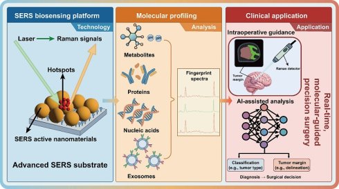
High-grade gliomas, especially glioblastoma, are the most malignant primary tumors of the central nervous system and have a poor prognosis due to their highly invasive growth. The precise detection and delineation of tumor margins are still significant challenges in present diagnosis and surgery. Surface-enhanced Raman spectroscopy (SERS) is a highly sensitive spectroscopic technique that leverages plasmonic enhancement, and thus it has been employed to perform molecular-level diagnosis of glioma. Although SERS has been used to some extent in the diagnosis of gliomas, an integrated, multifaceted platform that combines molecular detection, surgical guidance, and artificial intelligence (AI)-assisted analysis for gliomas has not been built yet. Here, we present the general contents of gliomas based on the combination of SERS-enabled liquid biopsy, molecular profiling, intraoperative navigation, and AI for spectral analysis. On this basis, we examine the main translation problems that have arisen in SERS, such as blood-brain barrier crossing, substrate consistency, AI explainability, and safety, and put forward plans for the clinical application of SERS in precision neuro-oncology. Collectively, these advances highlight the considerable potential of SERS to improve glioma diagnosis, intraoperative guidance, and precision neuro-oncology.
Keywords: glioma, surface-enhanced Raman spectroscopy (SERS), liquid biopsy, intraoperative navigation, artificial intelligence (AI)
1. Introduction
Although there have been many achievements in neuroimaging and molecular pathology, the precise diagnosis and accurate demarcation of glioma margins are still problems to be solved in clinical practice [1-3]. The highly invasive and heterogeneous nature of gliomas often leads to indistinct tumor boundaries, which significantly compromises the completeness of surgical resection and ultimately affects patient prognosis [4-9]. Therefore, the current problem is that there is a lack of analysis methods that can provide timely molecular information for diagnosis and other decisions during surgery.
Currently, the main way to assess gliomas before surgery is by means of magnetic resonance imaging (MRI) and histopathological examination; both are still required in actual clinical work [10,11]. In addition to the traditional structure of MRI, other high-end imaging methods are also applied, such as magnetic resonance spectroscopy (MRS), diffusion tensor imaging (DTI), and perfusion-weighted imaging (PWI), which can provide more details on tumor metabolic state, the organization of white matter tracts, and vascular characteristics [12-15]. The above methods have greatly advanced the diagnosis of gliomas radiologically, but they are unable to directly reveal molecular changes. Histopathological biopsy can provide definitive diagnostic information, but it is invasive and may not fully represent the marked spatial heterogeneity of gliomas due to sampling only a small part of the tumor [16,17]. Positron emission tomography (PET) offers additional metabolic insight, yet its routine use is constrained by limited spatial resolution, high cost, and restricted accessibility [18,19]. In short, these limitations underscore the need to integrate molecular information with traditional imaging to help us better diagnose and treat patients [20,21].
At present, the main treatments for gliomas are surgical resection, radiotherapy, and chemotherapy [22-24]. Glioblastoma is relatively diffuse, and it is easy for the cells to spread along white matter pathways and perivascular spaces in the surrounding brain tissue [25]. Consequently, the true tumor boundary is often indistinct during surgery, making complete resection difficult while increasing the risk of postoperative neurological deficits if excessive tissue is removed. In recent years, in an attempt to increase the precision of tumor localization before and during neurosurgery, many kinds of imaging guidance have begun to be used, including intraoperative magnetic resonance imaging (iMRI), intraoperative ultrasound and 5-aminolevulinic acid (5-ALA) fluorescence imaging [26-28]. Each mode of imaging has its own advantages and disadvantages. Although iMRI can compensate for intraoperative brain shift, update tumor residual status, and improve extent of resection (EOR), it is equipment-intensive and may extend the time for surgery. Intraoperative ultrasound has the advantage of real-time imaging, but it is subject to the skill of the operator and has weak tissue contrast. In addition, infiltrative tumor margins and many low-grade gliomas have reduced the diagnostic accuracy of 5-ALA-guided fluorescence because there is a weak or no fluorescence signal. Therefore, other kinds of technologies that can provide timely molecular information to help clarify the boundary of gliomas in surgery more precisely need to be developed.
Surface-enhanced Raman spectroscopy (SERS) has become a powerful analytical platform for ultrasensitive and multiplexed molecular detection, relying on plasmon-mediated signal amplification at nanostructured interfaces [29-31]. By means of the localized surface plasmon resonance (LSPR) effect of noble metal or semiconductor nanostructures [32,33], SERS can increase the Raman signal of molecules by several orders of magnitude. Many kinds of substances in the body, such as metabolites, proteins, nucleic acids, and extracellular vesicles, can be recognized specifically by SERS through the production of different vibrational fingerprints for each biomolecule [34-36]. Importantly, it is particularly attractive for intraoperative applications because it is less susceptible to photobleaching compared to fluorescence-based techniques and allows for real-time detection.
With the development of nanomaterials and data-driven analysis technology in recent years, some progress has also been achieved in the application of SERS to glioma research [37,38]. These advances have allowed for not only sensitive biomarker detection in complex biological matrices but also real-time tumor margin identification and intraoperative guidance. Recent advances in artificial intelligence (AI) have substantially expanded the analytical capability of SERS by enabling the extraction of diagnostically relevant information from complex, high-dimensional spectral datasets. Although many reviews have summarized the application of SERS in cancer diagnosis or pointed out developments in SERS technology, relatively few have specifically investigated its role at all stages of glioma diagnosis and treatment [39-41]. With the continuous development of nanotechnology, spectroscopy, and data-driven analytical methods, more and more attention is being paid to how these developments can be integrated into the clinical workflow for glioma treatment and how to address existing technical and translation problems. To date, there is no particular review article that has covered all the new developments in SERS-based liquid biopsy, molecular profiling, intraoperative guidance, AI-assisted spectral interpretation, and clinical application of gliomas from all these areas comprehensively.
Recently, some progress has been made in SERS for molecular diagnosis and surgical treatment of gliomas. This paper will briefly introduce the current situation of SERS in glioma research, such as its basic enhancement mechanism, the development of substrates, and typical applications in biomarker detection and tissue identification. We further critically evaluate the key barriers limiting clinical implementation and outline future directions for translating SERS from laboratory research to real-world neuro-oncology practice. This work aims to provide a systematic framework for the development of next-generation SERS platforms in precision neuro-oncology.
2. Mechanistic Basis of SERS Enhancement
To enhance the weak Raman signal, SERS has been applied to achieve sensitive molecular analysis by way of substrate mediation [42-44]. Signal amplification is generally the result of the combined effects of electromagnetic (EM) and chemical enhancement (CE) [45-47]. Therefore, in order to design a SERS platform for the detection of glioma biomarkers and guide surgery reasonably, we first need to know how the two mechanisms work together.
EM enhancement originates primarily from LSPR, which generates intense electromagnetic fields at nanoscale junctions known as hotspots. Molecules located within these hotspots will have Raman intensities that are several orders of magnitude higher [48,49]. Given that it can be applied to all kinds of molecules, EM enhancement is used as the main way in most biological SERS systems. However, the spatial distribution and stability of hotspots are still one of the main reasons for variations in signal reproducibility in complex biological samples [50,51]. Although there are defects mentioned above, EM-based substrates can also be used to detect all kinds of glioma-associated molecular targets, such as metabolites, proteins, and nucleic acids [52-54].
Unlike EM enhancement, CE is confined to molecules in direct contact with the substrate and arises from interfacial electronic interactions, predominantly charge-transfer processes [55-57]. Chemisorption can modify the molecular electronic structure and alter the molecular polarizability, thereby influencing Raman scattering intensity [58,59]. Although the enhancement factor of CE is generally lower than that of EM, it offers the advantage of being applicable to different systems based on semiconductors or hybrid materials, and the charge-transfer pathway can be deliberately tuned to increase some biomolecular signals. Such substrate-dependent selectivity is particularly valuable for distinguishing glioma tissues and analyzing complex biological fluids, where chemically similar components often coexist.
Hybrid substrates, particularly metal-semiconductor systems, provide a representative example of the synergy [60,61]. In such systems, the generation of strong electromagnetic fields in plasmonic metals and charge-transfer interactions in semiconductors lead to combined EM and CE enhancement [62,63]. The dual mechanism not only increases overall signal intensity but also can modulate spectral features, thereby improving molecular discrimination.
As shown in Table 1, the applications of each enhancement effect in glioma are presented. While EM enhancement ensures high sensitivity across a broad range of analytes, CE contributes to selective signal amplification for specific molecular species [64,65]. Thus, the balance between these mechanisms directly impacts the accuracy and reliability of SERS-based detection.
Relationship between SERS enhancement mechanisms and representative glioma applications.
| Enhancement mechanism | Main feature | Typical glioma application | Current challenge |
|---|---|---|---|
| Electromagnetic enhancement (EM) | Strong signal amplification generated by plasmonic hotspots | Label-free tissue analysis, tumor margin delineation, single-cell profiling | Signal variability caused by hotspot heterogeneity |
| Chemical enhancement (CE) | Charge-transfer-mediated signal enhancement with molecular selectivity | Metabolite sensing biomarker analysis in biofluids | Relatively low enhancement factor |
| Hybrid enhancement | Electromagnetic amplification and chemical selectivity | Molecular classification, multiplex biomarker detection | Complex substrate fabrication and optimization |
Overview of SERS strategies for glioma research: (A) SERS substrate materials and (B) applications of SERS in glioma research, encompassing (a) biomarker detection, (b) tissue differentiation and navigation, and (c) intraoperative navigation, with artificial intelligence (AI)-assisted spectral analysis and data interpretation facilitating SERS-based glioma diagnosis and clinical translation.
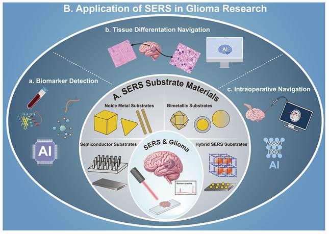
To sum up, the mechanisms of SERS enhancement can help us develop more reasonable substrate engineering strategies in the next part. It is necessary to better control the distribution of hotspots and improve the reproducibility and customization of molecular interactions. On the basis of reasonably regulating the above two enhancement mechanisms, new SERS substrates for glioma have been developed.
3. SERS Substrates for Glioma Applications
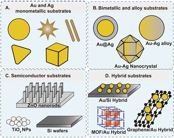
The performance of SERS in glioma research fundamentally depends on the design of the substrate material. Substrate design affects not only signal enhancement but also reproducibility, biocompatibility, and ultimately, clinical applications [66,67]. As shown in Figure 2, the commonly used SERS substrate materials are mainly noble metals, which are combined with other materials (e.g., semiconductors, graphene and metal-organic frameworks (MOFs)) to form composite materials, in order to further enhance the performance of the substrate in biosensing and improve biocompatibility [68-72].
Overview of SERS substrate materials: (A) Au and Ag monometallic substrates, (B) bimetallic and alloy substrates, (C) semiconductor substrates, and (D) hybrid substrates.
3.1 Hotspot Engineering for Signal Enhancement
The essence of SERS lies in the spatial confinement of electromagnetic fields into localized hotspots, which govern the overall signal amplification. These hotspots typically form in regions of strong plasmon coupling, such as nano-gaps, sharp tips, and interparticle junctions [73-75]. In recent years, researchers have conducted a series of studies in the biological field by designing specific hotspot regions using nanomaterials with different morphologies, including nanostars, nanorods, and nanoflowers [76-78]. For example, Au nanoflowers modified on the SERS substrate constructed from two-dimensional red phosphorus nanosheets enable enhanced detection of tumor markers; multi-branched Au nanostars provide superior stability and enhancement for the detection of biomolecules [79,80].
In addition to SERS substrates with different morphologies, constructing nanostructures with controllable interparticle gaps at smaller scales (such as sub-2 nm) has a stronger enhancement effect. For example, sub-2 nm quasi-two-dimensional plasma nano-SERS substrate composed of a nanoparticle-on-mirror (NPoM) geometry and microplate-on-foil (MPoF) nanogaps can achieve in situ spatiotemporal detection with sub-nanoscale resolution [81]. Furthermore, the development of ordered nanoparticle assemblies and hierarchical architectures significantly amplifies plasmonic coupling and enhances hotspot density [82-84]. These structural innovations enable the ultrasensitive detection of low-abundance biomarkers, including circulating tumor DNA, specialized metabolites, and exosomes [85-87]. As an illustration, a study reported a three-dimensional layered gold nanostructure SERS substrate formed by the self-assembly of head-cluster-shaped gold nanocolumns and DNA-modified gold nanoparticles, which can be used for label-free, ultra-sensitive detection of microRNAs [88]. Although the SERS substrate was developed for the detection of biomarkers in urine or serum, its excellent Raman enhancement effect provided a solid technical foundation for the detection of trace or ultra-trace biomarkers associated with gliomas in biological matrices [89-91]. However, the actual clinical applications of noble metal nanomaterials are not yet mature, and there are problems with long-term stability in the body, inconsistent signals, etc.
3.2 Hybrid and Multifunctional SERS Platforms
In recent years, hybrid SERS substrates have begun to be applied for the detection of biomarkers in that they can combine the various attributes of individual materials [92,93]. These systems can combine multiple functional components to achieve synergistic amplification and expanded analytical capabilities [94,95]. Metal-semiconductor heterostructures combine the strong electromagnetic enhancement effects of plasmonic metals with the chemical enhancement resulting from interfacial charge transfer interactions, thereby enhancing the amplification effect of the substrate material and meeting the requirements for high-sensitivity detection [96,97]. For example, by integrating inorganic perovskite-coated gold nanoparticles (Au NPs) onto nanocages to form a specific core-shell structure, which was then used as a SERS substrate, highly sensitive detection of neuron-specific enolases can be achieved [98,99].
Moreover, carbon-based materials, such as graphene and carbon nanotubes, are employed to enhance molecular adsorption and to inhibit fluorescence background, resulting in a significant improvement of signal-to-noise ratios in complex biological samples [100-102]. For example, Au NPs with graphene oxide (AuGO) can be used as a SERS substrate for ultra-trace detection of tumor markers [103]. Simultaneously, porous materials such as MOFs provide a tactical advantage in terms of selective enrichment and size-selective sieving of the analytes [104,105]. This is particularly beneficial for isolating low-abundance glioma markers from the protein-rich environment of cerebrospinal fluid. For instance, Au NPs-modified MIL-125(Ti) framework (Au/MIL-125(Ti)) can be assembled as a robust platform for rapid and non-invasive detection [106].
The above literature provides a brief description of common SERS substrate materials and their applications in the biological field. However, due to differences in their properties, the SERS substrate materials can be applied in various ways in glioma research. Surface plasmon substrates (such as Au nanostars, Au nanoflowers, and NPoM structures) typically provide the highest signal enhancement and are therefore more suitable for detecting low-abundance biomarkers, including ctDNA and microRNA. Hybrid substrates composed of graphene, MOFs, or semiconductors exhibit higher molecular selectivity and are particularly well-suited for metabolite and protein analysis in complex biological samples. For glioma tissue detection and intraoperative navigation, SERS substrate materials must demonstrate excellent uniformity and reproducibility. Consequently, ordered surface plasmon arrays and multifunctional nanoplatforms are more suitable. In general, the development of SERS substrate engineering has been shifting from simple material selection to application-oriented and multi-functional design strategies. Precise control of the architecture of hotspots can improve biocompatibility and the combination of multiple functions has significantly broadened the prospects of SERS in glioma research.
3.3 Biocompatibility and In Vivo Applicability
Biocompatibility of SERS substrates becomes a major consideration for applications with living systems, particularly for intraoperative navigation and in vivo sensing [107,108]. This is necessary since the central nervous system is immune-privileged and very sensitive to exogenous nanomaterials. To overcome these translational bottlenecks, surface modification and functionalization strategies have been extensively researched.
Biocompatibility, specific recognition ability and stability of biological samples can be greatly enhanced by modifying SERS substrates with polydopamine and surface molecularly imprinted polymers [109,110]. Polymer coatings such as polyethylene glycol (PEG) are commonly employed to prevent non-specific protein adsorption and to prolong the systemic circulation time in vivo [111]. In addition, biomimetic strategies such as cell membrane coating have provided a powerful tool to facilitate immune evasion and active transport across the blood-brain barrier (BBB) [112,113]. For instance, previous studies have reported the utilization of SERS nanomaterials coated with cell membranes derived from glioblastoma for the treatment of gliomas. This coating with autologous cell membranes enhances the ability of the material to cross the BBB, thereby improving the penetration of SERS nanoprobes into tumor regions to some extent [114].
Furthermore, in addition to the approaches mentioned above, SERS nanoprobes can be designed to specifically recognize tumor-associated biomarkers using biological recognition elements such as antibodies, peptides, and aptamers as targeting ligands, thereby achieving the goal of preferential enrichment of SERS nanoprobes within tumor tissue [115-117]. To illustrate, the biocompatibility and targeting specificity of the substrates can be effectively enhanced by functionalized Fe3O4/Au nanosatellite structures and Au/Ag nanorod SERS substrates with specific nucleic acid aptamers and bovine serum albumin (BSA) [118]. The above methods improve the biocompatibility of nanomaterials by modifying their surface properties, thereby ensuring that real-time molecular imaging and precise guidance can be performed during neurosurgical procedures.
4. SERS Strategies for Glioma: From Molecular Detection to Surgical Guidance
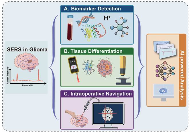
With the development of nano-synthesis and spectroscopic analysis technology, SERS has also evolved from a characterization method into an applied technology, such as SERS for glioma diagnosis and intraoperative guidance [119,120]. At present, not only can SERS detect particular categories of biomolecules, but it is also possible to simultaneously identify many different types of biomolecules for molecular profiling, tissue differentiation and surgical navigation. With the help of AI, spectral analysis, feature extraction and database construction can be carried out on the obtained molecular spectra for clinical application (Figure 3).
Overview of SERS-based approaches for glioma diagnosis and surgery: (A) biomarker detection, (B) tissue differentiation, and (C) intraoperative navigation assisted by AI-powered spectral analysis.
4.1 SERS for Molecular Profiling and Liquid Biopsy
Molecular detection using SERS plays a key role in glioma research, particularly in the fields of liquid biopsy and early diagnosis. Compared to traditional tissue biopsies, SERS offers a rapid, ultra-sensitive, and minimally invasive method for detecting tumor-associated molecular biomarkers in biological fluids [121].
4.1.1 Metabolic Profiling of Glioma Microenvironment
Metabolic reprogramming is one of the hallmark features of gliomas, including pH, glutathione (GSH), hydrogen peroxide (H2O2), and hydrogen sulfide (H2S), which reflect the redox state and metabolic activity of tumor cells [122-125]. Due to its fingerprint-like spectra and high sensitivity, SERS can be used to detect the above metabolic molecules and provide information related to the occurrence and development of gliomas [126].
For instance, a ratio-type pH-responsive SERS sensor chip was developed using silver nanoparticles (Ag NPs) modified with pH-sensitive reporter molecules (such as 4-mercaptopyridine (4-MPY)) [127]. This SERS chip was able to accurately identify tumor margins in nude mouse models, enabling maximal tumor resection and thereby improving patient prognosis (Figure 4A). Beyond pH sensing, the high concentration of GSH in GBM has been leveraged to develop active diagnostic tools. GSH-responsive SERS microneedle (DTNB@Au@Ag) as a novel diagnostic tool can be used for the rapid identification of GBM tissue (Figure 4B). Evaluations in orthotopic tumor models and ex vivo patient samples confirmed that the microneedles could accurately distinguish malignant regions from healthy brain tissue, providing a real-time molecular solution for precision surgery [128].
(A) Schematic diagram of the preparation of a pH-responsive SERS sensing chip and standard curve for microelectrode pH detection. Reproduced with permission from Ref. [127]. Copyright 2022, Elsevier. (B) Schematic diagram of the preparation and application of DTNB@Au@Ag microneedles for deep GBM tissue differentiation and Raman spectra and linearity plots of GSH measured using DTNB@Au@Ag microneedles. Reproduced with permission from Ref. [128]. Copyright 2024, American Chemical Society. (C) SERS nanosensors for monitoring endogenous H2S in living glioma cells. Reproduced with permission from Ref. [129]. Copyright 2015, Wiley-VCH.
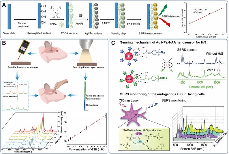
Furthermore, Li et al. designed an azide-modified Au NPs sensor for the specific detection of H2S [129]. By leveraging the H2S-mediated reduction of azides to amines to induce a distinct spectral shift, the sensor achieved a response time within 1 min, enabling the real-time monitoring of endogenous H2S flux within GBM cells (Figure 4C). Despite the high sensitivity of metabolic sensors, the specific detection of individual metabolites can be confounded by the spectral overlap inherent in complex biological matrices.
4.1.2 Proteomic Biomarker and Nucleic Acids Detection
Metabolic molecules provide limited effective information due to their relatively simple structures. Therefore, in order to detect metabolic small molecules, SERS is widely used to analyze proteins and nucleic acids associated with glioma, thereby providing more direct information about tumor genotype and phenotype.
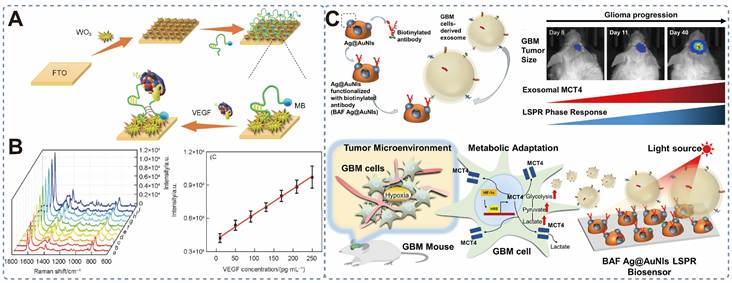
Compared to antibodies, aptamers exhibit greater stability and lower nonspecific binding. Consequently, SERS sensors constructed using aptamers have gradually been adopted for biomarker detection. For example, a pulse electro-deposited WO3 film modified with methylene blue-labeled VEGF aptamers can be used to detect angiogenic biomarkers in serum [130]. This platform detects VEGF by exploiting fluctuations in the Raman signal caused by conformational changes, with a limit of detection (LOD) as low as 8.7 pg/mL, providing a reliable method for monitoring angiogenic biomarkers in serum (Figures 5A and 5B). In another study, Liu et al. developed a site-specific, antibody-functionalized Ag@Au NIs LSPR biosensor for the label-free detection of MCT4, which is a key biomarker for GBM progression (Figure 5C). By exploiting the synergistic plasmonic enhancement between Ag NPs and Au nanodots, the sensor achieved ultrasensitive quantification of CD63 and MCT4 with detection limits as low as 0.38 ng/mL and 1.4 ng/mL, respectively, establishing a new platform for metabolism-related protein profiling in liquid biopsies [131].
(A) Construction of SERS aptasensor based on WO3 film for the determination of VEGF. (B) Raman spectra and linearity plots of VEGF at different concentrations detected using SERS aptamer sensors. Reproduced with permission from Ref. [130]. Copyright 2021, Springer-Nature. (C) Schematic illustration of the liquid biopsy of glioblastoma by the detection of MCT4 in blood-derived exosomes using a BAF Ag@Au NIs LSPR biosensor. Reproduced with permission from Ref. [131]. Copyright 2022, Elsevier.
Circulating nucleic acids are an important research subject in cancer detection, providing crucial information for tumor development and progression [132-134]. To harness the information for glioma screening, dual-signal amplification platforms have been engineered by integrating SERS with catalytic hairpin assembly (CHA) and magnetic enrichment strategies (Figure 6A) [135]. This method employs Au-Ag nanorods as an enhancement substrate and magnetic beads as capture carriers, thereby enabling ultra-high-sensitivity simultaneous detection of glioma-associated ctDNA (IDH1 R132H, BRAF V600E) in mouse serum with attomolar sensitivity, representing a significant leap in the early diagnosis of glioma (Figure 6B).
(A) Schematic diagram of SERS-assisted detection of glioma-associated ctDNA based on dual-signal amplification. (B) SERS spectra and corresponding linear calibration curves of IDH1 R132H and BRAF V600E at different concentrations in PBS buffer and serum. Reproduced with permission from Ref. [135]. Copyright 2023, Taylor & Francis Group, LLC. (C) Schematic of the multiplex SERS nanoplasmonic biosensor using head-flocked gold nanopillars for the label-free detection of miRNA. Reproduced with permission from Ref. [138]. Copyright 2019, The Royal Society of Chemistry.
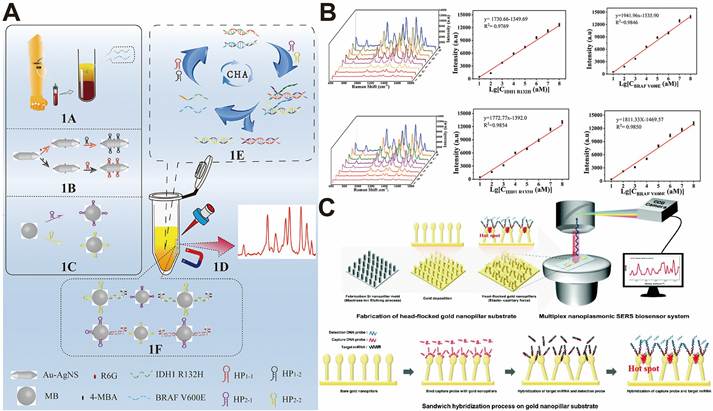
Micro-RNAs (miRNAs) are highly stable in human body fluids and exhibit significantly abnormal expression levels during tumorigenesis or metastasis, making them ideal non-invasive biomarkers for gliomas [136,137]. To analyze glioma-associated miRNAs, researchers prepared a head-flocked gold nanopillar SERS substrate, utilized liquid-induced aggregation to form high-density signal hotspots, and combined this with nucleic acid sandwich hybridization technology to achieve label-free, multiplex detection of glioma-associated miR-10b, miR-21, and miR-373 (Figure 6C). This method is sensitive to femtomolar concentrations, can distinguish single-base mismatches, and has excellent stability of the substrate in serum [138]. Hence, the method is suitable for non-invasive screening, metastasis assessment, and prognosis evaluation of gliomas and other tumors, offering a novel detection strategy for liquid biopsy in cancer. The development of the above sensing methods demonstrates the immense potential of SERS in the molecular diagnosis of gliomas. However, there are still some problems that need to be addressed in the next stage of clinical application of SERS detection, such as weak signals and interference in complex biological environments.
4.1.3 Extracellular Vesicles (EVs)
Extracellular vesicles (EVs) are membrane-bound vesicles that exist in the cells of all types of organisms, including prokaryotes, higher eukaryotes, and plants [139]. The goal of EVs is to pass information to other cells and alter the function of the recipient cells. EV-mediated signals can be carried by all types of biomolecules, such as proteins, lipids, nucleic acids, and carbohydrates [140]. This specific kind of enclosure is protective, and it can carry a large number of messenger molecules to different places far from the starting point of the vesicle. Therefore, through the analysis of SERS-tagged extracellular vesicles from glioma, we can obtain some useful information for the diagnosis or intraoperative navigation of glioma.
Since the cell-free vesicles still have membrane proteins that can recognize their origin cells, we can use these surface markers to select for them and then study how they change in the development and progression of gliomas. Based on the above idea, Zhang and others have developed the GEMPAC nanodiagnostic platform to detect brain-specific proteins (ATP1B2 and EAAT2) and stem cell markers (CD24 and CD44) in extracellular vesicles simultaneously by means of multiplex SERS [141]. Rather than providing a single static measurement, this platform enables longitudinal monitoring of tumor evolution and treatment-induced drug resistance, offering a practical strategy for evaluating therapeutic efficacy over the course of disease management.
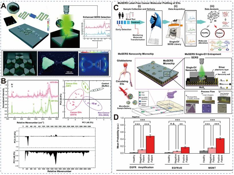
In addition, nanophotonic-integrated microfluidic platforms have been developed for the label-free detection of EVs [142]. Through principal component analysis (PCA), the platform successfully distinguished tumor-derived vesicles from normal glial EVs with high accuracy (Figures 7A and 7B). Similarly, Mahshid et al. introduced a MoSERS microfluidic device integrating MoS₂-plasmonic nanocavity arrays [143]. This setup allowed for molecular profiling at the single-EV level, identifying key alterations, such as EGFRvIII mutations and MGMT expression, with 87% diagnostic accuracy in GBM patient blood samples (Figures 7C and 7D).
(A) Schematic illustration of a surface-mounted microfluidic device with a floating nano-bowtie structure combined with SERS for the detection of glioma exosomes. (B) Comparison of ultra-sensitive SERS detection of exosomes from non-cancerous cells (NHA) and malignant gliomas (U87 and U373). Reproduced with permission from Ref. [142]. Copyright 2021, The Royal Society of Chemistry. (C) Schematic illustration of a SERS-based nano-surface microfluidic method for the molecular characterization of individual exosomes, combined with machine learning for the early detection and monitoring of cellular transformation during the progression and treatment of brain gliomas. (D) Probability distribution maps of MoSERS spectra from peripheral blood-derived exosomes in glioblastoma patients with different molecular alterations (EGFR amplification, EGFRvIII, and MGMT methylation). Reproduced with permission from Ref. [143]. Copyright 2023, American Chemical Society.
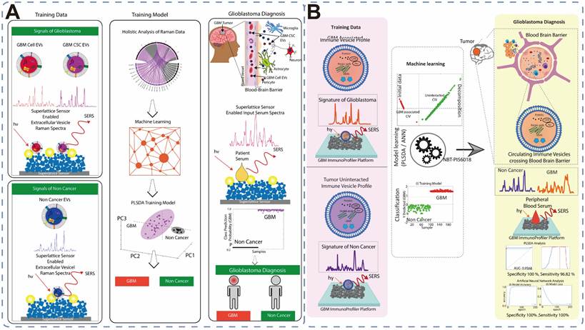
Furthermore, researchers have incorporated machine learning (ML) into SERS testing to analyze extracellular vesicle subtypes and predict tumor progression [144,145]. The machine learning-based SERS detection platform is capable of rapidly analyzing acquired SERS spectra, which significantly reduces the time required for data analysis during surgery and marks an important step forward in the transition of this technology from laboratory testing to clinical testing. For instance, self-functionalized Ni-Au/Pd superlattice sensors have been coupled with partial least squares discriminant analysis (PLS-DA) to achieve the direct detection of glioblastoma stem cell-derived EVs (GBM CSC-EVs) from serum without the need for prior isolation (Figure 8A). This platform achieved an amol-level sensitivity and 100% sensitivity in a limited cohort in clinical validation [146]. Furthermore, the characterization of circulating immune vesicles (CIVs) has been advanced through the use of femtosecond laser-engineered nickel quantum probes combined with artificial neural networks (ANN) (Figure 8B). This immunological analysis strategy achieved a sensitivity of 96.82% in glioblastoma diagnosis. By recognizing the specific expression of PD-L1 and CTLA-4, this platform can effectively distinguish glioblastoma from other malignant tumors of the central nervous system [147].
(A) Schematic illustration of a 3D superlattice sensor capturing extracellular vesicles and generating significantly enhanced SERS signals, combined with machine learning to distinguish between serum samples from healthy individuals and those with brain glioma. Reproduced with permission from Ref. [146]. Copyright 2023, American Chemical Society. (B) Schematic illustrating the utilization of SERS for the detection of extracellular vesicles associated with glioblastoma and the application of supervised machine learning classifiers for glioblastoma. Reproduced with permission from Ref. [147]. Copyright 2022, American Chemical Society.
Although SERS technology has been used to detect various glioma-related biomarkers, the progress of clinical translation varies significantly among different types of biomarkers. CtDNA and EVs-based biomarkers show the greatest potential for clinical application, as both provide tumor-specific molecular information and have been validated in clinical studies of liquid biopsy. Exosomes have become a research hotspot due to their ability to cross the BBB and protect tumor-derived components from degradation. In contrast, metabolic biomarkers such as GSH and H2S lack specificity for glioma and lack sufficient clinical validation data; therefore, they remain primarily in the basic research stage at this stage. Protein biomarkers contain a wealth of biological information and can provide insights into tumor angiogenesis and tumor progression. However, their low specificity and susceptibility to endogenous degradation limit their clinical application. Therefore, future SERS-based clinical assays could develop protein biomarkers with high specificity for gliomas and employ a series of modifications to reduce the likelihood of degradation. The above experiments can be used in conjunction with the platform for exosome detection in liquid biopsies to improve the sensitivity of the detection. In addition, advances in AI have greatly facilitated the application of SERS in liquid biopsy for gliomas. AI-powered spectral analysis can extract subtle molecular features from complex Raman data, thereby shortening diagnostic time. At the same time, AI-powered spectral analysis methods not only improve the accuracy of biomarker classification but also enable multiplex biomarker analysis and further facilitate the automated analysis of liquid biopsy data. The new SERS protocol will have good clinical value for the diagnosis of glioblastoma and changes over time.
Overall, SERS molecular analysis technology enables non-invasive, multiplex detection of glioma markers, but its clinical translation is still limited by the reproducibility of substrate materials and interference from complex substances in biological matrices.
4.2 Cellular-Level Analysis: Mechanistic Insights and Limitations
Liquid biopsy is generally used for the detection and analysis of biomarkers, while SERS can be used to examine individual cells or a small number of cells to obtain more information about the molecular condition of the tumor microenvironment by showing how nucleic acids, proteins, and lipids work together [148-150]. Single-cell and multicellular molecular profiling is essential to decipher intratumoral heterogeneity, identifying rare cell populations such as glioblastoma stem cells, and elucidating tumor development and progression.
4.2.1 Intracellular Molecular Mapping
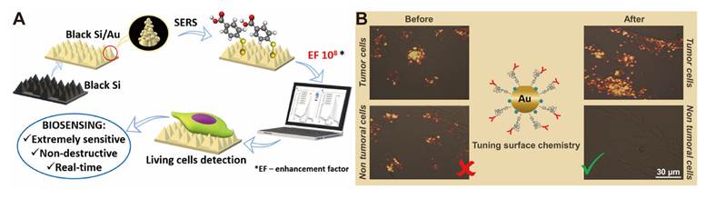
The development of high-performance substrates is crucial for amplifying the Raman characteristic peaks of intracellular substances. A gold-coated black silicon (bSi/Au) SERS substrate was engineered, which can achieve an enhancement factor as high as 10⁸ under 785 nm excitation (Figure 9A). This platform can successfully detect characteristic peaks of malignant transformation markers (e.g., nucleic acids, proteins, and lipids) in rat C6 glioma cells, providing a reliable solution for low-cost, scalable on-chip SERS biosensing [151]. Floriana Burgio and colleagues prepared SERS probes by modulating the surface chemistry of Au NPs and conjugating Raman reporter molecules (1,4-BDT) and polyethylene glycol (PEG) with anti-EGFR antibodies in an optimized ratio [152]. Probes can accurately recognize glioblastoma cells and quickly and accurately distinguish between tumor cells and normal cells in vitro (Figure 9B).
(A) Schematic illustration of SERS detection of trace amounts of small organic molecules in live C6 glioma cells from rats using a bSi substrate coated with a 25-50 nm Au layer. Reproduced with permission from Ref. [151]. Copyright 2020, American Chemical Society. (B) Schematic illustration of the specific detection of glioblastoma cells using SERS by modulating the surface chemistry of Au NPs. Reproduced with permission from Ref. [152]. Copyright 2020, American Chemical Society.
4.2.2 Immunological Profiling in Peripheral Blood
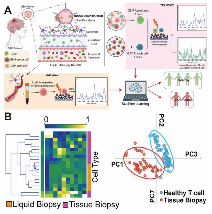
Beyond direct tumor cell sensing, SERS excels in profiling tumor-associated immune cells. Glioblastoma-associated T cells (GBMAT/GSCAT) crossing the BBB can serve as systemic diagnostic proxies (Figure 10A) [153]. By integrating SERS with ML algorithms, the platform achieved a sensitivity of up to 100% and a specificity of 98.7% in a limited cohort for GBM diagnosis from patient blood, highlighting the potential for non-invasive immunological monitoring (Figure 10B).
(A) Schematic illustration of SERS combined with machine learning for the diagnosis of glioblastoma using glioblastoma stem cell-associated T cells. (B) Comparison of SERS for GBM T-cell detection: PCA heatmaps of T cells from tissue biopsies and liquid biopsies, and PCA scatter plot showing differences between T cells from tissue biopsies and healthy T cells. Reproduced with permission from Ref. [153]. Copyright 2022, American Chemical Society.
Despite some progress in SERS detection for glioma cells, cellular-level SERS analysis still faces some inherent limitations, such as the heterogeneity of the intracellular environment, laser damage to cells, and the difficulty in obtaining individual cells during clinical surgery.
4.3 SERS for Intraoperative Tumor Margin Identification
Currently, SERS-based margin identification leverages two distinct yet complementary strategies: label-free intrinsic molecular fingerprinting and targeted functionalized nanoprobes.
4.3.1 Label-Free SERS Based on Molecular Fingerprinting
Label-free SERS utilizes the inherent vibrations of molecules to generate spectral fingerprints of biological tissues [154,155]. Compared to normal tissue, tumor tissue typically exhibits changes in its lipid, protein, and nucleic acid composition, resulting in spectral profiles that differ from those of normal tissue [156,157]. Based on the spectral differences between normal and tumor tissues, combined with statistical analysis methods such as principal component analysis (PCA) and support vector machines (SVM), effective differentiation can be achieved [158]. This method is simple to implement and avoids issues such as the biocotoxicity and biocompatibility associated with exogenous probes, making it more suitable for clinical applications.
Label-free confocal Raman spectroscopy combined with ML was used to distinguish between stem cells (GSCs) and differentiated cells. By analyzing the subcellular structures (such as nuclei and mitochondria) of 1,146 cells, an accuracy rate of 91.7% was achieved [159]. This method effectively addresses the issue of tumor heterogeneity and provides a rapid means of distinguishing highly oncogenic cells during surgery. Building upon this, Wang et al. developed SiO2@Au core-shell nanoprobes and Au NPs SERS substrates to perform SERS detection of gliomas at both the cellular and tissue levels (Figure 11A) [160]. At the cellular level, SVM algorithms precisely distinguish astrocytes from glioma cells. At the tissue level, OPLS-DA models enable differentiation between glioma and traumatic tissue, tumor grading, and IDH mutation identification, with 100% accuracy in a limited cohort (AUC > 0.99) (Figure 11B). The data obtained from the above study demonstrate that label-free SERS has certain potential for differentiating tumor tissues. However, this study is based on a small sample size and may not reflect its diagnostic performance in larger, more heterogeneous, multicenter clinical populations. The label-free approach provides a rapid strategy for real-time intraoperative diagnosis and molecular subtyping.
(A) Schematic diagram of the SERS detection process for glioma cells (a) and glioma tissue (b). (B) Average Raman spectra of different types of tissue samples and the corresponding OPLS-DA plots for the three groups. The shaded regions indicate the 90% confidence intervals. Reproduced with permission from Ref. [160]. Copyright 2024, Elsevier. (C) Schematic diagram of the processing of glioma tissue and the utilization of an Ag NPs@Ag NRs SERS substrate for tissue detection. Reproduced with permission from Ref. [161]. Copyright 2020, Elsevier. (D) Schematic diagram of Au@ZrO₂ plasmonic nanosensor combined with Raman spectroscopy for distinguishing brain tumor tissues. Reproduced with permission from Ref. [162]. Copyright 2024, Elsevier.
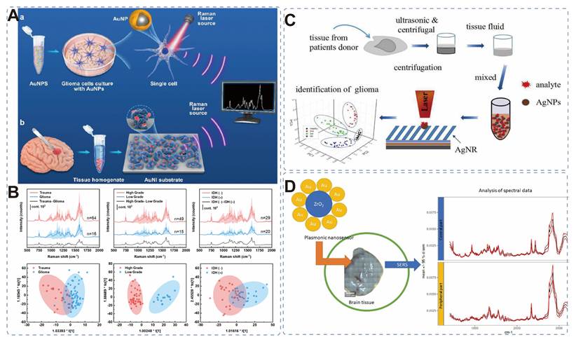
To further ensure reproducibility across different tumor grades, a SERS substrate consisting of Ag NPs modified with Ag nanorods (Ag NPs@Ag NRs) (with an enhancement factor of 1.37×10⁹) enabled the differentiation between healthy brain tissue and brain glioma tissue of various grades (Figure 11C) [161]. By analyzing characteristic peaks at 653 cm-1 and 724 cm-1 in the SERS spectra and combining with PCA, the method can achieve rapid, label-free differentiation between healthy tissue and grades I-IV, providing an efficient method for the non-invasive and rapid grading diagnosis of brain gliomas. This approach eliminates the need for complex labeling procedures and rapidly acquires comprehensive molecular information about the tissue, making it potentially valuable for real-time intraoperative detection. Similarly, an Au@ZrO2 plasmonic nanosensor can be used to distinguish tumor cores from peripheral regions intraoperatively (Figure 11D). The ZrO2 coating effectively suppresses fluorescence interference while enhancing Raman signals, achieving an LOD of 10-7 mol/L for Rhodamine 6G [162]. Analysis of the 178 spectral sets from the nine patients showed good discrimination at a certain threshold, and thus a new tool for precision neurosurgery has been obtained.
Based on the above, label-free SERS is currently one of the ways that is closer to clinical application. Label-free SERS does not need to be added to the sample, so the preparation steps before surgery are simpler. Label-free SERS detection has its own problems as well. Compared with the nanoprobe-assisted method, it is relatively more dependent on spectral differentiation for diagnosis, and it is also easy to be affected by tissue heterogeneity and fluorescence. Therefore, going forward, more efforts will need to be made in the field of label-free SERS detection to standardize spectral acquisition, uniform pretreatment protocols and multi-centre clinical validation to improve the reproducibility of clinical applications.
4.3.2 Nanoprobe-Based SERS for Enhanced Sensitivity
Although label-free methods are simple to operate and can achieve high classification performance through small-sample testing combined with multivariate analysis, their analytical performance mainly depends on the inherent spectral differences between tissues. In complex clinical samples, factors such as weak Raman scattering cross-sections, fluorescence background, tissue heterogeneity, and overlapping characteristic peaks can reduce spectral discrimination and limit stability across different patients and clinical centers. To overcome the problems of weak Raman signals and overlapping characteristic peaks in label-free detection, researchers used plasmonic nanoparticles composed of functionalized Raman reporter molecules and targeting ligands to detect tissues, thereby improving signal intensity and specificity.
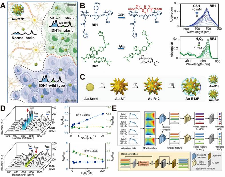
For instance, a SERS probe was exploited to simultaneously detect two metabolites in gliomas, with a deep learning model (DBCNet) to analyze Raman spectra (Figure 12A) [163]. By constructing an orthogonal response SERS probe (Au-R12P), they were able to simultaneously quantify GSH and H₂O₂ (Figures 12B and 12C). Combined with DBCNet, they completed intraoperative IDH1 genotyping of gliomas within 7 min, achieving an area under the curve (AUC) of 0.985 in 31 patients, thus providing a rapid new method for intraoperative molecular subtyping, precise diagnosis, and treatment of gliomas (Figures 12D and 12E).
(A) Identifying IDH1 genotypes of gliomas by determining redox-associated metabolites. (B) Chemical structures of Raman molecular reporters RR1 and RR2; absorbance of RR1 (5 × 10⁻⁶ M, upper panel) and RR2 (1 × 10⁻⁵ M, lower panel) as a function of GSH and H2O2 concentration, respectively. (C) Synthetic procedure of Au-R12P. (D) Raman spectra of Au-R12P in PBS solution as a function of GSH concentration (0.5-20 mM) and H2O2 concentration (5-200 μM). (E) Schematic diagram of the proposed DBCNet for analyzing the Raman spectra of Au-R12P in the presence of redox-associated metabolites. Reproduced with permission from Ref. [163]. Copyright 2025, Wiley-VCH.
4.4 From Margin Detection to Surgical Navigation
Compared with many other solid tumors, the surgery of glioma faces several unique challenges. Gliomas exhibit highly infiltrative growth patterns, resulting in poorly defined tumor margins that are difficult to distinguish from normal brain tissue using conventional visual inspection or neuronavigation alone [164,165]. In addition, because the brain contains many functional areas and gliomas often occur near critical regions responsible for language, motor, and sensory functions, it is essential to preserve neural function as much as possible while removing as much of the tumor as possible during surgical resection [166,167]. Also, substantial brain shift during the operation can decrease the precision of preoperative MRI-based navigation [168,169]. These factors collectively limit the effectiveness of conventional intraoperative guidance techniques.
Conventional 5-ALA fluorescence navigation is often compromised by photobleaching and limited sensitivity in low-grade gliomas, while MRI is hindered by high costs and poor real-time feedback [170,171]. SERS technology addresses these bottlenecks through its high spatial resolution, photostability, and superior capacity for precise margin delineation. As shown in Table 2, compared to traditional intraoperative navigation methods for gliomas (e.g., MRI, 5-ALA fluorescence and histopathology), SERS has significant potential in terms of resolution, molecular specificity, and real-time intraoperative navigation.
Comparison of SERS with conventional intraoperative and diagnostic modalities for glioma.
| Modality | Spatial Resolution | Sensitivity (%) | EOR | Intraoperative application | Key Limitations |
|---|---|---|---|---|---|
| MRI | ~1-2 mm | 80-95 (reported range) | Facilitates preoperative planning | Yes | Limited molecular information, brain shift |
| 5-ALA fluorescence | ~0.1-1mm | 75-95 (HGG); 20-60 (LGG) | Increases gross total resection rates by approximately 15-25% compared with white-light surgery | Yes | Photobleaching, low sensitivity in LGG |
| Histopathology | 1-10 μm | 90-99 | Provides definitive diagnosis | No | Time-consuming, sampling bias |
| SERS | <1 μm (subcellular) | Variable (85-99 in selected studies) | High accuracy for tumor margin; detection in preclinical and early translational studies | Yes | Reproducibility, standardization |
4.4.1 Metabolism-Guided Surgical Strategies
SERS-guided glioma surgical resection can be divided into two categories. One category directly uses SERS to detect normal and tumor tissues, and distinguishes tissues based on the differences in the characteristic peaks of metabolite molecules between the two types of tissues. The other category is based on the difference in the concentration of metabolites (such as extracellular acidosis) between normal and tumor tissues, and uses the response of specific SERS probes to metabolites to perform tissue resection.
For instance, Ag NPs as a substrate can be employed to differentiate between normal cells and glioblastoma cells based on the common characteristic peaks of two metabolites at 655 cm-1 and 717 cm-1 [172]. By combining neural networks with a multinomial regression model, this method can accurately delineate tumor boundaries during surgery, providing a new, rapid quantitative diagnostic method for the surgical resection of gliomas (Figure 13A).
(A) Schematic illustration for the detection of glioma cell proportion with an Ag NPs-based SERS method. Reproduced with permission from Ref. [172]. Copyright 2024, Elsevier. (B) Design and synthesis of pH-responsive SERS probe and schematic illustration of its application in guiding the surgical resection of gliomas. Reproduced with permission from Ref. [173]. Copyright 2020, The Royal Society of Chemistry. (C) Schematic diagram illustrating the use of SERS navigation system to automatically analyze Raman spectra using deep learning models, map the pH distribution at the tumor margin, and resect acidic tissue with a pH below 7.0. (D) Schematic illustration of the surgical procedure for acid-edge-guided glioma resection in an animal model, along with representative H&E, Ki67, and PCNA staining images of resected tissue under different pH conditions. Reproduced with permission from Ref. [174]. Copyright 2022, Wiley-VCH.
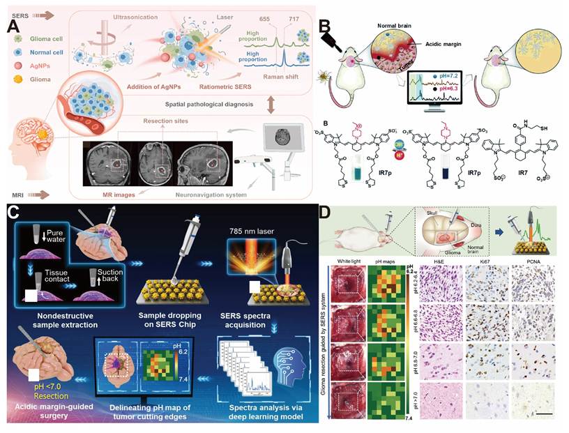
To address chemical gradients, Duan et al. developed a pH-responsive SERS probe (AuS-IR7p), based on the spatial orientation-induced intramolecular energy transfer (SOIET) mechanism (Figure 13B) [173]. This probe circumvents the BBB, identifying acidic tumor margins within 5 s and enabling guided resection up to a depth of 2.2 mm, which significantly improved survival in rat models.
Similarly, a probe-free intelligent SERS platform was utilized for surgical boundary navigation [174]. The system utilizes extracellular acidosis during glioma cell metabolism as a characteristic biomarker of gliomas. Combined with a one-dimensional convolutional neural network deep learning model, it can rapidly extract tumor margin metabolites during surgery and generate high-resolution pH maps, thereby accurately defining the boundaries of acidic infiltration. Furthermore, animal experiments demonstrated that surgery guided by this sensing platform significantly prolonged the overall survival of glioma models (Figures 13C and 13D).
4.4.2 Targeted Nanoprobe-Guided Surgery in Animal Models
Modifying the surface of SERS substrate materials with functionalized antibodies or peptides can specifically bind to tumor cells, enabling precise localization of tumor areas during surgery and significantly improving the contrast between tumors and normal tissues, thus facilitating more thorough tumor resection in animal models.
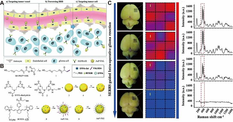
Qi et al. designed an EGFRvIII-targeting bimodal Au nanoprobe, AuP-FAL, with 60 nm gold nanospheres modified with MRI-enhanced Gd³⁺-DTPA, Raman reporter molecule IR783B, and targeting ligands FAL peptide and PEG (Figures 14A and 14B). In vitro validation showed that the probe had high targeting and uptake rates on EGFRvIII+ U87MG cells. In a nude mouse orthotopic glioma model, the tumor boundary could be delineated using preoperative MRI with a high target-to-background ratio, combined with intraoperative SERRS imaging using a handheld Raman scanner for precise resection (Figure 14C). Postoperative verification showed no tumor residue and it could also overcome false positives of brain displacement [175]. This provides an efficient tool for image-guided surgery of EGFRvIII+ glioblastoma and has clinical translational potential.
(A) Schematic diagram of AuP-FAL visualization of brain tumor margins by targeting EGFRvIII. (B) Synthesis of the nanoprobes. (i) FAL, (ii) 2-aminoethanethiol, (iii) H2O, (iv) GdCl3·6H2O, (v) 5, (vi) 1, PEG-SH and (vii) PEG-SH. (C) AuP-FAL guiding intra-operative brain tumor resection by handheld Raman detector. Reproduced with permission from Ref. [175]. Copyright 2017, The Royal Society of Chemistry.
4.4.3 Multimodal SERS Platforms
In addition to using the SERS method alone to guide surgical resection of gliomas, it can also be combined with other novel methods (such as fluorescence imaging, photothermal therapy, and photodynamic therapy) to simultaneously achieve diagnosis, intraoperative navigation, and treatment, showing broad prospects for the integrated development of diagnosis and treatment [176,177].
Xu et al. developed an integrated SPIT nanoprobe based on Au NSs (Figure 15A). The Au NSs were coated with macrophage membranes overexpressing the SIRPα variant, enabling their accumulation in tumor regions through BBB disruption and targeting strategies [178]. This strategy enables precise identification of tumor margins and micrometastases during surgery and eliminates residual tumors postoperatively through the photothermal effect. Using this approach, tumor recurrence in mice was significantly suppressed, while the survival was prolonged. The combination of intraoperative detection and postoperative treatment provides a new approach to the comprehensive diagnosis and treatment of glioblastoma.
(A) Schematic diagram of SPIT probe preparation and SPIT probe for glioma therapy integrating SERS and PTT. Reproduced with permission from Ref. [178]. Copyright 2025, Wiley-VCH. (B) Schematic illustration of the preparation of GBM-PDTCM@AuNRs and their application in GBM therapy. Reproduced with permission from Ref. [114]. Copyright 2023, American Chemical Society. (C) Schematic illustration of the preparation and application of hybrid CM-biomimetic NIR-II SERS probe for intraoperative resection guidance of in situ glioblastoma. Reproduced with permission from Ref. [179]. Copyright 2025, American Chemical Society.
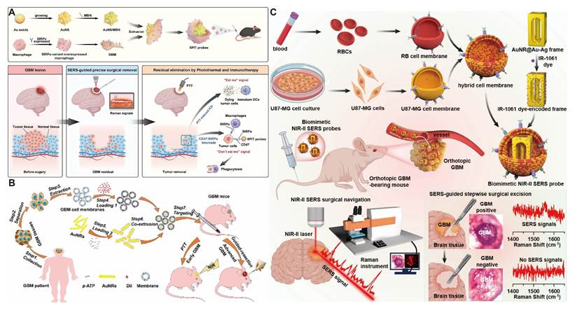
Furthermore, Au NRs with tumor cell membranes derived from glioblastoma patients (GBM-PDTCM) can create GBM-PDTCM@AuNRs (Figure 15B) [114]. Leveraging its homology, the probe efficiently crosses the BBB and precisely targets gliomas. It emits both fluorescence and SERS signals, enabling precise, complete resection via dual-modal navigation within 15 min. Additionally, it can destroy in situ tumors through photothermal therapy, extending the median survival of mice from 25 days to 52 days. This approach offers a new integrated surgical and non-surgical diagnostic and therapeutic strategy for glioblastoma at all stages.
In addition, an AuNR@Au-Ag framework plasmonic structure was coated onto a mixed cell membrane of erythrocytes and U87-MG cells to construct a near-infrared II (NIR-II) SERS probe (Figure 15C) [179]. This probe can effectively cross the BBB and target glioblastoma in situ. Utilizing the low scattering and high signal-to-noise ratio advantages of the NIR-II window, the probe can identify intraoperative microlesions (≈110 μm) in real time, thereby achieving precise stepwise resection. This study is the first to apply NIR-II SERS to glioma navigation, providing a high-performance optical solution for precise brain tumor surgery.
As shown in Table 3, we analyzed and compared the detection targets mentioned in the aforementioned literature. Metabolites, nucleic acids/proteins and exosomes possess significant clinical potential for various stages of glioma management, including preoperative diagnosis, intraoperative navigation and postoperative monitoring. Among these, exosomes are recognized as the most promising detection targets for glioma biomarkers due to their easily accessible nature and high information density. On the other hand, SERS detection of a single cell is limited by the high precision of instruments and the practical difficulties in obtaining a single-cell sample in clinical applications, which limit its general use. Label-free SERS has great clinical potential for direct detection of glioma tissue at the tissue level with easy specimen acquisition and simple sample preparation. For clinical applications of SERS-labeled probes, it is important to validate the biocompatibility and toxicity of the substrate materials. In summary, the SERS technology has extensive application prospects in the accurate surgical navigation of gliomas.
Comparison of SERS strategies for glioma detection and diagnosis.
| Strategy | Target | Development Status | Advantages | Limitations | Ref |
|---|---|---|---|---|---|
| Metabolite sensing | pH, GSH, H2S | Preclinical | Rapid response | Low specificity | 127-129 |
| Nucleic acid/protein | ctDNA, VEGF | Early clinical validation | High specificity | Complex preparation | 130,131, 135,138 |
| Exosomes | EVs | Early clinical validation | Non-invasive | Standardization needed | 141-143; 146, 147 |
| Cellular analysis | Glioma cells | Research stage | Mechanistic insight | Poor clinical translation | 151-153 |
| Label-free tissue SERS | Tissue fingerprint | Advanced translational stage | No labeling | Lower sensitivity | 159-161 |
| Nanoprobe-based imaging | Tumor markers | Preclinical | High contrast | Biosafety concerns | 163 |
| SERS-guided surgery | Tumor margin | Preclinical | Real-time guidance | Insufficient clinical evidence | 172-175; 114,178,179 |
Overall, nanoprobe-guided SERS provides superior sensitivity and molecular specificity, whereas label-free SERS offers greater simplicity and easier clinical integration. Despite encouraging preclinical performance, most navigation platforms have only been validated in small animal models or limited patient cohorts. Before SERS technology can be employed in actual neurosurgery, many more prospective clinical studies and rigorous prospective clinical trials must be carried out to confirm its reproducibility and long-term biosafety.
4.5 Artificial Intelligence for SERS-Based Glioma Diagnosis
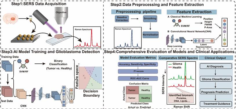
By means of combining artificial intelligence technology with SERS, all kinds of data related to tumors can be obtained from the analysis of complicated spectral information, thereby enabling highly sensitive detection of gliomas [180,181]. Unlike traditional spectral data analysis methods, AI-based data analysis models can rapidly identify and distinguish subtle spectral fluctuations that are complex and difficult to discern intuitively, thereby providing accurate information about gliomas in a short period of time to assist physicians in developing surgical resection plans (Figure 16). However, the effectiveness of AI models is closely related to the quality of SERS data. Therefore, it is necessary to establish an analytical framework guided by real-world clinical problems.
SERS-based artificial intelligence framework for glioblastoma detection: from data acquisition to clinical decision support.
4.5.1 Characteristics of SERS spectral data and implications for AI modeling
SERS spectra acquired from biological samples present challenges that are rarely encountered in conventional spectroscopic analyses, making the development of robust AI models considerably more difficult. One major limitation is the imbalance between the large number of spectral variables and the relatively small number of clinically annotated samples, a situation commonly referred to as the “small n, large p” problem, which substantially increases the risk of model overfitting [182,183]. Changes in the substrate, fluctuations in the excitation environment, and different compositional structures of biological samples can affect the performance of the model due to spectral changes. Background interference and fluorescence can further obscure diagnostically relevant spectral features, placing greater demands on feature extraction and model robustness rather than on classification accuracy alone. Consequently, in order to conduct reliable AI-assisted SERS analysis, improvements need to be made in the machine-learning algorithms, standardization of data acquisition and strictness of spectral preprocessing, and also careful control over experimental protocols.
4.5.2 Evolution of AI methodologies for SERS analysis
With the development of artificial intelligence technology over time, many studies have since been conducted to apply it to SERS-based analysis of gliomas, and deep learning (DL) methods have begun to replace traditional machine learning techniques [184,185]. Early studies primarily relied on classifiers such as principal component analysis-support vector machines (PCA-SVM) and partial least squares discriminant analysis (PLS-DA), where model performance depended heavily on feature extraction and dimensionality reduction before classification [186]. Due to the above reasons, the traditional machine learning model can still be employed for diagnosis with a small number of data samples. For example, Wang et al. combined SERS with multiple machine-learning algorithms to differentiate glioma from traumatic brain tissue and determine IDH mutation status, achieving classification accuracies above 97% and area under the curve (AUC) values exceeding 0.99 [160]. However, the above methods are often unable to capture the intricate, non-linear connections among the many pieces of information in high-dimensional spectra.
Recently, more research has begun to be conducted on how to use DL architectures, especially CNN and ResNet-based models, to solve the above problems [187]. By learning hierarchical feature representations directly from raw spectra, these networks reduce dependence on manual feature engineering while improving robustness to spectral variation and noise [188,189]. With the development of such technology, SERS is now being employed to recognize various types of molecules and investigate extracellular vesicles in glioblastoma. Jalali and others have developed a CNN-based framework for the analysis of single extracellular vesicle SERS spectra [143]. Based on this, it has been found that the genes of glioblastoma are altered in clinical samples with an accuracy of 87%, and at a very low frequency of 1.23%, mutant vesicles have also been identified. From the above results, it can be concluded that DL will be used to construct a high-sensitivity, label-free liquid biopsy assay.
Despite the above favorable developments, some problems have not been solved and thus cannot be applied to clinical work. Most of the reported models have only been trained and tested on data from a single hospital with a small number of cases, so it is unknown whether they can be reproduced and generalized to other groups of patients, instruments and sample-processing methods. In order to solve the problems mentioned above, more multi-centre datasets and independent external validation need to be obtained. In addition to the traditional machine learning method, new few-shot learning and domain adaptation approaches will also be employed to address the problem of small datasets and improve the consistency of performance in different clinical centres. In conjunction with standardization of spectral acquisition and preprocessing protocols, it is hoped that the new technology will be used in many clinics in the near future.
A key challenge currently facing the use of data models is the limited availability of data due to the difficulty in obtaining clinical samples. Therefore, in addition to traditional ML models that require extensive clinical data analysis, the development of emerging methods, such as those based on learning from small sample sizes, should be pursued to improve model accuracy when clinical samples are scarce. Furthermore, explainable artificial intelligence (XAI) frameworks are expected to play an increasingly important role. This approach improves model transparency and enhances clinical credibility by establishing links between spectral features and biologically meaningful molecular biomarkers. Leveraging the characteristics of Raman spectroscopy data, a standardized database can be constructed to further enhance cross-platform generalization capabilities and advance the standardization of molecular diagnostics.
Furthermore, the multimodal data fusion strategies involving SERS with histopathology, MRI, genomics and clinical information are anticipated to offer a more complete characterization of glioma biology and to improve the accuracy of diagnosis. Together, these developments will enable the transition of AI-assisted SERS systems from proof-of-concept studies to clinically deployable decision support tools.
Generally, AI and SERS have been used together to improve the analysis of large amounts of spectral data and enhance the accuracy of diagnostic information extraction for gliomas. According to the progress of algorithm development, construction of a standard spectral database, an interpretable analytical model and strict validation in many clinical centres will be conducted, and further development will be carried out. Due to the reasons listed above, a stable SERS-AI platform that can be used in the clinical application of neuro-oncology accurately will be built.
5. Challenges and Future Perspectives
5.1 Critical Challenges for Clinical Translation
SERS has shown some promise for the diagnosis and surgical guidance of glioma so far, but many problems have yet to be solved. One of the most critical challenges is that SERS nanoprobes generally exhibit limited ability to efficiently cross the BBB. Most of the platforms have reported so far that it is passive accumulation due to an increase in permeability and retention (EPR) effect or local disruption of the BBB caused by glioma progression, and active and selective transport across an intact BBB has not yet been achieved. This limitation is particularly relevant in early-stage gliomas, where BBB integrity is largely preserved, and nanoprobe uptake is consequently restricted.
The other critical question is that the SERS substrates are not reproducible. Even a small change in the shape of the nanostructure or in the distribution of hot spots will result in a relatively large shift of the spectrum and thus reduce the accuracy of quantification and interlaboratory consistency. The AI-assisted SERS system also has the problem that it is not working well due to a small amount of low-quality spectral data. Many deep learning models are not easy to interpret, and thus they have not been widely used in serious cases of medical treatment. Another translation issue is that we do not know how safe plasmonic nanomaterials are in the long run. Extensive research has been conducted on the biodistribution, pharmacokinetics, metabolism, and excretion of these materials, as well as stringent regulatory approvals; only then will they be widely used in the clinical treatment of gliomas.
Importantly, future progress is unlikely to rely solely on increasingly complex deep learning architectures. Instead, standardized SERS data acquisition and high-quality multicenter datasets are expected to contribute more substantially to clinical performance than algorithmic complexity alone.
5.2 Future Perspectives: Toward Precision Neuro-oncology
5.2.1 Technological Advances for SERS-Based Glioma Research
As nanotechnology, spectral analysis techniques and AI algorithms continue to advance, SERS has great potential in glioma research applications. Despite the rapid development of SERS for glioma diagnosis and intraoperative navigation, there are still several challenges to be solved for its extensive clinical application. Future work may be directed to the development of smart nanoprobes capable of crossing the BBB and accumulating specifically in invasive tumor regions. Furthermore, combining SERS with NIR-II imaging may enhance tissue penetration depth and minimize background noise, enabling real-time imaging of infiltrative tumor borders during molecularly guided surgery.
Another key direction is to improve the reproducibility of the substrate. The future SERS substrate will move away from the dependence on randomly generated hotspots and utilize photolithographically engineered surface plasmon arrays, DNA origami-assisted assembly of nanoparticles, and microfabrication techniques to realize highly uniform electromagnetic enhancement and batch-to-batch consistency. Such approaches could allow for large-scale production and regulatory approval.
Biosafety in the long term is also a prerequisite for clinical translation. Future studies should focus on short-term toxicity studies and systematic studies of biodistribution, metabolism, clearance pathways and chronic biological effects of nanoparticles. Long-term safety concerns can be reduced by using biodegradable plasmonic materials and nanostructures that are cleared by the kidneys.
There are issues such as the substrate materials for current Raman analysis, and thus no particular quantitative standards have been established yet, except for multiplexing. Future strategies, including internal standards, ratio-based SERS and AI-assisted spectral calibration, may improve the analytical accuracy and reproducibility, thus facilitating the clinical applications.
5.2.2 Emerging Applications and Clinical Translation in Precision Neuro-oncology
Beyond technological improvements, emerging interdisciplinary approaches are expected to further expand the applications of SERS in precision neuro-oncology. In particular, advances in single-cell analysis and AI are expected to further broaden the capabilities of SERS. The combination of single-cell analysis with high-resolution SERS imaging will allow us to characterize the heterogeneity of gliomas at the cellular level, thus providing new insights into glioma evolution, treatment resistance and tumor microenvironment interactions. Moreover, the combination of SERS with AI may enable the development of digital pathology platforms for automatic tumor classification, molecular subtype recognition, and intraoperative decision making.
The integration of multi-omics liquid biopsy with SERS is likely to broaden the scope of molecular characterization in glioma by enabling the concurrent analysis of circulating tumor DNA (ctDNA), microRNAs, proteins, metabolites, and extracellular vesicles. Thus, one of the reasons for the good spectral resolution of Raman spectroscopy has been exploited. Raman bands are generally only about 10-20 cm-1 wide, much narrower than fluorescence emission bands, and thus many molecular signals can be identified in one measurement. Therefore, SERS is suitable for multiplex biomarker detection, and it will be used to study the molecular heterogeneity of gliomas in more detail.
New combinations of neurotechnology and SERS will be developed in the future. By means of SERS and electroencephalography (EEG) coupled with neural recording platforms, changes in both molecules and electricity in the brain can be observed simultaneously. All the above many-format approaches will likely help improve the environment for early warning of all kinds of diseases and advance the construction of intelligent diagnostic workflows and other related technologies.
Although there have been some good developments, there are currently no FDA-approved SERS products for the treatment of gliomas, and most studies are still in the preclinical stage. Nevertheless, tissue-based diagnostics and liquid biopsy platforms are generally considered the most promising candidates for near-term clinical translation. Further advances are required to translate SERS from a promising research tool to a practical clinical technology for precision neuro-oncology, including substrate standardization, large-scale clinical validation, regulatory approval pathways and interpretable artificial intelligence.
In conclusion, although SERS still faces some problems in glioma research, the application potential of SERS in precision diagnosis, intraoperative navigation and personalized treatment will be further realized with continuous technological development. With further technological validation and clinical studies, SERS is expected to be a key tool for glioma diagnosis and therapy through interdisciplinary collaboration and expanded clinical research.
Acknowledgements
This work was financially supported by the project ZR2025MS257, supported by the Shandong Provincial Natural Science Foundation.
Authorship contributions
Min Zhang: Investigation and Writing. Ya-jun Luo: Investigation and Figures. Ya-nan Liu: Investigation. Ji-sen Li: Proofreading and Supervision.
Artificial intelligence (AI) usage statement
During the writing of the paper, AI tools (ChatGPT) were used solely for language polishing and grammar checking to enhance the academic quality of the paper. AI tools were not used for drafting the manuscript, analyzing data, creating figures and tables, or drawing conclusions. All content, including the interpretation of literature, presentation of data, and revision of the final draft, was independently completed, reviewed, and approved by the author. The author assumes full responsibility for the accuracy, originality, and completeness of this study.
Competing Interests
The authors have declared that no competing interest exists.
References
1. Weller M, Wen PY, Chang SM, Dirven L, Lim M, Monje M. et al. Glioma. Nat Rev Dis Primers. 2024;10:33
2. van den Bent MJ, Geurts M, French PJ, Smits M, Capper D, Bromberg JEC. et al. Primary brain tumours in adults. Lancet. 2023;402:1564-79
3. Alghamri MS, McClellan BL, Hartlage CS, Haase S, Faisal SM, Thalla R. et al. Targeting neuroinflammation in brain cancer: Uncovering mechanisms, pharmacological targets, and neuropharmaceutical developments. Front Pharmacol. 2021;12:680021
4. Horbinski C, Berger T, Packer RJ, Wen PY. Clinical implications of the 2021 edition of the WHO classification of central nervous system tumours. Nat Rev Neurol. 2022;18:515-29
5. Weller M, van den Bent M, Preusser M, Le Rhun E, Tonn JC, Minniti G. et al. EANO guidelines on the diagnosis and treatment of diffuse gliomas of adulthood. Nat Rev Clin Oncol. 2021;18:170-86
6. Song W-S, Chang P-C, Hueng D-Y, Li Y-F. TMEM106A as a macrophage-associated biomarker of prognosis in IDH-wildtype glioma: Integrative multi-omics and spatial analyses. Cancer Med. 2025;14:e71454
7. Verdugo E, Puerto I, Medina MÁ. An update on the molecular biology of glioblastoma, with clinical implications and progress in its treatment. Cancer Commun. 2022;42:1083-111
8. Park JH, Hothi P, de Lomana ALG, Pan M, Calder R, Turkarslan S. et al. Gene regulatory network topology governs resistance and treatment escape in glioma stem-like cells. Sci Adv. 2024;10:eadj7706
9. Sethi P, Ghosh S, Singh KK, Han SS, Bhaskar R, Sinha JK. Nanoparticle-based therapeutics for glioblastoma multiforme treatment. Adv Ther. 2025;8:2400546
10. Xie R, Fu S, Zhu G, Ai H, Song B, Lui S. et al. Iron oxide nanoclusters formed by acid-induced in situ calcium ion cross-linking for targeted magnetic resonance imaging of glioblastoma. Chem Eng J. 2024;484:149410
11. Singh K, Hotchkiss KM, Parney IF, De Groot J, Sahebjam S, Sanai N. et al. Correcting the drug development paradigm for glioblastoma requires serial tissue sampling. Nat Med. 2023;29:2402-5
12. Caro C, Paez-Muñoz JM, Pernía Leal M, Carayol M, Feijoo-Cuaresma M, García-Martín ML. Metabolically-driven active targeting of magnetic nanoparticles functionalized with glucuronic acid to glioblastoma: Application to MRI-tracked magnetic hyperthermia therapy. Adv Healthcare Mater. 2025;14:2404391
13. Lemarié A, Lubrano V, Delmas C, Lusque A, Cerapio J-P, Perrier M. et al. The STEMRI trial: Magnetic resonance spectroscopy imaging can define tumor areas enriched in glioblastoma stem-like cells. Sci Adv. 2023;9:eadi0114
14. Cozzi FM, Mayrand RC, Wan Y, Price SJ. Predicting glioblastoma progression using MR diffusion tensor imaging: A systematic review. J Neuroimaging. 2025;35:e13251
15. Wang F, Zhou X, Chen R, Kang J, Yang X, Lin J. et al. Improved performance of non-preloaded and high flip-angle dynamic susceptibility contrast perfusion-weighted imaging sequences in the presurgical differentiation of brain lymphoma and glioblastoma. Eur Radiol. 2023;33:8800-8
16. Weller M, Wick W, Aldape K, Brada M, Berger M, Pfister SM. et al. Glioma. Nat Rev Dis Primers. 2015;1:15017
17. Armakolas A, Kotsari M, Koskinas J. Liquid biopsies, novel approaches and future directions. Cancers. 2023;15:1579
18. Dillemuth P, Ayo A, Airenne TT, Lövdahl P, Bakay E, Zhuang X. et al. Utilizing monocarboxylate transporter 1-mediated blood-brain barrier penetration for glioblastoma positron emission tomography imaging with 6-[18F]fluoronicotinic acid. Mol Pharmaceutics. 2025;22:4819-30
19. Albert NL, Galldiks N, Ellingson BM, van den Bent MJ, Chang SM, Cicone F. et al. PET-based response assessment criteria for diffuse gliomas (PET RANO 1.0): A report of the RANO group. Lancet Oncol. 2024;25:29-41
20. Trivedi R, Bhat KP. Liquid biopsy: Creating opportunities in brain space. Br J Cancer. 2023;129:1727-46
21. Zheng M-M, Zhou Q, Chen H-J, Jiang B-Y, Tang L-B, Jie G-L. et al. Cerebrospinal fluid circulating tumor DNA profiling for risk stratification and matched treatment of central nervous system metastases. Nat Med. 2025;31:1547-56
22. Karschnia P, Gerritsen JKW, Teske N, Cahill DP, Jakola AS, van den Bent M. et al. The oncological role of resection in newly diagnosed diffuse adult-type glioma defined by the WHO 2021 classification: A review by the RANO resect group. Lancet Oncol. 2024;25:404-19
23. Li R, Wang H, Liang Q, Chen L, Ren J. Radiotherapy for glioblastoma: Clinical issues and nanotechnology strategies. Biomater Sci. 2022;10:892-908
24. Wood H. Epigenetic reprogramming of glioblastoma to overcome chemotherapy resistance. Nat Rev Neurol. 2025;21:234
25. He Q, Yang M, Wang P, Graca L, Sun H, Li C. Tailoring imaging probes for defining glioma invasive margins: Tracing the molecular footprints of cancer cells. Biomaterials. 2026;327:123779
26. Baptista JM, Brenner LBO, Henrique A, Ito LA, Nabarro PH, Santos LP. et al. Efficacy and safety of intraoperative magnetic resonance imaging for low-grade and high-grade gliomas: An updated systematic review and meta-analysis. Neurosurg Rev. 2025;48:465
27. Sadrinasab S, Aminiyan A, Saket S, Khosravi F, Pourmohammadi N, Saadat Fakhr M. Navigating the challenges: Ultrasound innovations in brain glioma surgery. Front Neurol. 2025;16:1553018
28. Chen J-S, Young JS, Berger MS. Current and future applications of 5-aminolevulinic acid in neurosurgical oncology. Cancers. 2025;17:1332
29. Lee S, Dang H, Moon J-I, Kim K, Joung Y, Park S. et al. SERS-based microdevices for use as in vitro diagnostic biosensors. Chem Soc Rev. 2024;53:5394-427
30. Cialla-May D, Bonifacio A, Bocklitz T, Markin A, Markina N, Fornasaro S. et al. Biomedical SERS-the current state and future trends. Chem Soc Rev. 2024;53:8957-79
31. Li Q, Huo H, Wu Y, Chen L, Su L, Zhang X. et al. Design and synthesis of SERS materials for in vivo molecular imaging and biosensing. Adv Sci. 2023;10:2202051
32. Yao L, Chen S, Yang S, Qiu T, Hao Q. Plasmonic nanoarrays as SERS substrates: Advances, challenges, and perspectives. Acc Chem Res. 2026;59:1244-55
33. Guselnikova O, Lim H, Kim H-J, Kim SH, Gorbunova A, Eguchi M. et al. New trends in nanoarchitectured SERS substrates: Nanospaces, 2D materials, and organic heterostructures. Small. 2022;18:2107182
34. Shin H-J, Choi K, Son J, Kim M, Park SS, Nam J-M. Metal-organic framework/Au core/shell nanocubes for spatially aligned molecular analysis and molecular-fingerprint sensing with surface-enhanced Raman scattering. J Am Chem Soc. 2025;147:29792-800
35. Wang X, Shen S, Sun N, Zhu Y, Zhang J. Neural network-assisted dual-functional hydrogel-based microfluidic SERS sensing for divisional recognition of multimolecule fingerprint. ACS Sens. 2025;10:1197-205
36. Sinha SS, Jones S, Pramanik A, Ray PC. Nanoarchitecture based sers for biomolecular fingerprinting and label-free disease markers diagnosis. Acc Chem Res. 2016;49:2725-35
37. Cutshaw G, Uthaman S, Hassan N, Kothadiya S, Wen X, Bardhan R. The emerging role of Raman spectroscopy as an omics approach for metabolic profiling and biomarker detection toward precision medicine. Chem Rev. 2023;123:8297-346
38. Liu Y, Liu H, Kang C, Wang C. Cancer diagnosis using label-free SERS-based exosome analysis. Theranostics. 2024;14:1966-81
39. Liu H, Gao X, Xu C, Liu D. SERS tags for biomedical detection and bioimaging. Theranostics. 2022;12:1870-903
40. Kaur B, Kumar S, Kaushik BK. Recent advancements in optical biosensors for cancer detection. Biosens Bioelectron. 2022;197:113805
41. Richards-Kortum R, Lorenzoni C, Bagnato VS, Schmeler K. Optical imaging for screening and early cancer diagnosis in low-resource settings. Nat Rev Bioeng. 2024;2:25-43
42. Han XX, Rodriguez RS, Haynes CL, Ozaki Y, Zhao B. Surface-enhanced Raman spectroscopy. Nat Rev Methods Primers. 2022;1:87
43. Lian S, Li X, Lv X. Recent developments in SERS microfluidic chips: From fundamentals to biosensing applications. ACS Appl Mater Interfaces. 2025;17:10193-230
44. Cai Q, Li LH, Mateti S, Bhattacharjee A, Fan Y, Huang S. et al. Boron nitride nanosheets: Thickness-related properties and applications. Adv Funct Mater. 2024;34:2403669
45. Chaudhry I, Hu G, Ye H, Jensen L. Toward modeling the complexity of the chemical mechanism in SERS. ACS Nano. 2024;18:20835-50
46. Le Ru EC, Auguié B. Enhancement factors: A central concept during 50 years of surface-enhanced Raman spectroscopy. ACS Nano. 2024;18:9773-83
47. Zhang M, Liu A, Zhang J, Meng X, Wang Y, Song X. et al. Chemical engineering of heterojunction SERS substrates: Emerging tools for disease diagnosis and health monitoring. SmartMat. 2025;6:e70045
48. Schuknecht F, Kołątaj K, Steinberger M, Liedl T, Lohmueller T. Accessible hotspots for single-protein SERS in DNA-origami assembled gold nanorod dimers with tip-to-tip alignment. Nat Commun. 2023;14:7192
49. Zhang J, Song C, He X, Liu J, Chao J, Wang L. DNA-mediated precise regulation of SERS hotspots for biosensing and bioimaging. Chem Soc Rev. 2025;54:5836-63
50. Ma H, Pan S-Q, Wang W-L, Yue X, Xi X-H, Yan S. et al. Surface-enhanced Raman spectroscopy: Current understanding, challenges, and opportunities. ACS Nano. 2024;18:14000-19
51. Yang Y, Wu S, Chen Y, Ju H. Surface-enhanced Raman scattering sensing for detection and mapping of key cellular biomarkers. Chem Sci. 2023;14:12869-82
52. Zhang K, Wang Y, Wu M, Liu Y, Shi D, Liu B. On-demand quantitative SERS bioassays facilitated by surface-tethered ratiometric probes. Chem Sci. 2018;9:8089-93
53. Yu Y, Wang Y, Peng Y, Liu Y, Li M, Chen R. et al. Activity-based nanoprobes for multiscale functional analysis of proteases in medulloblastomas. ACS Sens. 2025;10:6981-90
54. Liu Y, Cai C, Xu W, Li B, Wang L, Peng Y. et al. Interpretable machine learning-aided optical deciphering of serum exosomes for early detection, staging, and subtyping of lung cancer. Anal Chem. 2024;96:16227-35
55. Sun T, Wu Y, Ma H, Zhang C, Li C, Man B. et al. The design of WTe2/Graphene/Ag NPs heterostructure for the improvement of the chemical enhancement in SERS. Nano Lett. 2024;24:15324-30
56. Wang R, Ma J, Dai X, Gao Y, Gu C, Jiang T. Highly active SERS chip with both structure-ameliorated chemical enhancement and nanocavity-mediated electromagnetic enhancement. Sens Actuators, B. 2023;374:132782
57. Li J, Chen X, Zhu J-J. DNA functionalized plasmonic nanoassemblies as SERS sensors for environmental analysis. Aggregate. 2023;4:e271
58. Zhao J, Chen Y, Tang Y, Li B, Wang Q, Wang J. et al. Tandem dual-ratiometric SERS probe enables Raman imaging of neurological pH fluctuations in epilepsy. Angew Chem, Int Ed. 2025;64:e202504822
59. Wang X, Huang S-C, Hu S, Yan S, Ren B. Fundamental understanding and applications of plasmon-enhanced Raman spectroscopy. Nat Rev Phys. 2020;2:253-71
60. Ben-Shahar Y, Stone D, Banin U. Rich landscape of colloidal semiconductor-metal hybrid nanostructures: Synthesis, synergetic characteristics, and emerging applications. Chem Rev. 2023;123:3790-851
61. Tan S, Argondizzo A, Ren J, Liu L, Zhao J, Petek H. Plasmonic coupling at a metal/semiconductor interface. Nat Photonics. 2017;11:806-12
62. Liu D, Xue C. Plasmonic coupling architectures for enhanced photocatalysis. Adv Mater. 2021;33:2005738
63. Wang M, Wang T, Ojambati OS, Duffin TJ, Kang K, Lee T. et al. Plasmonic phenomena in molecular junctions: Principles and applications. Nat Rev Chem. 2022;6:681-704
64. Schlücker S. Surface-enhanced Raman spectroscopy: Concepts and chemical applications. Angew Chem, Int Ed. 2014;53:4756-95
65. Tan EX, Zhong Q-Z, Ting Chen JR, Leong YX, Leon GK, Tran CT. et al. Surface-enhanced Raman scattering-based multimodal techniques: Advances and perspectives. ACS Nano. 2024;18:32315-34
66. Lin S, Cheng Z, Li Q, Wang R, Yu F. Toward sensitive and reliable surface-enhanced Raman scattering imaging: From rational design to biomedical applications. ACS Sens. 2021;6:3912-32
67. Hang Y, Boryczka J, Wu N. Visible-light and near-infrared fluorescence and surface-enhanced Raman scattering point-of-care sensing and bio-imaging: A review. Chem Soc Rev. 2022;51:329-75
68. Li F, Mu X, Tang X, Song G, Sun H, Zha X. et al. Semiconductor SERS on colourful substrates with Fabry-Pérot cavities. Angew. Chem. 2023;135:e202218055
69. Wang X, Guo L. SERS activity of semiconductors: Crystalline and amorphous nanomaterials. Angew Chem, Int Ed. 2020;59:4231-9
70. Cao Z, He P, Huang T, Yang S, Han S, Wang X. et al. Plasmonic coupling of Ag NPs near graphene edges: A cross-section strategy for high-performance SERS sensing. Chem Mater. 2020;32:3813-22
71. Zhang J, Wu J, Wang B, Geng Y, Wang Z. A new strategy for constructing 3D hybrid graphene/Au/rectangular pyramids PMMA on a flexible SERS substrate for trace molecule detection. Sens Actuators, B. 2024;410:135711
72. Lafuente M, De Marchi S, Urbiztondo M, Pastoriza-Santos I, Pérez-Juste I, Santamaría J. et al. Plasmonic MOF thin films with Raman internal standard for fast and ultrasensitive SERS detection of chemical warfare agents in ambient air. ACS Sens. 2021;6:2241-51
73. Yan S, Zhang Z, Chen J, Wang Q, Wu Y, Sui Y. et al. Cavity-like silver aggregates-based colloidal SERS microfluidic platform for highly reproducible online reaction process analysis. Small. 2025;21:2501338
74. Jiang T, Zeng B-F, Zhang B, Tang L. Single-molecular protein-based bioelectronics via electronic transport: Fundamentals, devices and applications. Chem Soc Rev. 2023;52:5968-6002
75. Ying Y, Tang Z, Liu Y. Material design, development, and trend for surface-enhanced Raman scattering substrates. Nanoscale. 2023;15:10860-81
76. Tukova A, Nguyen NTT, Garcia-Bennett A, Rodger A, Wang Y. Plasmonic nanostars: Unique properties that distinguish them from spherical nanoparticles from a biosensing perspective. Adv Opt Mater. 2024;12:2401183
77. Pearce AK, Wilks TR, Arno MC, O’Reilly RK. Synthesis and applications of anisotropic nanoparticles with precisely defined dimensions. Nat Rev Chem. 2021;5:21-45
78. Lin Y, Hou J, Li B, Shu W, Wan J. Advancements in nanomaterials and molecular probes for spatial omics. ACS Nano. 2025;19:11604-24
79. Atta S, Zhao Y, Yampolsky S, Sanchez S, Vo-Dinh T. A simple sensitive solution-based “Mix-and-Detect” SERS platform utilizing highly stable multibranched gold nanostars for bilirubin detection in urine. Anal Chem. 2025;97:13930-9
80. Li X, Ma J, Zhang Y, Xu L, Gu C, Wei G. et al. Reusable dual-functional SERS sensor based on gold nanoflowers-modified red phosphorus nanoplates for ultrasensitive immunoassay and degradation of CA19-9. Biosens Bioelectron. 2022;207:114148
81. Liu H, Jiang Z, Hu Z, Zhang B, He T, Dong X. et al. Spatiotemporal Raman probing of molecular transport in sub-2-nm plasmonic quasi-2D nanochannels. Sci Adv. 2026;12:eaec3641
82. Wy Y, Jung H, Hong JW, Han SW. Exploiting plasmonic hot spots in Au-based nanostructures for sensing and photocatalysis. Acc Chem Res. 2022;55:831-43
83. Kim J, Zhao Q, Choi I, Oh MJ, Kwon S, Park S. Ensemble hot-spots in 3D supercrystals of plasmonic octahedral nanoparticles in tip-to-tip configured superlattices. Nat Commun. 2025;16:2762
84. Peng Y, Zhang W, Xu M, Zhao S, Yang L, Li D. et al. Morphology engineering of SnS2 nanostructures to stimulate PICT resonance for ultra-sensitive SERS sensors. Exploration. 2025;5:270016
85. Zhu R, Li J, Lin L, Song J, Yang H. Emerging plasmonic assemblies triggered by DNA for biomedical applications. Adv Funct Mater. 2021;31:2005709
86. Wang C, Wu Y, Cen P, Chen Q, Shao H. Plasmonic interfaces for advanced profiling of cell-free circulating biomarkers. ACS Appl Mater Interfaces. 2025;17:66203-20
87. Wang Z, Zhou X, Kong Q, He H, Sun J, Qiu W. et al. Extracellular vesicle preparation and analysis: A state-of-the-art review. Adv Sci. 2024;11:2401069
88. Kim WH, Lee JU, Jeon MJ, Park KH, Sim SJ. Three-dimensional hierarchical plasmonic nano-architecture based label-free surface-enhanced Raman spectroscopy detection of urinary exosomal miRNA for clinical diagnosis of prostate cancer. Biosens Bioelectron. 2022;205:114116
89. Al Shboul S, Singh A, Kobetic R, Goodlett DR, Brennan PM, Hupp T. et al. Mass spectrometry advances in analysis of glioblastoma. Mass Spectrom Rev. 2025;44:630-64
90. Cao S, Yang Z, Yuan J, He L, Sun X, Ding X. et al. Dual-modal serum and urine SERS metabolic fingerprint for the diagnosis and activity assessment of childhood lupus nephritis. Anal Chem. 2026;98:14204-16
91. Li H, Chen Y-Y, Wu L, Chen Y, Fu S-Y, Lin J-S. et al. Label-free SERS strategy for simultaneous detection of triplex renal biomarkers in serum toward precise renal function evaluation. Anal Chem. 2026;98:799-808
92. Fu B-B, Tian X-D, Song J-J, Wen B-Y, Zhang Y-J, Fang P-P. et al. Self-calibration 3D hybrid SERS substrate and its application in quantitative analysis. Anal Chem. 2022;94:9578-85
93. Cardellini J, Dallari C, De Santis I, Riccio L, Ceni C, Morrone A. et al. Hybrid lipid-AuNP clusters as highly efficient SERS substrates for biomedical applications. Nat Commun. 2024;15:7975
94. Liu Z, Jiang H, Lv X, Lian S, Li X. Performance enhancement of SERS-based lateral flow assays: Progress and prospective. Anal Chem. 2025;97:23701-20
95. Hu C, Guan M, Mi F, Zhang S, Geng P. Multimodal SERS biosensing platforms: Emerging opportunities for ultrasensitive biomarker detection and intelligent diagnostics. ACS Sens. 2026;11:1794-830
96. Liu Y, Ma H, Han XX, Zhao B. Metal-semiconductor heterostructures for surface-enhanced Raman scattering: Synergistic contribution of plasmons and charge transfer. Mater Horiz. 2021;8:370-82
97. Zhou H, Li D, Lv Q, Lee C. Integrative plasmonics: Optical multi-effects and acousto-electric-thermal fusion for biosensing, energy conversion, and photonic circuits. Chem Soc Rev. 2025;54:5342-432
98. Miao J, Liu Y, Xiao Y, Yuan C, Xu Q, Chen P. et al. Water-stable perovskite/metallic nanocomposites-based SERS aptasensor for detection of neuron-specific enolase. Biosens Bioelectron. 2025;280:117462
99. Lien M-C, Yeh IH, Tadepalli S, Liu K-K. ZnO nanocages decorated with Au@AgAu yolk-shell nanomaterials for SERS-based detection of hyperuricemia. ACS Omega. 2024;9:16160-7
100. Timilsina SS, Jolly P, Durr N, Yafia M, Ingber DE. Enabling multiplexed electrochemical detection of biomarkers with high sensitivity in complex biological samples. Acc Chem Res. 2021;54:3529-39
101. Yang L, Zhang B, Xu B, Zhao F, Zeng B. Ionic liquid functionalized 3D graphene-carbon nanotubes-AuPd nanoparticles-molecularly imprinted copolymer based paracetamol electrochemical sensor: Preparation, characterization and application. Talanta. 2021;224:121845
102. Ganesh S, Venkatakrishnan K, Tan B. Quantum scale organic semiconductors for SERS detection of DNA methylation and gene expression. Nat Commun. 2020;11:1135
103. Basu S, Rana N, Morgan D, Sen K. Gold nanoparticle incorporated graphene oxide as a SERS platform for ultratrace antibody free sensing of the cancer biomarker CEA. Langmuir. 2025;41:7886-901
104. Mohan B, Najafov B, Sharma PK, Pal A, Garazade IM, Pombeiro AJL. et al. Electrochemiluminescent COFs and HOFs as porous material engineering systems for bioanalysis and environmental monitoring. Small. 2026;22:e14406
105. Sun Y, Li Y, Ru Z, Leng D, Geng H, Wu T. et al. Heterolinker MOFs synergized with entropy-driven DNA amplification for precision dual-mode sensing of Microcystin-LR. Biosens Bioelectron. 2026;306:118712
106. Liu Y, Li M, Zhao Z, Zhao Z, Li M. A rapid and sensitive SERS-based Au/MIL-125(Ti) biosensor for hyperuricemia diagnosis via saliva analysis. Anal Chem. 2025;97:6438-46
107. Cheng H, Chen R, Zhan Y, Dong W, Chen Q, Wang Y. et al. Novel ratiometric surface-enhanced Raman scattering (SERS) biosensor for ultrasensitive quantitative monitoring of human carboxylesterase-1 in hepatocellular carcinoma cells using Ag-Au nanoflowers as SERS substrate. Anal Chem. 2024;96:18555-63
108. Lu Y, Shi D-W, Pan S-F, Liao Y-Q, Liu M-Q, Zhou L. et al. In situ spatial profiling of cytochrome C release at the single-cell level under photothermal stress utilizing a 3D bifunctional SERS substrate. Biosens Bioelectron. 2025;288:117818
109. Tang S, Hao Q, Huang Y, Zhao S, Hu K. Highly selective and sensitive histamine and tryptamine analysis using SiO2@AuNPs@PDA molecularly imprinted polymer coupled with SALDI-TOF MS. Talanta. 2024;270:125538
110. Li Y, Fan Z, Wei Z, Zhang H, Li Q, Yang K. Epitope-imprinted polymers: Fabrication technologies and emerging applications. Anal Chem. 2026;98:12177-200
111. Chen B, Gao J, Sun H, Zhao Y, Liu Y, Qiu X. Carboxyl-PEG modified Fe3O4 nanoparticles as an ultrasensitive SERS substrate for multiplex detection of exogenous hormones related to endometrial cancer. Methods. 2026;245:35-52
112. Rampado R, Caliceti P, Agostini M. Latest advances in biomimetic cell membrane-coated and membrane-derived nanovectors for biomedical applications. Nanomaterials. 2022;12:1543
113. Liao J, Gong L, Xu Q, Wang J, Yang Y, Zhang S. et al. Revolutionizing neurocare: Biomimetic nanodelivery via cell membranes. Adv Mater. 2024;36:2402445
114. Zhang H, Guan S, Wei T, Wang T, Zhang J, You Y. et al. Homotypic membrane-enhanced blood-brain barrier crossing and glioblastoma targeting for precise surgical resection and photothermal therapy. J Am Chem Soc. 2023;145:5930-40
115. Terzapulo X, Kassenova A, Bukasov R. Immunoassays: Analytical and clinical performance, challenges, and perspectives of SERS detection in comparison with fluorescent spectroscopic detection. Int J Mol Sci. 2024;25:2080
116. Dallari C, Lenci E, Trabocchi A, Bessi V, Bagnoli S, Nacmias B. et al. Multilayered bioorthogonal SERS nanoprobes selectively aggregating in human fluids: A smart optical assay for β-amyloid peptide quantification. ACS Sens. 2023;8:3693-700
117. Zheng M, Xiao Y, Zhang W, Lv S, Sun L, Wang Q. et al. Sensitive SERS platform for the detection of Staphylococcus aureus via freezing SERS tags and core-shell aptamer-Au@Fe3O4 nanoparticles. ACS Omega. 2025;10:30651-9
118. Muhammad M, Liu C, Yang G, Shao C-S, Xiong L, Xia H. et al. Early-stage Alzheimer’s disease profiling in blood achieved by multiplexing aptamer-SERS biosensors. Biosens Bioelectron. 2025;268:116907
119. Thenuwara G, Curtin J, Tian F. Advances in diagnostic tools and therapeutic approaches for gliomas: A comprehensive review. Sensors. 2023;23:9842
120. Behbahani M, Keisham B, Rosinski CL, Berry V, Mehta AI. Intraoperative imaging device for glioblastoma multiforme surgery: Review of Raman-based intraoperative imaging and introduction of a novel handheld probe technology. J Raman Spectrosc. 2021;52:1228-36
121. Huda NU, Bari RZA, Javed MA, Kiani MN, Jin Y. SERS meets artificial intelligence: A new frontier in cancer diagnosis and prognosis. Anal Chem. 2026;98:8757-80
122. Miska J, Rashidi A, Lee-Chang C, Gao P, Lopez-Rosas A, Zhang P. et al. Polyamines drive myeloid cell survival by buffering intracellular pH to promote immunosuppression in glioblastoma. Sci Adv. 2021;7:eabc8929
123. Du K, Xia Q, Sun J, Feng F. Visible light and glutathione dually responsive delivery of a polymer-conjugated temozolomide intermediate for glioblastoma chemotherapy. ACS Appl Mater Interfaces. 2021;13:55851-61
124. Musaie K, Abbaszadeh S, Marais K, Nosrati-Siahmazgi V, Rezaei S, Xiao B. et al. H2O2-generating advanced nanomaterials for cancer treatment. Adv Funct Mater. 2025;35:2425866
125. Li S, Liu Y, Yang T, Deng M, Cheng D, He L. Lysosome-specific near-infrared fluorescent probe with large Stokes shift for H2S imaging in U87 cells and brain glioma mice. Sens Actuators, B. 2025;426:137109
126. Nair JB, Mohapatra S, Joseph MM, Maniganda S, Gupta V, Ghosh S. et al. Tracking the footprints of paclitaxel delivery and mechanistic action via SERS trajectory in glioblastoma cells. ACS Biomater Sci Eng. 2020;6:5254-63
127. Yang G, Zhang K, Qu X, Xu W, Xu S. Ratiometric pH-responsive SERS strategy for glioma boundary determination. Talanta. 2022;250:123750
128. Zhang M, You Y, Zhang H, Zhang J, Yang F, Wang X. et al. Rapid glutathione analysis with SERS microneedles for deep glioblastoma tissue differentiation. Anal Chem. 2024;96:10200-9
129. Li D-W, Qu L-L, Hu K, Long Y-T, Tian H. Monitoring of endogenous hydrogen sulfide in living cells using surface-enhanced Raman scattering. Angew Chem. 2015;127:12949-52
130. Liu X, Zhou Y, Zheng T, Tian Y. Surface-enhanced Raman scattering technology based on WO3 film for detection of VEGF. Chem Res Chin Univ. 2021;37:900-5
131. Liu L, Thakur A, Kar Li W, Qiu G, Yang T, He B. et al. Site specific biotinylated antibody functionalized Ag@AuNIs LSPR biosensor for the ultrasensitive detection of exosomal MCT4, a glioblastoma progression biomarker. Chem Eng J. 2022;446:137383
132. Vaughan HJ, Green JJ, Tzeng SY. Cancer-targeting nanoparticles for combinatorial nucleic acid delivery. Adv Mater. 2020;32:1901081
133. Sanz-Garcia E, Zhao E, Bratman SV, Siu LL. Monitoring and adapting cancer treatment using circulating tumor DNA kinetics: Current research, opportunities, and challenges. Sci Adv. 2022;8:eabi8618
134. Wang H, Liu Q, Lan X, Jiang D. Framework nucleic acids in nuclear medicine imaging: Shedding light on Nano-Bio interactions. Angew Chem, Int Ed. 2022;61:e202111980
135. Wang Y, Yang Y, Cao X, Liu Z, Chen B, Du Q. et al. Simple and ultrasensitive detection of glioma-related ctDNAs in mice serum by SERS-based catalytic hairpin assembly signal amplification coupled with magnetic aggregation. Int J Nanomed. 2023;18:3211-30
136. Ragab MA, Fathy N, Attia YM, Amer EA, Fahim SA. The paradox of miR-107 in oncology: Dual roles as a tumor suppressor and oncomir. Cancer Gene Ther. 2026;33:888-96
137. Kciuk M, Yahya EB, Mohamed MM, Abdulsamad MA, Allaq AA, Gielecińska A. et al. Insights into the role of LncRNAs and miRNAs in glioma progression and their potential as novel therapeutic targets. Cancers. 2023;15:3298
138. Kim WH, Lee JU, Song S, Kim S, Choi YJ, Sim SJ. A label-free, ultra-highly sensitive and multiplexed SERS nanoplasmonic biosensor for miRNA detection using a head-flocked gold nanopillar. Analyst. 2019;144:1768-76
139. Kalluri R, LeBleu VS. The biology, function, and biomedical applications of exosomes. Science. 2020;367:eaau6977
140. Tenchov R, Sasso JM, Wang X, Liaw W-S, Chen C-A, Zhou QA. Exosomes─nature’s lipid nanoparticles, a rising star in drug delivery and diagnostics. ACS Nano. 2022;16:17802-46
141. Zhang Z, Lobb RJ, Tooney P, Wang J, Lane R, Zhou Q. et al. Monitoring glioblastoma extracellular vesicle evolution using a nanodiagnostic platform to detect glioma stem cells driving recurrent disease. Sci Adv. 2026;12:eadt2804
142. Jalali M, Isaac Hosseini I, AbdelFatah T, Montermini L, Wachsmann Hogiu S, Rak J. et al. Plasmonic nanobowtiefluidic device for sensitive detection of glioma extracellular vesicles by Raman spectrometry. Lab Chip. 2021;21:855-66
143. Jalali M, del Real Mata C, Montermini L, Jeanne O, Hosseini II Gu Z. et al. MoS2-plasmonic nanocavities for Raman spectra of single extracellular vesicles reveal molecular progression in glioblastoma. ACS Nano. 2023;17:12052-71
144. Joshi R, Ahmad R, Gardner K, Ahmadi H, Chen C-C, Stott SL. et al. Functional biomaterials and machine learning approaches for phenotyping heterogeneous tumor cells and extracellular vesicles. Biomater Sci. 2025;13:6239-69
145. Veliz L, Cooper TT, Grenier-Pleau I, Abraham SA, Gomes J, Pasternak SH. et al. Tandem SERS and MS/MS profiling of plasma extracellular vesicles for early ovarian cancer biomarker discovery. ACS Sens. 2024;9:272-82
146. Premachandran S, Haldavnekar R, Ganesh S, Das S, Venkatakrishnan K, Tan B. Self-functionalized superlattice nanosensor enables glioblastoma diagnosis using liquid biopsy. ACS Nano. 2023;17:19832-52
147. Ishwar D, Haldavnekar R, Das S, Tan B, Venkatakrishnan K. Glioblastoma associated natural killer cell EVs generating tumour-specific signatures: Noninvasive GBM liquid biopsy with self-functionalized quantum probes. ACS Nano. 2022;16:10859-77
148. Hwang I-J, Choi C, Kim H, Lee H, Yoo Y, Choi Y. et al. Confined growth of Ag nanogap shells emitting stable Raman label signals for SERS liquid biopsy of pancreatic cancer. Biosens Bioelectron. 2024;248:115948
149. Zieren RC, Zondervan PJ, Pienta KJ, Bex A, de Reijke TM, Bins AD. Diagnostic liquid biopsy biomarkers in renal cell cancer. Nat Rev Urol. 2024;21:133-57
150. Zhan C, Gao R, Wang Y, Tan H, Wang F, Chen X. et al. SERS-based microfluidic liquid biopsy of prostate cancer: New bioinspired dual recognition interface for single-cell detection. Chem Eng J. 2025;516:164059
151. Golubewa L, Karpicz R, Matulaitiene I, Selskis A, Rutkauskas D, Pushkarchuk A. et al. Surface-enhanced Raman spectroscopy of organic molecules and living cells with gold-plated black silicon. ACS Appl Mater Interfaces. 2020;12:50971-84
152. Burgio F, Piffaretti D, Schmidt F, Pieles U, Reinert M, Ritz M-F. et al. Tuning the surface chemistry of gold nanoparticles to specifically image glioblastoma cells using surface-enhanced Raman spectroscopy. ACS Appl Nano Mater. 2020;3:2447-54
153. Dhinakaran AK, Dharmalingam P, Ganesh S, Venkatakrishnan K, Das S, Tan B. Molecular crosstalk between T cells and tumor uncovers GBM-specific T cell signatures in blood: Noninvasive GBM diagnosis using immunosensors. ACS Nano. 2022;16:14134-48
154. He Q, Koster HJ, O'Sullivan J, Ono SG, O'Toole HJ, Leiserowitz GS. et al. Integration of label-free surface enhanced Raman spectroscopy (SERS) of extracellular vesicles (EVs) with Raman tagged labels to enhance ovarian cancer diagnostics. Biosens Bioelectron. 2025;288:117800
155. Krafft C, Schmitt M, Schie IW, Cialla-May D, Matthäus C, Bocklitz T. et al. Label-free molecular imaging of biological cells and tissues by linear and nonlinear Raman spectroscopic approaches. Angew Chem, Int Ed. 2017;56:4392-430
156. Danenberg E, Bardwell H, Zanotelli VRT, Provenzano E, Chin S-F, Rueda OM. et al. Breast tumor microenvironment structures are associated with genomic features and clinical outcome. Nat Genet. 2022;54:660-9
157. Nia HT, Munn LL, Jain RK. Physical traits of cancer. Science. 2020;370:eaaz0868
158. Elsheikh S, Coles NP, Achadu OJ, Filippou PS, Khundakar AA. Advancing brain research through surface-enhanced Raman spectroscopy (SERS): Current applications and future prospects. Biosensors. 2024;14:33
159. Wurm LM, Fischer B, Neuschmelting V, Reinecke D, Fischer I, Croner RS. et al. Rapid, label-free classification of glioblastoma differentiation status combining confocal Raman spectroscopy and machine learning. Analyst. 2023;148:6109-19
160. Wang C, Huang L, Wang S, Wu L, Wang Y, Dong J. A distinction of gliomas at cellular and tissue level by surface-enhanced Raman scattering spectroscopy. Chin Chem Lett. 2024;35:109383
161. Li J, Wang C, Yao Y, Zhu Y, Yan C, Zhuge Q. et al. Label-free discrimination of glioma brain tumors in different stages by surface enhanced Raman scattering. Talanta. 2020;216:120983
162. Ranc V, Pavlacka O, Kalita O, Vaverka M. Discrimination of resected glioma tissues using surface enhanced Raman spectroscopy and Au@ZrO2 plasmonic nanosensor. Spectrochim Acta, Part A. 2024;305:123521
163. Yin H, Zhang X, Zhao Z, Cao C, Xu M, Zhou S. et al. Artificial intelligent-enhanced metabolite profiling for intraoperative IDH1 genotyping in glioma using an orthogonally responsive SERS probe. Adv Sci. 2025;12:2503360
164. Fernandez NR, Chang Y, Nunes NM, Dimayacyac JR, Levine A, Ringel A. et al. Patterns of hypermutation shape tumorigenesis and immunotherapy response in mismatch-repair-deficient glioma. Nat Genet. 2026;58:132-42
165. Adnani L, Kassouf J, Meehan B, Spinelli C, Tawil N, Nakano I. et al. Angiocrine extracellular vesicles impose mesenchymal reprogramming upon proneural glioma stem cells. Nat Commun. 2022;13:5494
166. Sipos D, Raposa BL, Freihat O, Simon M, Mekis N, Cornacchione P. et al. Glioblastoma: Clinical presentation, multidisciplinary management, and long-term outcomes. Cancers. 2025;17:146
167. Taylor KR, Monje M. Neuron-oligodendroglial interactions in health and malignant disease. Nat Rev Neurosci. 2023;24:733-46
168. Huang S, He Z, Chen Y, Chen J, Hong S, Zhang Y. et al. Hybrid pneumatic-hydraulic actuation for MRI-guided robotic stereotactic neurointervention. Sci Adv. 2025;11:eady3624
169. He Z, Dai J, Ho JD-L, Tong H-S, Wang X, Fang G. et al. Interactive multi-stage robotic positioner for intra-operative MRI-guided stereotactic neurosurgery. Adv Sci. 2024;11:2305495
170. Valerio JE, de Jesús Aguirre Vera G, Zumaeta J, Rea NS, Fernandez Gomez MP, Mantilla-Farfan P. et al. Comparative analysis of 5-ALA and fluorescent techniques in high-grade glioma treatment. Biomedicines. 2025;13:1161
171. Tian L, Peng N, Qian Z, Hu J, Cheng W, Xia Y. et al. Clinical evaluation of resection of functional area gliomas guided by intraoperative 3.0 T MRI combined with functional MRI navigation. BMC Surg. 2024;24:216
172. Sun J, Cheng W, Guo S, Cai R, Liu G, Wu A. et al. A ratiometric SERS strategy for the prediction of cancer cell proportion and guidance of glioma surgical resection. Biosens Bioelectron. 2024;261:116475
173. Duan W, Yue Q, Liu Y, Zhang Y, Guo Q, Wang C. et al. A pH ratiometrically responsive surface enhanced resonance Raman scattering probe for tumor acidic margin delineation and image-guided surgery. Chem Sci. 2020;11:4397-402
174. Jin Z, Yue Q, Duan W, Sui A, Zhao B, Deng Y. et al. Intelligent SERS navigation system guiding brain tumor surgery by intraoperatively delineating the metabolic acidosis. Adv Sci. 2022;9:2104935
175. Yue Q, Gao X, Yu Y, Li Y, Hua W, Fan K. et al. An EGFRvIII targeted dual-modal gold nanoprobe for imaging-guided brain tumor surgery. Nanoscale. 2017;9:7930-40
176. Hosseini A, Ashraf H, Rahimi F, Alipourfard I, Alivirdiloo V, Hashemi B. et al. Recent advances in the detection of glioblastoma, from imaging-based methods to proteomics and biosensors: A narrative review. Cancer Cell Int. 2023;23:98
177. Kalashgrani MY, Mousavi SM, Akmal MH, Gholami A, Omidifar N, Chiang W-H. et al. Gold fluorescence nanoparticles for enhanced SERS detection in biomedical sensor applications: Current trends and future directions. Chem Rec. 2025;25:e202300303
178. Xu Q, Yu M, Guo J, Liu X, Ding Y, Wang Y. et al. SERS-guided precision resection combined with photothermal-immunotherapy for glioblastoma recurrence prevention. Adv Funct Mater. 2025;35:2425168
179. Lu Y, Shan B, Li L, Jiang R, Li M. Tumor-homing biomimetic near-infrared Ⅱ SERS probes for targeted intraoperative resection guidance of orthotopic glioblastoma. Nano Lett. 2025;25:2325-33
180. Chen X, Wu T, Chen W, Lin Z, Zhang Q, Zhou B. et al. From nanotags to precision biomedicine: SERS-driven progress and innovation in tumor biomarker profiling, dynamic bioimaging, AI-enhanced diagnostics and therapy. Theranostics. 2026;16:3447-87
181. Hassan YM, Wanas A, Ali AA, El-Sayed WM. Integrating artificial intelligence with nanodiagnostics for early detection and precision management of neurodegenerative diseases. J Nanobiotechnol. 2025;23:668
182. Wagner MW, Namdar K, Biswas A, Monah S, Khalvati F, Ertl-Wagner BB. Radiomics, machine learning, and artificial intelligence-what the neuroradiologist needs to know. Neuroradiology. 2021;63:1957-67
183. Bouveyron C. Probabilistic model-based discriminant analysis and clustering methods in chemometrics. J Chemom. 2013;27:433-46
184. Evangelou K, Kotsantis I, Kalyvas A, Kyriazoglou A, Economopoulou P, Velonakis G. et al. Artificial intelligence in the diagnosis and treatment of brain gliomas. Biomedicines. 2025;13:2285
185. Bi X, Ai X, Wu Z, Lin LL, Chen Z, Ye J. Artificial intelligence-powered surface-enhanced Raman spectroscopy for biomedical applications. Anal Chem. 2025;97:6826-46
186. Xue B, Bi X, Dong Z, Xu Y, Liang M, Fang X. et al. Deep spectral component filtering as a foundation model for spectral analysis demonstrated in metabolic profiling. Nat Mach Intell. 2025;7:743-57
187. Cai Y, Lin Y, Cai H, Ni H. Deep learning in vibrational spectroscopy: Benefits, limitations, and recent progress. J Chin Chem Soc. 2025;72:611-26
188. Ahmed YK, Naji ANA. Smart feature extraction using deep learning for early diagnosis of chronic diseases in next-generation medical decision support systems. Netw Model Anal Health Inform Bioinform. 2025;14:140
189. Boateng D. Advances in deep learning-based applications for Raman spectroscopy analysis: A mini-review of the progress and challenges. Microchem J. 2025;209:112692
Author contact
Corresponding author: Ji-sen Li (senjilicom).