Impact Factor




Theranostics 2027; 17(1):199-231. doi:10.7150/thno.142321 This issue Cite
Review
Rational design of mRNA delivery systems: balancing delivery efficiency and safety toward clinical translation
Ping Chen1,*, Zhongyuan Cai1,*, Jian Zhang1, Bing Wang1,  , Qiyong Gong1,2,3, , Kui Luo1,2,
1. Department of Radiology, Institution of Radiology and Medical Imaging, Huaxi MR Research Center (HMRRC), Frontiers Science Center for Disease-Related Molecular Network, National Clinical Research Center for Geriatrics, State Key Laboratory of Biotherapy, West China Hospital, Sichuan University, Chengdu 610041, China.
2. Psychoradiology Key Laboratory of Sichuan Province, West China Hospital, Sichuan University, Research Unit of Psychoradiology, Chinese Academy of Medical Sciences, Chengdu 610041, China.
3. Xiamen Key Lab of Psychoradiology and Neuromodulation, Department of Radiology, West China Xiamen Hospital of Sichuan University, Xiamen 361021, China.
* Ping Chen and Zhongyuan Cai contributed equally to this work.
Received 2026-8-10; Accepted 2026-9-24; Published 2027-1-1
Abstract
Messenger RNA (mRNA) therapeutics are expanding from vaccination to cancer immunotherapy, protein replacement, and in vivo gene editing. Across these applications, delivery systems must provide sufficient efficacy in diverse tissues and treatment settings while maintaining acceptable safety. For both lipid nanoparticles (LNPs) and polymeric carriers, delivery efficiency and safety are closely intertwined through charge exposure, membrane activity, and material biopersistence. These material properties facilitate mRNA loading, cellular uptake, and endosomal escape. However, the presence of excessive charges or membrane disruption can induce cell injury and immune activation, whereas prolonged material persistence can increase tissue accumulation and cumulative toxicity. This review presents a shared materials framework for both LNPs and polymeric carriers for mRNA therapeutics. It examines how engineered lipids, alternative surface coatings, and biodegradable or charge-shifting polymers maintain delivery efficiency while reducing carrier-associated safety risks. We further propose practical principles for evaluating mRNA carriers under clinically relevant treatment conditions and for defining an acceptable therapeutic window. Together, this review provides a shared design framework for developing LNPs and polymeric carriers with effective delivery, acceptable safety, and sustained therapeutic benefits.
Keywords: mRNA delivery, lipid nanoparticles, polymeric carriers, carrier-associated toxicity, material biopersistence, therapeutic performance
1. Introduction
Messenger RNA (mRNA)-based therapeutics have emerged as a versatile and scalable modality with growing impact on the prevention and treatment of diverse diseases [1-3]. Compared with protein drugs and DNA-based gene therapies, mRNA drugs offer several biological and manufacturing advantages [4, 5]. mRNA is translated directly in the cytoplasm without a procedure for nuclear entry and therefore does not carry the same risk of genomic integration and insertional mutagenesis as DNA-based approaches [6-9]. Moreover, mRNA is assembled from four chemically similar nucleotides, facilitating platform-based and standardized manufacturing. This relative compositional uniformity may resolve formulation and production challenges associated with the complex higher-order structures of protein drugs [10-12]. mRNA can be produced by cell-free in vitro transcription with considerable process flexibility, scalability, and control over manufacturing costs [13, 14]. Together, these advantages have motivated the development of mRNA therapeutics for infectious disease vaccines [15, 16], cancer immunotherapy [17, 18], protein replacement therapy [19], and gene editing [20, 21]. As the application indications of mRNA therapeutics expand, more rigorous safety evaluation is required.
The physicochemical properties of mRNA remain major barriers to effective clinical translation, and they may also contribute to safety-associated risks [22, 23]. Its large molecular size, hydrophilicity, and susceptibility to ribonucleases can substantially compromise its stability in vitro and in vivo. In addition, the polyanionic mRNA backbone has low permeability across the anionic phospholipid bilayer, resulting in inefficient cellular uptake of naked mRNA [24-26]. Foundational demonstrations of lipid-mediated RNA transfection and polyethylenimine (PEI)-based nucleic acid complexation established the feasibility of nonviral nucleic acid delivery [27, 28], and subsequent advances in modular ionizable lipid design have established and refined structure-activity relationships (SARs) that continue to guide LNP optimization [29]. Accordingly, delivery systems capable of protecting and transporting mRNA in vivo have been extensively developed. In principle, an effective platform for delivering mRNA should preserve its integrity during administration and extracellular transport [30]. It should also modulate tissue and cellular exposure, promote cellular uptake and endosomal release, and ultimately support sufficient protein expression [31-33]. Clinically established lipid nanoparticles (LNPs) [34] and emerging platforms, including polymeric nanoparticles [35] and lipid-polymer hybrid nanoparticles [36], have substantially improved mRNA delivery and therapeutic performance. However, these advances also highlight a central design challenge: the same carrier properties that facilitate mRNA complexation, cellular uptake, and intracellular release may also induce adverse biological responses. Therefore, balancing delivery efficiency with carrier-associated safety risks is central to the design and clinical development of mRNA delivery systems.
The global deployment of COVID-19 mRNA vaccines has increased attention to the safety of mRNA delivery systems. Reported concerns include carrier-associated cytotoxicity, complement-mediated hypersensitivity, and inflammatory responses, although their occurrence and severity depend on formulation composition and exposure conditions [37, 38]. Ionizable lipids can promote cellular uptake and endosomal escape through membrane interactions. These interactions may disrupt membranes or activate innate immune signaling, including TLR4/NF-κB, and they are also associated with increased proinflammatory cytokine production [39]. PEG-lipids can improve colloidal stability and prolong circulation, but pre-existing or induced anti-PEG antibodies may accelerate particle clearance or promote complement activation. PEG-containing formulations have also been linked to complement activation-related pseudoallergy and, in rare cases, severe hypersensitivity reactions. For many cationic polymer formulations, sufficient protonatable charge promotes mRNA condensation and cellular uptake. Excessive or constitutively exposed positive charge may nevertheless strengthen nonspecific interactions with membranes and blood components, contributing to membrane injury, cytotoxicity, or hematological effects [40, 41]. These safety-associated risks have been ascribed to the carrier architecture, molecular weight, degradability, and other physicochemical properties. Such safety concerns become particularly important during systemic, high-dose, or repeated administration, which may narrow the therapeutic window of mRNA therapeutics [42-44].
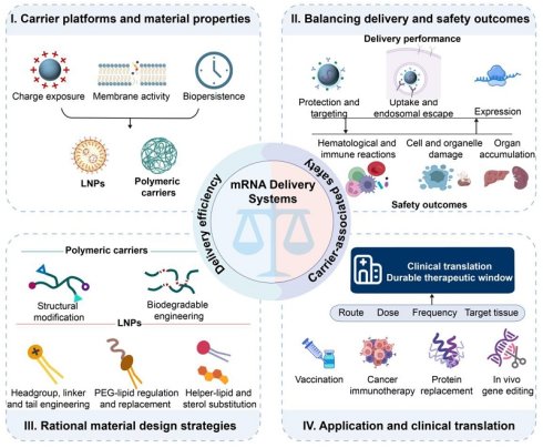
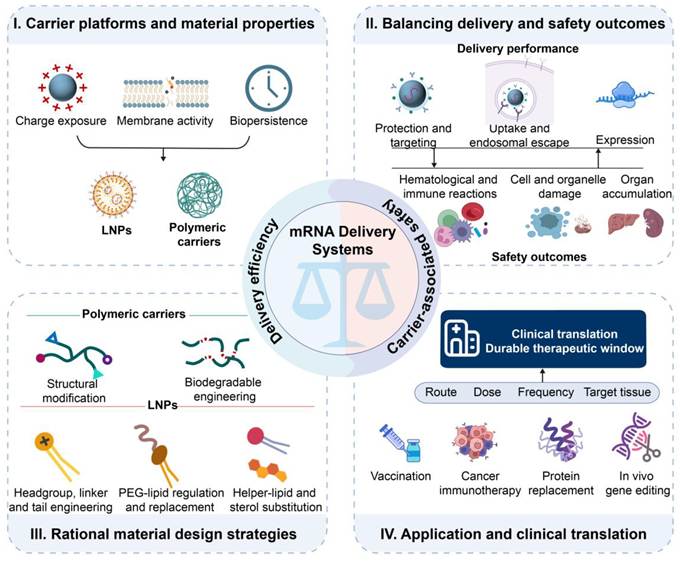
Recent reviews have examined LNP formulation, polymeric modifications, tissue targeting, inflammatory mechanisms, and clinical development [35, 45-48]. Cross-platform analyses have revealed how nanocarrier design influences biological transport and translational feasibility across different RNA modalities [49]. However, these reviews are often dedicated to a single topic or a combination of a few topics, while very few of them have comprehensively addressed how carrier design balances mRNA delivery and its safety. Recent advances in carrier design by modulating charge exposure, membrane activity, and material persistence have demonstrated that a balance in supporting mRNA loading and intracellular delivery, and mitigating harmful biological interactions and excessive tissue accumulation could be achieved. This review examines LNPs and polymeric carriers within a shared materials framework and elaborates rational design strategies for balancing delivery efficiency and safety across different therapeutic applications (Figure 1). We used keywords to identify studies on mRNA carrier design by evaluating both delivery performance and safety in Web of Science and Crossref-indexed publication records with dates up to August 2026. The search combined (“mRNA” OR “messenger RNA”) with (“lipid nanoparticle” OR “LNP” OR “polymer” OR “polyplex”) and (“delivery” OR “transfection” OR “toxicity” OR “safety” OR “immunogenicity” OR “complement” OR “reactogenicity” OR “biodegradation” OR “clearance”). After duplicate removal, records underwent title and abstract screening followed by full-text assessment. English-language original studies identifying the carrier and reporting relevant delivery or safety outcomes were eligible, whereas unverifiable abstracts, studies lacking carrier identity, and non-mRNA studies without direct mechanistic relevance were excluded. Clinical and regulatory information was verified through ClinicalTrials.gov and official FDA, EMA, and PMDA records.
A framework for balancing delivery efficiency and safety in mRNA delivery systems. Charge exposure, membrane activity, and material biopersistence jointly shape the delivery performance and safety outcomes of LNPs and polymeric carriers. Rational material design should be tailored to the therapeutic indication, administration route, dose, target tissue, and dosing frequency. Created in https://BioRender.com.
2. Material properties that couple delivery efficiency and safety
The balance between delivery performance and safety in mRNA delivery systems is predominantly governed by three interrelated material properties. First, cationic and ionizable groups facilitate mRNA complexation, cellular uptake, and endosomal escape, whereas excessive charge exposure can damage membranes and activate innate immunity. Second, membrane destabilization promotes cytosolic release, but extensive endosomal or lysosomal injury can trigger inflammation and cell death. Third, material stability protects mRNA, whereas excessive biopersistence may promote carrier or metabolite accumulation after systemic or repeated administration. In this review, safety is used as an overarching concept, whereas toxicity refers to harmful biological effects, immunogenicity describes the ability to elicit an immune response, and reactogenicity refers to expected local or systemic reactions following administration. The following sections discuss efficiency-safety challenges in LNPs and polymeric carriers.
2.1 Lipid nanoparticles
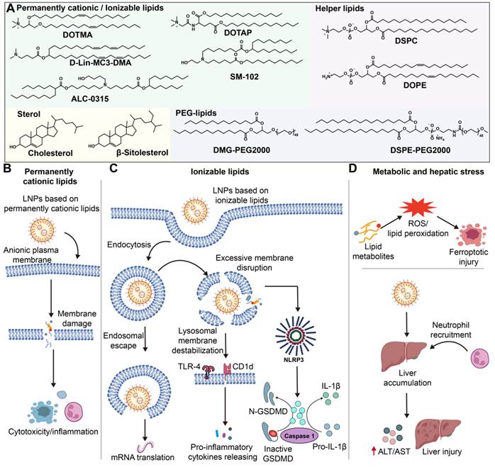
LNPs are the most clinically established platform for mRNA delivery, particularly for vaccines. A typical formulation contains an ionizable lipid or, less commonly in current mRNA products, a permanently cationic lipid, together with a PEGylated lipid, a sterol, and a helper phospholipid (Figure 2A). Together, these components determine the three material properties considered throughout this review. Lipid protonation and surface presentation govern charge exposure, lipid packing and fusogenicity determine membrane activity, and chemical stability and metabolic fate influence biopersistence.
(A) Representative charge-bearing lipids, including permanently cationic lipids DOTMA and DOTAP and ionizable lipids DLin-MC3-DMA (MC3), SM-102, and ALC-0315, together with helper lipids (DSPC and DOPE), sterols (cholesterol and β-sitosterol), and PEGylated lipids (DMG-PEG2000 and DSPE-PEG2000). (B) Charge-mediated membrane damage associated with permanently cationic lipids. (C) Endosomal escape and inflammatory signaling reported for selected ionizable-lipid formulations. (D) Lipid metabolic disturbance, peroxidation, ferroptotic injury, and hepatotoxicity observed under formulation- and exposure-specific conditions. Created in https://BioRender.com.
2.1.1 Permanently cationic versus ionizable lipids
Charge-bearing lipids used for nucleic acid delivery can be divided into permanently cationic and ionizable lipids according to their protonation behavior. Permanently cationic lipids retain a net positive charge across physiological and endosomal pH, whereas ionizable lipids are designed to remain neutral near physiological pH and become protonated at a low pH in endosomes. These charge behaviors lead to different patterns of protein adsorption, nonspecific membrane interaction, cellular uptake, pharmacokinetics, and toxicity [50, 51].
Permanently cationic lipids, such as N-[1-(2,3-dioleyloxy)propyl]-N,N,N-trimethylammonium chloride (DOTMA) and 1,2-dioleoyl-3-trimethylammonium-propane chloride (DOTAP), retain a positive charge across the physiological pH range. Their strong electrostatic interactions with anionic cell membranes can improve cellular uptake but also destabilize the plasma membrane, increase nonspecific uptake, and provoke cytotoxic or inflammatory responses (Figure 2B) [52-54]. These limitations motivated the development of ionizable lipids, which are designed to reduce extracellular charge exposure while retaining protonation-dependent endosomal escape. This charge-switching mechanism may reduce nonspecific membrane interactions, but it does not by itself establish lower in vivo toxicity, which also depends on lipid structure, formulation, dose, route, and tissue exposure.
Accordingly, ionizable lipids have been widely used in clinically approved mRNA vaccines. Nevertheless, several studies indicate that specific ionizable-lipid formulations can raise safety concerns, as illustrated in Figure 2C. Protonation within endosomes can promote membrane destabilization, but excessive disruption of endosomal or lysosomal membranes may induce cytotoxicity and inflammatory signaling [55, 56]. In some cases, a distinct swelling-associated form of cell death has been reported [57]. Chaudhary et al. found that lipids bearing hydrophobic moieties in their amine headgroups can penetrate more deeply toward the bilayer core and induce membrane phase separation. In the experimental system, phase separation was ascribed to lipid-raft formation, strong interactions with TLR4 and CD1d, and enhanced secretion of pro-inflammatory mediators, such as TNF, IFN-γ, IL-6, and IL-2 [58]. In addition, ionizable lipids can provide both the priming and activation signals required for the assembly of NOD-like receptor family pyrin domain-containing 3 (NLRP3) inflammasomes. Through caspase-1 cleavage and gasdermin D (GSDMD)-mediated pore formation, they can promote IL-1β maturation and pyroptotic cell death [59, 60]. These inflammatory responses may also suppress intracellular mRNA translation [61, 62]. For example, LNPs formulated with MC3 or SM-102 induce marked IL-1 and IL-1ra release in human peripheral blood mononuclear cells (PBMCs). Notably, empty SM-102-based LNPs can trigger robust IL-1β secretion even without mRNA cargoes, indicating that ionizable lipids themselves can induce sufficient signals for inflammasome activation [63].
Beyond charge exposure and membrane activity, the metabolic products of LNP lipids raise additional safety concerns related to biopersistence (Figure 2D). In this review, biopersistence is broadly categorized into the retention of intact carrier lipids and the accumulation of slowly cleared or biologically active metabolites in the human body. Lipids and their metabolites frequently play a role in energy metabolism and immune regulation, while disruption of these pathways can disturb lipid homeostasis, promote lipid peroxidation, and contribute to ferroptosis-related cellular injury [64, 65]. Intracellular accumulation of esterified fatty acids, for example, can increase lipid peroxidation [66]. If the resulting reactive lipid species are not adequately cleared, sustained oxidative stress may lead to tissue damage [67]. Fatty acids can also modulate inflammatory signaling through peroxisome proliferator-activated receptor (PPAR), further supporting the role of lipid metabolism in immune responses. These effects are particularly relevant after systemic administration because the majority of LNP formulations preferentially accumulate in the liver. In one example, high-dose intravenous administration of YSK13-containing LNPs results in an increase in serum aspartate aminotransferase (AST) and alanine aminotransferase (ALT) levels. Lipid accumulation in liver sinusoidal endothelial cells may also be associated with neutrophil-mediated inflammation and subsequent liver injury. The comparable hepatotoxicity observed with empty and RNA-loaded LNPs further suggests that the carrier itself may contribute substantially to the hepatic response [68]. Therefore, although liver tropism can enhance hepatic mRNA expression, lipid-associated metabolic stress and inflammation may be concentrated in the same organ, increasing the risk of local toxicity.
2.1.2 PEGylated lipids
PEGylated lipids usually account for a small fraction of an LNP formulation, yet they strongly influence the particle size, colloidal stability, aggregation, and circulation behavior of LNPs. Surface PEG chains create a hydrated steric barrier that prevents protein adsorption and uptake by the mononuclear phagocyte system (MPS) [69, 70]. Excessive or persistent PEG coverage, however, can reduce cellular uptake and endosomal escape, and repeated exposure can induce anti-PEG immune responses. Therefore, the PEG density, chain length, lipid anchor, and shedding kinetics must strike a balance between maintaining circulation stability and ensuring cellular delivery while minimizing immunogenicity (Figure 3).
Adverse immunological reactions to PEGylated LNPs include pre-existing or induced anti-PEG antibody-mediated accelerated blood clearance (ABC) and complement activation-related pseudoallergy (CARPA). CARPA is driven by complement activation products, particularly C3a and C5a, which promote mast-cell and basophil degranulation and histamine release, thereby triggering acute hypersensitivity. Created in https://BioRender.com.
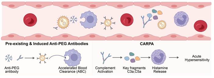
Anti-PEG antibodies can be generated after repeated administration, inducing accelerated blood clearance (ABC) of PEGylated products. PEG has been recognized as an immunogenic material that can stimulate the production of anti-PEG immunoglobulin M (IgM) and immunoglobulin G (IgG) antibodies. Interestingly, pre-existing anti-PEG antibodies have been detected in a substantial proportion of the general population due to long-term exposure to PEG-containing cosmetics, pharmaceuticals, and consumer products. After PEG chains on the LNP surface are recognized by these pre-existing antibodies, rapid clearance of LNPs by the reticuloendothelial system can be triggered, which has been described as the ABC phenomenon [71, 72]. As a consequence, a reduction in the mRNA delivery efficiency and an enhancement in immune activation have been observed upon repeated dosing of LNPs. In an indirect clinical comparison, greater increases in anti-PEG IgG and IgM were observed following mRNA-1273 vaccination than following BNT162b2 vaccination. A 30 μg mRNA dose of BNT162b2 contains approximately 50 μg of ALC-0159. For the original 100 μg mRNA-1273 formulation, the total lipid content is approximately 1.93 mg per dose, with PEG2000-DMG accounting for 1.5 mol% of the lipid formulation, corresponding to an estimated 117 μg per dose [73, 74]. Although the formulations, vaccination regimens, and participant characteristics were not directly matched, the observed differences support a possible association between PEG-lipid amount and anti-PEG antibody responses under specific conditions. In addition, PEG-lipid structure, membrane retention and shedding, overall LNP composition, dosing schedule, and pre-existing anti-PEG immunity may also influence these responses.
PEGylated LNPs may also contribute to complement activation-related pseudoallergy (CARPA), which is one of the most serious acute adverse reactions associated with LNP administration. Unlike classical immunoglobulin E (IgE)-mediated type I hypersensitivity, CARPA can be induced upon first exposure to LNPs without prior sensitization and, in severe cases, the first exposure may lead to anaphylactic shock or even death [75]. Mechanistically, activation of the classical complement pathway can be initiated after anti-PEG antibodies bind to PEG chains on the nanoparticle surface, while PEG itself may additionally enhance complement activation through the alternative pathway. Consequently, rapid generation of complement activation products, particularly the anaphylatoxins C3a and C5a, together with formation of the terminal complement complex sC5b-9, can trigger mast-cell and basophil degranulation and promote the release of histamine and other inflammatory mediators. Acute hypersensitivity symptoms, such as bronchospasm and hypotension, may develop [76, 77]. In addition, amplification of Th2-type inflammatory responses through activation of TLR2/TLR7 signaling pathways has been implicated in the progression of PEG-associated hypersensitivity reactions [78, 79]. These reactions suggest that a surface modification introduced to stabilize particles can alter immune recognition, particularly during repeated or systemic administration.
After contact with blood, LNPs acquire a dynamic protein corona, which may alter circulation, biodistribution, cellular uptake, and immune recognition of LNPs. PEGylation can modulate protein corona formation but cannot prevent it completely. Pre-existing or induced anti-PEG antibodies may remodel the protein corona, thus promoting complement activation and phagocytic uptake, accelerating blood clearance, inducing hypersensitivity, and reducing efficacy after redosing [80-82]. These outcomes are highly contingent upon the antibody profile, PEG architecture, administration route, and exposure history. Therefore, the protein corona and anti-PEG immunity should be considered to achieve the delivery efficiency-safety balance.
2.1.3 Cholesterol
Cholesterol molecules are usually embedded in the lipid bilayer of LNPs, where they regulate the lipid bilayer fluidity and permeability, thereby improving nanoparticle structural stability, preventing premature mRNA leakage, and facilitating membrane fusion and endosomal escape [83]. Although cholesterol is an endogenous molecule in the body, cholesterol-containing formulations are not metabolically inert. In ROS-enriched inflammatory microenvironments, macrophages can convert cholesterol into oxysterols, such as 7-KC and 25-HC, via cholesterol hydroxylase. These oxysterols are potent endogenous immune modulators that directly activate pro-inflammatory signaling pathways, significantly upregulating the expression of pro-inflammatory cytokines, including IL-8, IL-1β, and IL-6, and amplifying LNP-induced inflammatory responses. Meanwhile, abnormal accumulation of cholesterol and its oxidation products in the liver and blood vessels may disturb lipid metabolism homeostasis and increase the risk of metabolism-related disease after long-term administration [84]. Notably, current evidence does not yet establish the long-term contribution of LNP-associated cholesterol to metabolic disease. Studies should be conducted to distinguish administered cholesterol from endogenous sterol pools and assess the biological effects of its metabolites after repeated dosing.
2.1.4 Helper lipids
Helper lipids are essential components in LNP formulations. Classical helper lipids, such as DSPC and DOPE, help maintain LNP structural integrity and facilitate mRNA encapsulation and endosomal escape by regulating the lipid bilayer fluidity and phase transition behavior. Although the safety challenges of helper lipids have been characterized less extensively than those of ionizable lipids, increasing evidence suggests that their chemical structures can influence the biological behavior and toxicity of LNPs. Within the same comparative study, DOPE- and 1,2-dioleoyl-sn-glycero-3-phosphocholine (DOPC)-containing LNPs exhibited distinct immune profiles. DOPE-containing formulations showed lower inflammatory activity, whereas DOPC-containing formulations were associated with greater cytokine production and immune-cell infiltration under the tested conditions [85]. In addition, once LNPs enter biological fluids such as blood, proteins and other biomolecules rapidly adsorb onto the nanoparticle surface, resulting in the formation of protein corona. Neutral helper lipids with relatively large hydrophilic headgroups, such as DSPC, may be exposed on the outer surface of LNPs. Consequently, their physicochemical properties can substantially influence the protein corona composition, thereby affecting the pharmacokinetics, biodistribution, and toxicity profiles of LNPs in vivo [86, 87]. Therefore, helper lipids should be selected by balancing between maintaining particle integrity and membrane fusion and mitigating nonspecific biological interactions.
2.2 Polymeric carriers
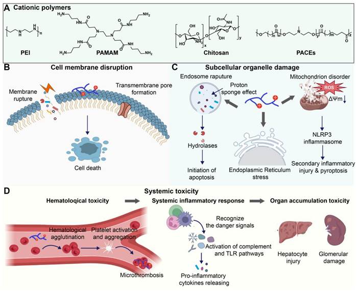
Polymeric carriers have been employed to build structurally diverse mRNA delivery systems. Representative materials include PEI, poly(amidoamine) (PAMAM) dendrimers, and poly(amine-co-ester)s (PACEs) (Figure 4A). The amine content, protonation behavior, and charge accessibility govern mRNA complexation and extracellular charge exposure. A branching architecture, the buffering capacity, and the hydrophobicity level shape membrane interactions and endosomal escape. The molecular weight, backbone degradability, and degradation products influence material biopersistence and clearance. Although these features can be tuned for the optimization of the mRNA efficacy, an improvement in the delivery performance may be accompanied by additional safety concerns.
(A) Chemical structures of representative cationic polymer carriers, including PEI, PAMAM, chitosan, and PACEs. (B) Positively charged polymers can interact with negatively charged cell membranes, inducing membrane rupture or transmembrane pore formation. This acute membrane injury can subsequently activate cell death pathways. (C) The proton sponge effect may lead to endosome rupture and release of hydrolytic enzymes, initiating apoptosis. Some cationic polymers also induce endoplasmic reticulum stress and impair mitochondrial function, leading to ROS overproduction, mitochondrial membrane potential (ΔΨm) collapse, and activation of the NLRP3 inflammasome, driving secondary inflammatory injury and pyroptosis. (D) Selected polymeric carriers can produce hematological effects, systemic inflammatory responses, and organ accumulation under formulation- and exposure-specific conditions. Created in https://BioRender.com.
2.2.1 mRNA condensation versus charge-mediated membrane damage
In many polymeric carriers, protonatable amines become positively charged under physiological or endosomal conditions. This positive charge facilitates mRNA condensation and cellular association, while it may also increase nonspecific interactions with anionic membrane components. The polymer structure, accessible charge, carrier-to-mRNA ratio, and exposure level in highly cationic formulations have been found to play a role in membrane deformation, pore formation, and the loss of membrane integrity (Figure 4B) [88, 89]. Extensive membrane disruption may cause acute leakage of intracellular contents and necrotic injury, whereas sublethal damage may initiate cellular stress and apoptotic signaling. Therefore, cationic charge should support mRNA complexation during formulation and become selectively exposed during cellular uptake and endosomal escape, while remaining largely shielded during circulation.
The molecular weight, branching structure, amine chemistry, and charge density can jointly influence delivery efficiency and toxicity of the delivery system, but their contributions are very specific across polymers. A higher molecular weight or a higher degree of branching is often associated with an increased local charge density and multivalent membrane interactions, although the resulting effects are also determined by the backbone chemistry, degradability, and formulation [40, 88]. Strong electrostatic interactions may improve mRNA condensation and transfection, while they may impede cargo release and increase membrane-associated toxicity. Therefore, cationic polymer design should strike a balance between complex stability, timely intracellular release and acceptable charge-mediated injury [90-92]. For example, 25-kDa PEI is an efficient commercial transfection polymer, but its high charge density and poor degradability are associated with dose-dependent cytotoxicity that hampers its systemic application [93].
2.2.2 Intracellular transport versus subcellular organelle damage
The membrane activity required for endosomal escape may disturb intracellular organelle homeostasis. Endosomal release by cationic polymers is often attributed partially to buffering-induced ion influx, osmotic stress, and membrane destabilization, which are commonly described as the proton sponge effect. However, the proton sponge effect varies among polymers, and it is not universally established. Direct polymer-membrane interactions and a unique polymer architecture may also contribute to endosomal escape of mRNA from the polymeric delivery system. However, when membrane destabilization is excessive, late-endosomal or lysosomal membrane permeabilization can release hydrolases, including cathepsins, into the cytosol and promote apoptotic or inflammatory signaling (Figure 4C) [94].
After endosomal escape, highly cationic polymers may interact with mitochondrial membranes in the cytosol. In formulation- and dose-dependent studies, such interactions have been associated with an impairment in electron transport, the loss of mitochondrial membrane potential, an alteration in energy metabolism, and an increase in reactive oxygen species production [95]. Sustained mitochondrial stress may damage intracellular structures. Under some specific conditions, the stress contributes to NLRP3 inflammasome activation and downstream inflammation [96]. Endoplasmic-reticulum stress and unfolded-protein responses have also been reported for certain polymeric formulations, potentially promoting calcium dysregulation, mitochondrial depolarization, and apoptosis [97, 98]. Notably, these mechanisms have been found to be associated with polymer carriers and they should not be generalized to other mRNA carriers.
2.2.3 Systemic delivery versus organ toxicity
Following systemic, particularly intravenous administration, polymeric carriers encounter plasma proteins, blood cells, clearance organs and the immune system (Figure 4D). Cationic polymers may adsorb anionic plasma proteins and interact with erythrocytes or platelets, potentially promoting opsonization, cell aggregation, or platelet activation. Upon high or poorly tolerated experimental exposure, these interactions have been associated with thromboembolic and microcirculatory disturbances [99, 100]. However, their clinical relevance depends on the polymer identity, formulation, doses, infusion conditions, and the experimental model.
Some polymeric formulations can also activate complement or pattern-recognition pathways, thus stimulating the production of inflammatory mediators [101]. The corresponding mechanisms may not be intrinsic to the polymer class. The activation of complement or pattern-recognition pathways may be influenced by molecular heterogeneity, residual components, surface charge, protein-corona formation, and administration conditions. Biopersistence and organ accumulation of polymeric carriers introduce additional concerns, particularly during and after systemic or repeated treatment. The formulations specifically designed for delivery to the liver and kidneys have been associated with histological injury and an increase in serum transaminases, creatinine, or blood urea nitrogen in many preclinical or high-dose studies. [102, 103] Although CALAA-01 delivered siRNA rather than mRNA, this first-in-human study provides an informative clinical precedent for evaluating the biodistribution and tolerability of targeted polymeric nanoparticles [104]. Nondegradable, high-molecular-weight polymers may also persist in tissues, which may result in an increase in delayed or cumulative toxicity [105, 106]. These findings are useful for hazard identification, but the clinical risk under therapeutic exposure conditions cannot be established.
2.3 Sources of adverse effects in multicomponent mRNA formulations
Adverse effects observed during the development or clinical use of mRNA therapeutics may arise from multiple interacting sources beyond the delivery carrier [22]. They may reflect interacting contributions from the mRNA cargo, manufacturing-related impurities, exposure conditions, and host factors, including mRNA sequence and structure, nucleoside modification, encoded protein, double-stranded RNA (dsRNA), residual solvents and free formulation components, dose, administration route, infusion rate, dosing interval, and patient immune status. Nucleoside modification and IVT purification reduce mRNA-induced immune activation through different but complementary mechanisms. Substitution of uridine with N1-methylpseudouridine can reduce uridine-sensitive innate recognition while preserving translation, whereas dsRNA removal limits activation of dsRNA-sensing pathways and downstream type I interferon responses [107]. These cargo-level interventions interact with carrier design because the carrier determines biodistribution, the cell populations exposed, the route of cellular uptake, endosomal residence, and the timing and extent of cytosolic release. When modified, highly purified mRNA is delivered by a carrier that restricts off-target exposure and supports efficient intracellular release, a greater proportion of the administered cargo can undergo translation rather than activate innate immune sensors. The resulting increase in potency may permit lower doses of both mRNA and carrier, thereby reducing cargo-mediated immune stimulation and carrier-associated complement activation, membrane injury, inflammatory signaling, and tissue accumulation. Conversely, prolonged endosomal retention or delivery to immune-rich tissues may amplify sensing of residual RNA impurities, whereas an intrinsically inflammatory or persistent carrier may offset the benefits of optimized mRNA. Nucleoside modification, RNA purification, and carrier engineering can therefore act synergistically to shift the formulation-specific immunogenic threshold, but they are not interchangeable and cannot establish a universal safe exposure level. This interaction is particularly relevant to repeated administration, in which cargo- and carrier-associated immune responses may impair tolerability and subsequent delivery performance. Accordingly, conclusions from individual formulations or high-dose studies require controls that distinguish carrier-associated risks from other contributors [108]. Recognizing these multifactorial contributors, the present review focuses on the rational design of delivery carriers to balance mRNA delivery performance with carrier-associated safety risks.
3. Rational design of LNPs to balance delivery efficiency and safety
Charge exposure, membrane activity, and material biopersistence may arise from interactions among formulation of LNPs. Ionizable and permanently cationic lipids have distinct charge profiles, thus influencing mRNA delivery, membrane injury, and inflammatory signaling. PEGylated lipids, cholesterol, and helper lipids modulate particle assembly, biological identity, trafficking, and tissue exposure. The following sections examine engineering strategies for individual components and their combination to achieve a balance between delivery performance and safety under the intended clinical conditions.
3.1 Chemical engineering of lipid modules
Although permanently cationic and ionizable lipids have different ionization profiles, both lipid classes generally have a modular architecture comprising a headgroup, linker, and hydrophobic tails. Headgroups primarily govern ionization and charge exposure, tails shape membrane interactions and metabolic fate, and linkers influence stability, degradation, and biopersistence. It is noted that these functions are intertwined. Modifications of multiple modular components are often conducted for optimizing the performance and safety of lipids in LNPs. For clarity, each individual module component is discussed below to delineate its contribution to the overall SARs; cross-component interactions are interpreted as synergistic only when appropriate controls are available.
3.1.1 Engineering cationic and ionizable lipid headgroups
The headgroup governs lipid protonation, interactions with mRNA, and membrane activity. A headgroup that protonates efficiently in acidic endosomes can improve cytosolic delivery, whereas the residual charge or strong membrane affinity at physiological pH can increase nonspecific uptake, hemolysis, and inflammatory activation. Some common headgroups and novel functional headgroups are shown in Figure 5A and Figure 5B. Current strategies include tuning apparent pKa, introducing zwitterionic or clinically characterized motifs, incorporating anti-inflammatory function, and reducing dependence on electrostatic mRNA binding.
(A) Representative lipid headgroups commonly used in LNP formulations. (B) Representative functional headgroups developed to improve lipid biocompatibility. (C) Schematic overview of headgroup pKa regulation using thioamide-containing lipids. (D) Hemolysis of turkey red blood cells induced by LNPs containing SAM-3-Adm, OAM-3-Adm, or MC3 as an ionizable lipid at pH 7.4. Data are presented as mean ± SD; n = 3. Statistical significance was analyzed by one-way ANOVA by comparing SAM-3-Adm LNPs with OAM-3-Adm and MC3 LNPs. *P < 0.05, **P < 0.01, ***P < 0.001, ****P < 0.0001. (E) Bioluminescence imaging at 4 h after intravenous administration of SAM and OAM LNPs. SAM, thioamide-containing; OAM, amide-containing; Adm, dimethylaminoethylamine. Adapted with permission from Ref. [109]. Copyright 2025 Wiley-VCH GmbH. (F) Schematic illustration of headgroup pKa regulation using pyridine-based carboxybetaine derivatives to enhance melanoma immunotherapy and reduce reactogenicity. MeDZ, membrane-destabilizing zwitterionic; PyCB, pyridine-based carboxybetaine. (G) Schematic overview of the anti-inflammatory design, characterization, and in vivo delivery performance of HL LNPs. Created in https://BioRender.com.
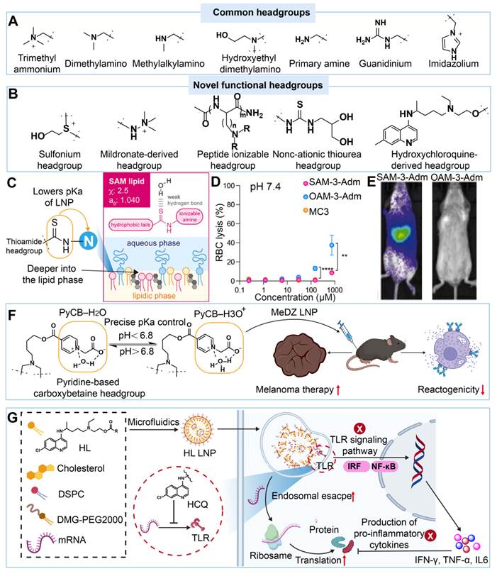
One important strategy is regulation of the headgroup pKa. Chen et al. introduced thioamide groups adjacent to ionizable amines through a one-pot three-component reaction to obtain thioamide-containing (SAM) LNPs, thereby increasing local hydrophobicity and promoting deep insertion of the headgroup into the lipid bilayer during circulation (Figure 5C). In a matched head-to-head comparison, the thioamide modification maintained the apparent pKa of LNPs within the favorable range of 6.0-7.0, reducing nonspecific membrane disruption (Figure 5D) and cytotoxicity under physiological conditions while preserving protonation-driven endosomal escape. At 4 h after injection, SAM LNPs achieved significantly greater delivery efficiency than amide-containing (OAM) LNPs, in which the thioamide was replaced with a conventional amide (Figure 5E) [109]. Similarly, Zhao et al. developed a membrane-destabilizing zwitterionic (MeDZ) ionizable lipid containing a pyridine-based carboxybetaine (PyCB) headgroup. Under physiological conditions, the PyCB headgroup formed a stable zwitterionic PyCB-water complex, which mitigated nonspecific membrane interactions and improved biocompatibility. After endosomal acidification (pH < 6.8), protonation increased the positive charge of the lipid and promoted endosomal escape. Against the clinically established BNT162b2 LNP benchmark, MeDZ LNPs produced higher OVA expression and stronger MHC-I antigen presentation in BMDCs under the reported experimental conditions. In the same study, MeDZ LNPs also induced less injection-site inflammation and neutrophil infiltration (Figure 5F) [110]. These findings support an improved delivery-reactogenicity profile in this model but do not establish broader or repeated-dose safety.
Clinically characterized small molecules have been used as metabolically recoverable building blocks for lipids, which offers a strategy for mitigating safety concerns over lipid degradation products. Mildronate, a clinically used cardioprotective drug with a quaternary ammonium group, has been incorporated as a lipid headgroup through an ester-containing structure. LNPs containing approximately 1 mol% of a mildronate-derived lipid retained the mRNA delivery efficiency, but local and systemic inflammatory markers were downregulated under the tested conditions [111]. Upon degradation of the lipid, mildronate-derived products were generated with an established tolerability profile, which could be advantageous compared to entirely unknown metabolites from other lipids. Nevertheless, the intact lipid remains a new chemical entity, and its pharmacokinetics, complete metabolite profiles, and repeated-dose tolerability should be evaluated and established.
Peptide-based ionizable lipids (PILs) offer another strategy for tailoring headgroup chemistry. Their delivery and safety profiles can be tuned by varying the amino-acid side-chain structure and the number of ionizable Fmoc-protected amino acid (AIFA) units. In general, increasing the number of AIFA building blocks or extending the side chain enhances hepatic mRNA delivery but also increases hepatotoxicity. After initial in vivo screening, PILs based on lysine or 2,4-diaminobutyric acid (Dab), with a saturated C12 alkyl chain and three to five AIFA units, showed superior protein expression. Against the established ALC-0315 benchmark, a12Dab4 PIL produced higher hepatic protein expression in the reported head-to-head study. At the tested high mRNA dose, the two formulations showed comparable acute tolerability across the measured endpoints [112, 113].
Replacing protonatable amines with alternative cationic centers, such as sulfonium groups, offers another strategy for balancing delivery efficacy and safety. Previous studies have shown that replacing the charge-bearing amine groups of cationic lipids with sulfonium cations can enable lung-targeted delivery. Compared with MC3-based LNPs, sulfonium lipid-based LNPs elicited lower TNF-α secretion and showed no histologically detectable lung injury during the observation period [114, 115]. Mechanistically, although not yet validated in vivo, replacing protonatable amines with sulfonium centers may alter membrane interactions and inflammatory signaling. Further studies are needed to determine whether this mechanism accounts for the observed reduction in TNF-α.
Non-cationic or weakly cationic headgroups can be used to mitigate the exposure of positive charges in lipid-based mRNA delivery systems [116]. Wang et al. synthesized biomimetic non-cationic lipids with thiourea-functionalized headgroups that primarily bind to mRNA through hydrogen bonding, rather than electrostatic interactions. The design can help reduce positive charge-associated safety concerns. Consistent with this rationale, negligible inflammatory responses and cytotoxicity were observed after subcutaneous, intramuscular, or intravenous administration of the noncationic thiourea-lipids nanoparticles (NC-TNP). In contrast, SM-102 LNPs elicited pronounced inflammatory and cytotoxic effects under the tested conditions [117].
Beyond reducing carrier-associated toxicity through structural design, a complementary strategy is to incorporate pharmacological agents in LNPs to attenuate the resulting inflammatory response. Hydroxychloroquine (HCQ)-functionalized ionizable lipids (HLs) provide a representative example of this strategy [118]. When formulated as LNPs, selected HL LNPs promoted endosomal escape and mRNA expression while reducing TLR-associated cytokine production. During the reported 13-day repeat-dosing study, HL-3 and HL-6 LNPs also showed a smaller decline in mRNA expression than SM-102-based LNPs (Figure 5G) [119]. Notably, lower cytokine levels support pharmacological modulation of innate signaling but do not demonstrate that membrane injury, carrier biopersistence, or organ exposure has been reduced.
Overall, engineering of headgroups of lipids could strengthen mRNA binding and facilitate endosomal escape while mitigating excessive charge exposure, membrane damage, and innate immune activation. These outcomes could be achieved through the optimization of the headgroup chemistry and the overall formulation. The relationship between the headgroup structure and the delivery efficiency and in vivo toxicity can be established to guide the design of safe, effective LNPs.
3.1.2 Engineering lipid linkers for controlled stability and degradation
The linker connecting the headgroup and hydrophobic tails influences lipid conformation, chemical stability, and metabolic degradation, thereby shaping material biopersistence. Introducing chemically labile or environment-responsive bonds may promote degradation after delivery and reduce toxicity associated with accumulation during repeated administration, after cleavage of these bonds and metabolite clearance have been experimentally verified. Representative linker chemistries commonly used in carrier design, including ester, phosphate, carbonate, and amide linkages, are illustrated in Figure 6A. Ester bonds are the most widely used degradable linkers because they can be cleaved by endogenous carboxylesterases in vivo. Clinically used ionizable lipids, including SM-102 and MC3, contain ester-based linkers. Phosphate and carbonate linkers can also introduce hydrolytic or enzymatic liability [120, 121]. Amide groups can act as hydrogen-bond donors and interact with the phosphate groups of mRNA, thereby strengthening mRNA encapsulation through noncovalent interactions. LNPs formulated with the amide-containing 2C-PILs showed favorable tolerability endpoints in vivo. Furthermore, this formulation simplified the LNP composition, facilitating a more convenient and efficient preparation process [122].
Schematic illustration of representative linker design strategies in charge-bearing lipids, including (A) common linkages, (B) stimuli-responsive linkers, and (C) headgroup-tunable linkers. (D) Plasma concentrations of MC3, SM-102, and (4S)-KEL12 in mice after intravenous administration at 0.5 mg kg-1. (E) Expression of EGFP mRNA delivered by KEL LNPs in dendritic cells (CD11c+, CD45+) from inguinal lymph nodes, as measured by flow cytometry after intravenous injection. Data are presented as mean ± SEM; n = 5-10. Statistical significance was analyzed by one-way ANOVA followed by the Student-Newman-Keuls post hoc test. ns, not significant; *P < 0.05, **P < 0.01, ***P < 0.001. Adapted from Ref. [125] under the terms of the Creative Commons Attribution License. Copyright 2024 The Authors, published by Wiley-VCH GmbH. (F) Rectal temperatures measured 6 h after booster immunization with HA-LNPs. Data are presented as mean ± SD; n = 5 per group. Statistical significance was analyzed using Tukey’s multiple-comparisons test. ns, not significant; **P < 0.01, ***P < 0.001. (G) Mice were injected intramuscularly with luciferase mRNA-loaded LNPs (Luc-LNPs). Bioluminescence was measured at the indicated time points using an in vivo imaging system; representative whole-body images are shown at 6 h post-injection. Reproduced from Ref. [123] under the terms of the Creative Commons Attribution-NonCommercial-NoDerivatives License. Copyright 2025 The Authors, published by Elsevier Inc. on behalf of the American Society of Gene and Cell Therapy. (H) GSH-triggered cleavage and degradation of disulfide-containing ssPalmO. (I) Schematic illustration of the formulation and immune-safety profile of the MOP-1-based influenza A mRNA-LNP vaccine. Created in https://BioRender.com.
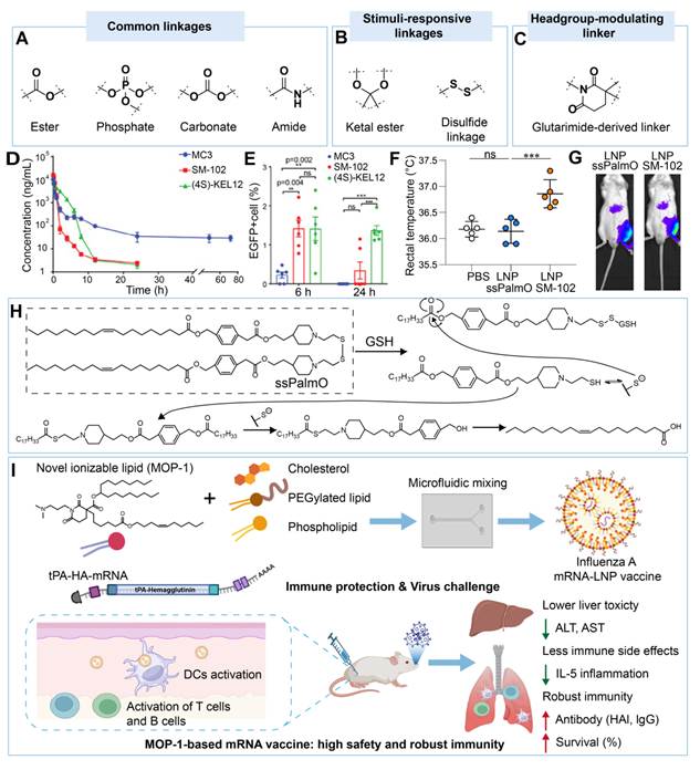
The differences among extracellular, intracellular, and pathological microenvironments have been harnessed for rational design of advanced linkers to achieve selective degradation of charge-bearing lipids in LNPs [123-125]. Stimuli-responsive designs include ketal and disulfide linkages (Figure 6B), as well as linkers that tune headgroup presentation (Figure 6C) [118, 126]. pH-responsive bonds can undergo structural changes in acidic endosomes, facilitating endosomal escape and intracellular mRNA release [127]. For example, after intravenous administration, (4S)-KEL12, which contained ketal ester linkers, displayed a decline in the plasma exposure. Its plasma levels fell below 10 ng/mL at 12 h and became undetectable after 24 h, a clearance profile comparable to SM-102 and faster than MC3 at later time points (Figure 6D). This design reduced hepatotoxicity and maintained normal serum ALT and AST levels. In inguinal lymph nodes, (4S)-KEL12 LNPs sustained dendritic-cell mRNA expression from 6 to 24 h (Figure 6E), whereas SM-102 LNPs showed a time-dependent decline, suggesting improved dendritic-cell delivery and immune-organ tropism [125].
Redox-responsive linkers take advantage of a high glutathione concentration in the cytoplasm to promote lipid cleavage and mRNA release. Kawai et al. developed ssPalmO, a lipid containing both disulfide bonds and phenyl ester linkers. In the reductive intracellular environment, disulfide cleavage generated sulfhydryl groups that intramolecularly attacked the phenyl ester linker, triggering cascade self-degradation (Figure 6H). This degradation pathway converted ssPalmO lipids into endogenous oleic acid and hydrophilic amine derivatives, potentially limiting lipid accumulation. Its biological effects were evaluated in a matched head-to-head comparison with SM-102 LNPs. At 6 h after both prime and booster immunizations, antigen mRNA-loaded ssPalmO LNPs did not significantly elevate circulating pro-inflammatory cytokines, including IFN-α, relative to PBS controls, whereas SM-102 LNPs induced marked cytokine elevation. Following high-dose intramuscular priming, ssPalmO-treated mice showed less body-weight loss than SM-102-treated mice. After boosting, their rectal temperatures remained comparable to those of PBS-treated controls, whereas SM-102 LNPs caused a clear increase (Figure 6F). Notably, both formulations produced comparable luciferase expression at the injection site when loaded with Luc mRNA (Figure 6G). Together, these findings indicate that the degradable linker reduced acute inflammatory responses and improved tolerability while maintaining local mRNA delivery [123, 128, 129].
In addition to controllable degradation, linker chemistry can be optimized to orient the headgroup and modulate membrane interactions. For example, a glutarimide-derived MOP-1 ionizable lipid displayed lower in vitro cytotoxicity, a 2.3-fold increase in the mean lymph-node signal after administration, and less severe liver and kidney histopathology at the reported time point compared with ALC-0315. MOP-1 also induced a smaller increase in cytokines and less body-weight loss (Figure 6I) [130]. These findings support improved delivery and tolerability of MOP-1 under the tested conditions. Further pharmacokinetic, metabolite-safety, and repeated-dose studies are needed to define its broader safety and clinical relevance.
Overall, linker engineering enables the maintenance of structural stability during formulation and circulation and facilitates timely intracellular disassembly and post-delivery clearance. Linker chemistry influences not only structural changes but also lipid conformation, cargo release, tissue retention, metabolite formation, and inflammatory responses. Its impact on delivery and safety should be evaluated over the intended dosing interval.
3.1.3 Engineering hydrophobic tails for membrane activity and metabolic control
Hydrophobic tail engineering can balance delivery efficiency and safety by regulating membrane activity. The tail length, branching, unsaturation, and composition have been reported to play a role in lipid packing and membrane fusion, as well as degradation, metabolic clearance, and tissue retention (Figure 7A) [113]. For example, Ge et al. showed that the total degree of hydrophobic-tail unsaturation was correlated with the membrane-disruptive activity of ionizable lipids under acidic conditions (Figure 7D). Within selected structural series, greater unsaturation of the R3 tail improved mRNA loading and in vivo protein expression, and optimized LNPs enabled efficient IL-27 mRNA delivery with minimal inflammatory responses. However, the degree of unsaturation alone did not consistently predict delivery efficiency or immunogenicity across the lipid library. Other structural features, including the position and distribution of double bonds, also critically influenced LNP performance [131].
Structural optimization strategies for charge-bearing lipid tails, including (A) modulation of the alkyl architecture, such as tail length, unsaturation, and branching, (B) incorporation of antioxidant moieties, such as vitamin E, and (C) conjugation with anti-inflammatory agents, such as dexamethasone. (D) Ugi-4CR synthesis and in vivo screening of ionizable lipids with varied tail unsaturation for mRNA delivery efficiency and immunogenicity. (E) Schematic illustration of H2O2-responsive vitamin E release, ROS scavenging, reduced lipid peroxidation, and enhanced mRNA translation and antigen presentation. Created in https://BioRender.com.
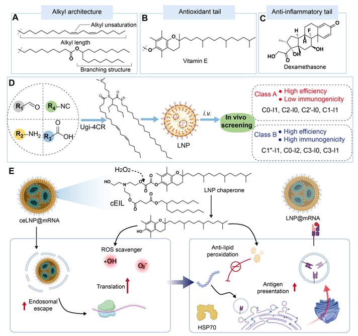
Beyond modulating the membrane activity, hydrophobic tails can be functionalized with antioxidants to reduce transfection-induced oxidative stress and improve mRNA delivery. For example, vitamin E has been used to modify the tails of ionizable lipids (Figure 7B). Yu et al. designed a cleavable vitamin E-containing ionizable lipid (cEIL) with an H2O2-responsive oxalate ester linker (Figure 7E). During transfection, elevated intracellular H2O2 triggered linker cleavage and released vitamin E in situ. The released vitamin E scavenged ROS through the hydroxyl group on its chromanol ring, helping restore redox homeostasis, mitigate endoplasmic reticulum stress, and suppress inflammatory activation in DCs. This antioxidant design also enhanced mRNA translation and promoted antigen presentation through the HSP70-mediated MHC-I pathway. Conventional LNP@mRNAOVA induced strong lipid peroxidation and sustained overproduction of intracellular H2O2 and oxygen radicals in DCs, whereas ceLNP@mRNAOVA markedly reduced lipid peroxidation, scavenged excess ROS, and restored intracellular redox balance to a level comparable to that in the PBS-treated control. Consistent with this antioxidant effect, ceLNP@mRNAOVA showed stronger antigen presentation than LNP@mRNAOVA and eLNP@mRNAOVA; flow cytometric analysis showed the proportion of SIINFEKL-H-2Kb+ DCs in the ceLNP@mRNAOVA group was 7.5-fold and 1.7-fold higher than that in the LNP@mRNAOVA and eLNP@mRNAOVA groups, respectively [132]. In addition, lipids with polyphenol-based tails, such as C-a16 prepared through the Mannich reaction, have similar antioxidant properties. Their phenolic hydroxyl groups scavenge free radicals, downregulate genes associated with oxidative stress and immune activation, reduce pro-inflammatory cytokine secretion, and prolong mRNA-encoded protein expression. Accordingly, C-a16 has been shown to reduce toxicity while improving delivery performance [133]. These systems use the hydrophobic domain as an active regulator of intracellular stress rather than as a passive membrane anchor.
Furthermore, dexamethasone (Dex) (Figure 7C), a widely used anti-inflammatory corticosteroid, was incorporated into an ionizable lipid-containing prodrug. In these lipids, Dex-derived units were covalently integrated into the scaffold, enabling glutathione-responsive release and suppression of TLR4-NF-κB signaling while facilitating mRNA delivery and endosomal escape. In an endogenous PCSK9 base-editing model, a12K4 SHIELD (the Safe and Highly Efficient Lipid-pro-dexamethasone) LNPs achieved higher liver editing efficiency than a12K4 LNPs, while the levels of IL-6 and CCL2 were maintained at the baseline after four injections [134]. It is noted that although Dex in the lipids can suppress inflammatory signaling, a lower level of cytokines is not well correlated with reduced membrane or tissue injury. Dex loading and release should be tailored to the application. Limited and localized Dex exposure may better preserve antigen-presenting-cell activation in vaccines and cancer immunotherapy, whereas strong, controlled suppression of inflammation should be implemented in the repeated systemic applications, such as protein replacement or gene editing.
Adjusting tail hydrophobicity, branching, and unsaturation can improve lipid packing and endosomal escape, but excessive membrane activity may increase oxidative stress and inflammation. Incorporation of antioxidant or anti-inflammatory groups may help reduce these effects while facilitating mRNA delivery. These benefits should not compromise metabolite safety or carrier clearance.
During rational design of lipids, multiple structural components are often modified simultaneously to achieve the optimized effects. For example, a pKa-tuned headgroup preserves acid-responsive endosomal activation, a cleavable linker facilitates carrier clearance after delivery, and an antioxidant tail mitigates transfection-induced stress. However, the contribution of each individual modification is challenging to determine from this simultaneous approach. We therefore recommend factorial experiments in which delivery efficiency and safety are evaluated simultaneously under matched doses, routes, and sampling times.
3.2 Engineering PEGylated lipids and PEG alternatives
PEG-associated challenges can be addressed through the molecular architecture, linker chemistry, the chain length, terminal groups, and lipid-anchor stability (Figure 8A). Branched PEG lipids have been developed to replace conventional linear PEG lipids, and the branched architecture can markedly reduce their binding to anti-PEG antibodies and mitigate the generation of new antibodies [135]. To address the challenges of repeated administration, cleavable linkers have also been introduced between the PEG chain and the lipid anchor, allowing stimulus-responsive PEG detachment [70]. This design maintains steric shielding during systemic circulation, but promotes PEG shedding at the target site, which can improve immune evasion and alleviate ABC. Furthermore, the PEG chain length affects ABC in a biphasic manner. Both long- and short-chain PEG conjugates can induce stronger ABC responses than their medium-chain analogues. In the reported formulation context, PEG2000-lipid conjugates provided a favorable balance among steric stabilization, targeting, and the ABC response [136]. Additionally, the terminal chemistry of PEG can influence the biological behavior of LNPs. Hydroxyl-terminated PEG generally exhibits lower anti-PEG antigenicity than methoxy-terminated PEG; however, it may also alter surface hydration and protein adsorption patterns, leading to rapid systemic clearance in some cases. This effect may be associated with differences in complement activation and foreign-body recognition [137, 138].
(A) Modular design of PEGylated lipids, highlighting key structural variables, including the PEG chain length, the molecular architecture, and linker chemistry. (B) Representative structures of synthetic polymers investigated as alternatives to PEG for LNP stabilization. (C) Poly(carboxybetaine) (PCB) lipids as PEG alternatives for improved mRNA delivery and reduced immunogenicity. Created in https://BioRender.com.
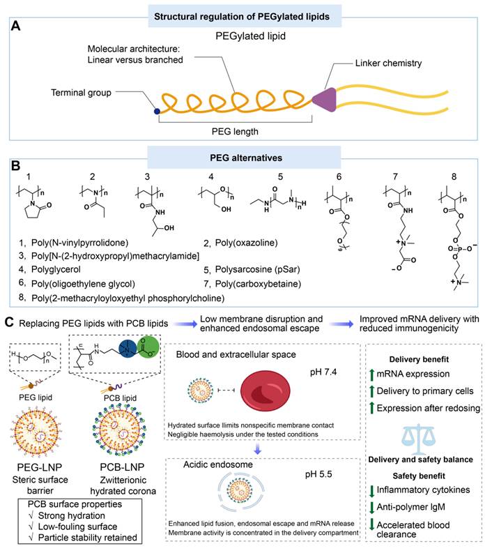
The immunological responses to PEG conjugates have driven the search for alternative stealth polymers, including poly(N-vinylpyrrolidone), poly(oxazoline), poly(N-propionyl methacrylamide), poly[N-(2-hydroxypropyl)methacrylamide], polyglycerol, polyzwitterions, and polysarcosine (Figure 8B) [139-142]. For example, Xiao et al. prepared high-density brush polymer lipids from poly(ethylene glycol) methyl ether methacrylate and used the resulting BPLs to replace conventional DMG-PEG2000 lipids. In a matched head-to-head comparison using the same repeated-dose protocol, optimized BPL LNPs showed lower anti-PEG IgG and IgM binding, better preservation of FAH expression, and improved survival than DMG-PEG2000 LNPs [143]. These findings are promising, but the contributions from altered antibody binding, protein corona composition, complement engagement, and clearance should be accurately defined.
Zwitterionic polymers have also attracted attention because their highly hydrated ionic solvation layers can provide stronger antifouling activity than conventional hydrophilic coatings, which are predominantly based on hydrogen bonding [144, 145]. Among them, Poly(carboxybetaine) (PCB) is a promising PEG alternative. Luozhong et al. synthesized PCB lipids with different carboxybetaine repeat units and acyl chain lengths through reversible addition-fragmentation chain-transfer polymerization and incorporated them into mRNA LNPs as PEG-lipid substitutes (Figure 8C). In a matched head-to-head comparison, PCB-based SM-102-M2 LNPs produced higher tdTomato expression than SM-102-PEG LNPs in primary human T cells after 24 h. They also induced lower IL-1β, IL-6, and TNF-α secretion under the tested conditions. Following repeated administration of PCB-LNPs, FLuc expression was maintained, whereas reduced expression was seen after the second dose of SM-102-PEG LNPs, which was consistent with accelerated blood clearance. PCB-LNPs induced negligible polymer-specific IgM responses, and serum AST and ALT levels were not significantly elevated [146]. However, longer and more intensive repeated-dose studies should assess complement activation, renal and hepatic function. These studies should include complement products such as C3a, C5a, and sC5b-9, together with appropriate organ-function markers, including blood urea nitrogen and creatinine for renal function.
Rational design of stealth materials should consider their behavior in human blood and substantial variabilities among individual patients. In 19 adults receiving an mRNA-1273 booster, mRNA and SM-102 exhibited similar blood-decay kinetics, while there were marked interindividual variations in the residual serum mRNA levels. mRNA remained detectable for up to 14-15 days in some participants [147]. A subsequent preprint involving 73 participants likewise reported vaccine platform-dependent differences in mRNA kinetics [148]. Furthermore, person-specific plasma coronas and anti-PEG antibody profiles have been reported to have an impact on nanoparticle-immune-cell interactions [149-151]. These findings suggest that PEG alternatives should be evaluated beyond antifouling activity and transfection efficiency. Recent reviews also suggest that PEG alternatives should be treated as new nano-bio interfaces, rather than as simple PEG mimics, because the polymer chemistry, anchor architecture, and grafting density reshape protein adsorption, complement activation, biodistribution, and cellular uptake [80-82]. Serial pharmacokinetics, induced anti-carrier antibodies, complement and inflammatory responses, and patient-level variability should be evaluated for repeated-dose development.
3.3 Non-covalent sterol/helper-lipid substitution
Direct substitution of sterols or helper lipids represents a distinct strategy from covalent lipid-prodrug engineering. In non-covalent substitution, the incorporated component primarily modifies lipid packing, membrane organization, and intracellular trafficking. By contrast, SHIELD-type lipids covalently incorporate Dex-derived units through cleavable linkers, making their activity and clearance dependent on linker cleavage and metabolite fate. These approaches therefore require different structural characterization and safety assessment.
Sterol substitution aims to reduce oxysterol formation and lipotoxicity while preserving the membrane fluidity and structural stability required for LNP function [75]. Owing to its structural similarity to cholesterol, Dex can serve as a sterol analogue while also providing anti-inflammatory activity [152]. In the reported formulation, replacing cholesterol with Dex reduced inflammatory markers and increased hepatic mRNA expression by approximately 1.5-fold relative to matched cholesterol-containing LNPs. Substitution with β-sitosterol similarly reduced MCP-1 and related acute inflammatory markers in a matched comparison [153].
Helper-lipid substitution follows a similar rationale by promoting cytosolic mRNA release while limiting excessive membrane disruption and secondary inflammation [154]. For example, replacing conventional DSPC with sphingomyelin can enhance cellular uptake and endosomal escape by promoting active lipid raft formation, while reducing nonspecific membrane disruption and lipid-associated cytotoxicity [155, 156].
Collectively, rational LNP design enables coordinated control of charge behavior, membrane activity, stability, and biodegradation throughout the delivery process. Ionizable lipids can minimize extracellular charge exposure and facilitate endosomal escape, and optimized helper lipids, sterols, surface polymers, and cleavable structures can improve particle stability, tissue delivery, and post-delivery clearance. Integration of these engineering strategies during LNP design may facilitate mRNA delivery while reducing systemic exposure and cumulative safety risks, and the final formulation could be tailored to the cargo, route, dosing schedule, target tissue, and indication.
4. Rational design of polymeric carriers to balance delivery efficiency and safety
Polymeric carriers face the same three efficiency-safety challenges as LNPs, although they arise from different molecular features. The charge density and accessibility govern mRNA binding and nonspecific electrostatic interactions. Hydrophobicity, buffering capacity, and the structural architecture influence membrane activity and endosomal escape. The molecular weight, backbone stability, and degradation products affect biopersistence. These features are interconnected: reducing the charge density can weaken mRNA condensation, increasing hydrophobicity can enhance cellular uptake while intensifying membrane interactions, and premature degradation can compromise delivery efficiency.
4.1 Structural modification of polymeric carriers
Structural modification can enhance the delivery efficiency of low-molecular-weight polymers without introducing the persistent charge and toxicity associated with high-molecular-weight counterparts. Hydrophobic modification, fluorination, amine conversion, and biomimetic functionalization can tune mRNA binding, self-assembly, membrane interactions, and pharmacokinetics. These effects should be assessed across the entire delivery process. For example, a reduction in the surface charge may improve cytocompatibility but weaken mRNA condensation or endosomal escape.
Proper hydrophobic engineering can partially shield the positive charge of the polymer backbone, thereby reducing nonspecific electrostatic interactions with cell membranes and alleviating membrane damage and cytotoxicity (Figure 9A). Meanwhile, hydrophobic interaction can stabilize polymer/mRNA nanocomplexes, thereby improving mRNA binding and delivery efficiency while maintaining the measured tolerability endpoints. For example, deoxycholic acid-modified PEI substantially reduced the intrinsic cytotoxicity of PEI while improving skin penetration. Local administration of this system markedly reduced systemic exposure and enabled efficient gene editing in a local melanoma model [157]. This benefit should not be generalized to highly hydrophobic polymers, which may aggregate or interact strongly with membranes. The combination of zwitterionic and hydrophobic groups has been reported to reduce nonspecific serum protein adsorption. Building on this principle, the widely used cationic polymer PEI can be modified with both phospholipid and alkyl groups to generate phospholipidated and alkylated polymers (PAPs). PAPs retained high delivery efficacy across different fetal bovine serum (FBS) concentrations and significantly outperformed their parent cationic and alkylated counterparts. By contrast, MC3 LNPs showed a rapid decline in efficacy at higher FBS concentrations. Importantly, the improved delivery performance of PAPs did not compromise cellular tolerance [158]. In addition, side-chain modification of polylysine with ionizable hydrophobic isothiocyanates produced thiourea-modified polylysine carriers (PLL-T) (Figure 9E). The thiourea groups provide two hydrogen-bond donor sites, while electrostatic and hydrophobic interactions jointly strengthen mRNA binding and polyplex stability. This multimodal interaction improved mRNA loading and transfection without increasing cytotoxicity. PLL-1A1B(T)@mRNA also induced negligible TNF-α and IL-6 responses and supported antigen expression, presentation, and antitumor activity. These findings suggest that combining complementary interaction modes can improve delivery while limiting inflammatory activation [159]. Together, these systems show that cooperative moderate interactions can reduce reliance on a high cationic charge density.
Representative structural modification strategies for polymeric carriers, including (A) hydrophobic modification, (B) conjugation with biocompatible moieties, and (C) fluorination. (D) Synthetic route for preparing a functional polymer through the split Ugi multicomponent reaction. (E) Schematic illustration of PLL-T-based mRNA vaccine delivery. Multiple interactions between thiourea-modified poly(l-lysine) and mRNA through cooperative hydrogen bonding, electrostatic interaction, and hydrophobic forces enable precise mRNA encapsulation, reduced inflammation, and efficient vaccination. Created in https://BioRender.com. (F) Representative H&E-stained images of paraffin-embedded lung sections. Red dashed outlines indicate inflammatory cell infiltration and possible microabscess formation. Scale bar = 200 μm (left panels) and 20 μm (right panels). (G) Quantification of the tdTomato-positive area in lung sections. Data are presented as means ± SD (n = 3 biological replicates; unpaired two-tailed t-test, P = 0.0392). Adapted from Ref. [163]. Copyright 2024 Springer Nature Limited. (H) TUNEL staining images of the retinal tissue. DAPI in blue and TUNEL-positive cells in red. Scale bar = 75 μm. Adapted from Ref. [166] under the terms of the Creative Commons Attribution License. Copyright 2025 The Authors, published by Wiley-VCH GmbH.
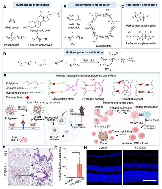
Endogenous or biomimetic moieties can reduce the number or accessibility of primary amines while introducing alternative interactions (Figure 9B). For example, imidazole-acetic acid (IAA), an endogenous histamine metabolite, was used to convert primary amines on branched PEI and chitosan into secondary and tertiary amines. This modification reduced in vivo hematological and immunological toxicity by attenuating TLR4-related inflammatory activation in bPEI systems and complement-associated neutrophil responses in chitosan systems, while preserving mRNA delivery activity [101]. Incorporation of naturally derived or biomimetic molecules, such as cyclodextrin and polysarcosine (pSar), can also improve the safety of polymeric carriers [160]. For example, conjugation of cyclodextrin to PEI 2k (CP 2k) reduced the charge density of the polyamine backbone, thereby alleviating PEI-associated cytotoxicity while preserving adequate protonatable groups for mRNA condensation and intracellular delivery. In a repeated intranasal dosing model, coadministration of CP 2k/mRNA with lipopolysaccharide (LPS) did not increase serum ALT or AST levels or cause detectable liver histopathology compared with LPS alone. By contrast, PEI 25k/mRNA promoted nasal absorption of LPS, resulting in elevated ALT levels and hepatocellular swelling, suggesting that CP 2k provides a safer alternative to PEI 25k for intranasal mRNA delivery [161, 162].
Multicomponent reactions enable multiple polymer properties to be tuned within a single synthetic step. By incorporating hydrophobic segments, degradable linkages, bioactive groups and charge-regulating motifs, these polymers can reduce cytotoxicity while improving mRNA condensation, cellular uptake, endosomal escape and tissue-selective delivery. Vlasova et al. developed an mRNA delivery platform with greater performance and lower reported acute toxicity than PEI by using the Ugi multicomponent reaction to generate a PEI-based cationic polymer. In a head-to-head comparison, the lead candidate, U155, efficiently delivered mRNA to endothelial and immune cells, including T cells, in the lung in vivo, while showing lower values for the measured acute toxicity endpoints than PEI. Histological evaluation at 24 h after intravenous administration of 5 μg nanoparticle-formulated mRNA showed no significant increase in immune-cell infiltration or tissue damage in the U155@lipids-treated group compared with the PBS-treated group (Figure 9F). Consistently, serum levels of acute inflammatory cytokines, including IL-1, IL-6 and TNF-α, remained comparable to those in the control group throughout the study. U155@lipids also enabled repeated mRNA dosing without a substantial loss of transfection efficiency and facilitated CRISPR-Cas9 delivery to the lung in Ai9 mice (Figure 9G). Mechanistically, the tertiary amine units introduced into U155 may contribute to its lower acute toxicity endpoints. Compared with primary and secondary amines, tertiary amines have lower proton-buffering capacity at physiological pH, which may reduce osmotic stress during intracellular trafficking and thereby alleviate cellular toxicity [163].
Fluorination can modulate polymer self-assembly, membrane interaction, and circulation, but the delivery efficiency and safety of fluorinated materials remain formulation-specific (Figure 9C). A low surface energy and unique physicochemical properties of fluorinated materials can improve nanoparticle self-assembly and promote endosomal escape [164]. For example, fluorinated low-molecular-weight PEI (FPEI) nanoparticles were used for systemic IL-2 mRNA delivery, and incorporation of 15% or 20% fluorinated copolymers prolonged detectable circulation of mRNA@FPEI-FCP beyond 72 h while cell viability was maintained at the tested concentrations [165]. Prolonged circulation may improve tumor exposure; however, the biopersistence of the carrier and its fragments may pose an explicit safety risk. In a separate ocular model, fluoropolymer carriers delivered mRNAs encoding genome-editing proteins, and a therapeutic effect was achieved without detectable ocular damage within the endpoints and follow-up studies (Figure 9H) [166]. These findings support advantages of fluorination of polymers for mRNA delivery, but do not confirm favorable safety for fluoropolymers. Repeated-dose tolerability, systemic distribution, and the fate of fluorinated fragments should be systematically evaluated.
Polymer modification often simultaneously modulates charge exposure, membrane activity, and biopersistence. Amine shielding can improve hemocompatibility, but it may weaken mRNA condensation. Introduction of hydrophobic or fluorinated groups may enhance cellular uptake and endosomal escape, while these groups may influence protein corona formation, membrane injury, and tissue retention. Biomimetic motifs may introduce additional challenges associated with immunogenicity, material heterogeneity, and manufacturing. Therefore, modified polymers should be evaluated against their unmodified counterparts under identical experimental conditions in terms of delivery efficiency and safety endpoints, including organ accumulation, repeated-dose toxicity, and structure-associated toxicity.
4.2 Engineering biodegradable and charge-shifting polymeric carriers
Biodegradable polymeric carriers are designed to reduce material biopersistence through fragmentation into smaller products after delivery at the target site, and these small fragments can be cleared more readily. Representative systems include poly(amine-co-ester)s (PACEs), poly(β-amino ester)s (PBAEs), and poly(amino acid)-based polymers (Figure 10A) [167]. Suberi et al. optimized biodegradable PACE mRNA polyplexes for pulmonary delivery by combining a modified E14 end group with 10 wt% PACE-PEG. In head-to-head comparisons, PACE-E14 polyplexes maintained higher cell viability than LipoMM (Figure 10D). Formulations containing PACE-E14 and 10 wt% PACE-PEG produced robust pulmonary mRNA expression and outperformed jetPEI and GenVoy-ILM at 24 h (Figure 10E). Higher PACE-PEG contents reduced expression, indicating that PEG coverage must be optimized to preserve delivery performance. Histology showed no acute epithelial damage apart from mild focal neutrophil infiltration, while no sustained leukocyte accumulation or changes in serum creatinine and BUN were observed (Figure 10F). These findings support tolerability under the tested conditions, but long-term and repeated-dose studies are still needed to assess cumulative pulmonary safety [168]. In addition, PBAE-E63 enabled lung editing after repeated systemic administration, and no evident pulmonary injury was seen in the reported histological assessments through nine doses [169]. These findings indicate favorable tolerability of biodegradable polymeric carriers within the tested models and observation periods, although further studies are needed to define their systemic and long-term safety. Hydrophobic-segment engineering can tune degradation kinetics [170], but clinical translation requires efficient clearance of both the parent carrier and its degradation products without compromising mRNA delivery.
(A) Representative biodegradable polymer backbones. Representative polymer designs responsive to (B) pH and (C) reducing conditions. (D) In vitro cytotoxicity of PEGylated PACE-E14 polyplexes compared with LipoMM. Asterisks indicate statistically significant differences between the PACE formulations and LipoMM. (E) Luciferase expression in lung tissues after delivery of 5 μg FLuc mRNA via PEGylated PACE-E14 or in vivo-jetPEI. RLU, relative light units. (F) Blood urea nitrogen (BUN) and creatinine (Cr) levels as indicators of kidney function on day 14 after delivery. Data are presented as mean ± SD with individual measurements. Statistical significance was assessed using two-way ANOVA with Sidak’s multiple-comparisons test. ns, not significant. Reproduced with permission from Ref. [168]. Copyright 2023, AAAS. (G) pH-dependent charge conversion of Orn-CART nanoparticles for lung endothelial mRNA delivery. Created in https://BioRender.com.
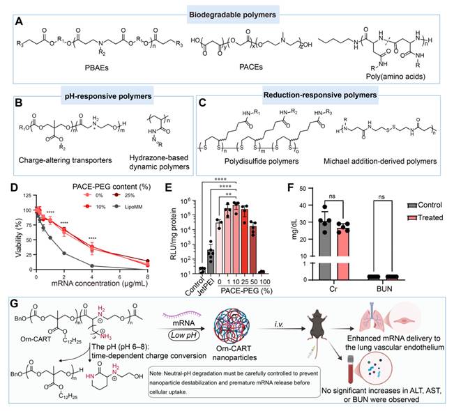
The challenges of LNPs and polymeric carriers for mRNA delivery. (A) Toxicity mechanisms are often examined separately, leaving pathway interactions, delayed effects, and interindividual susceptibility incompletely understood. (B) Material properties that enhance delivery may also contribute to toxicity, complicating the simultaneous optimization of efficacy and safety. (C) Conventional in vitro and animal models do not fully reproduce human physiological and immune environments, limiting their clinical predictive value. (D) Long-term repeated-dose data remain limited, particularly for cumulative toxicity, anti-carrier immune responses, and chronic inflammation relevant to chronic disease treatment. Created in https://BioRender.com.
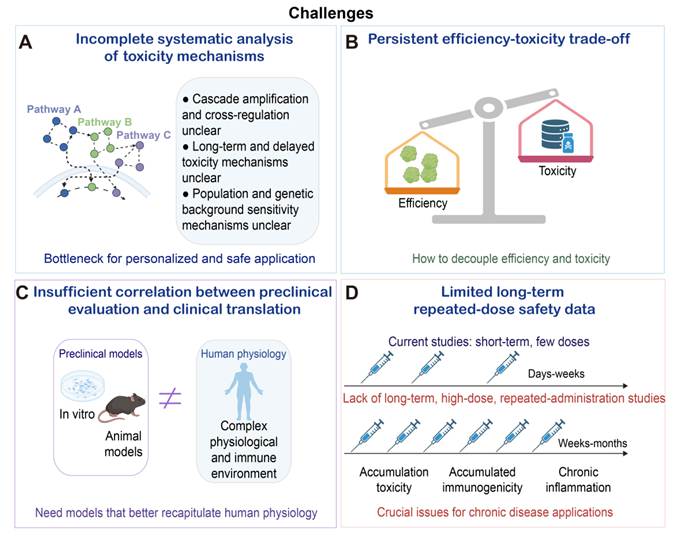
Intracellular environment-responsive linkages have been employed for polymeric carriers by harnessing intracellular microenvironmental cues to induce degradation or charge conversion (Figure 10B) [171-173]. Charge-altering releasable transporters (CARTs) containing β-amide carbonate linkages undergo time- and pH-dependent conversion from cationic structures to products with lower charge, thereby promoting cargo release and limiting persistent charge exposure. In the reported studies, this transition reduced hemolysis while maintaining delivery performance [174]. Moreover, Orn-CARTs condensed mRNA at low pH and enabled ligand-free delivery to the pulmonary vascular endothelium following intravenous administration. At near-neutral pH, degradation of the cationic block generated lower-charge products. At the tested dose, serum ALT, AST, and BUN remained unchanged after both single and repeated administration (Figure 10G) [175]. Importantly, near-neutral degradation may promote mRNA release and reduce persistent cationic charge. The conversion rate must nevertheless be controlled to prevent nanoparticle destabilization and premature mRNA release before cellular uptake. Dynamic covalent biodynamers based on pH-responsive acylhydrazone and imine bonds exhibited substantially lower cytotoxicity than PEI and PLL in the reported in vitro assays and no distinct toxicity signal in the zebrafish assays [176].
Redox-responsive polymers have been developed for mRNA delivery by harnessing intracellular glutathione gradients (Figure 10C). Disulfide-containing polymers remain relatively stable in blood but rapidly degrade in the reductive cytosolic environment, thereby promoting mRNA release while mitigating cytotoxicity [177]. Ben-Akiva et al. reported a bioreducible lipophilic PBAE platform. Disulfide bonds were incorporated into the polymer backbone to induce cytosol-responsive degradation. To evaluate the systemic safety of an optimized R18D polymer, serum ALT and AST were measured after administration of PBAE-mRNA or its combination with adjuvants such as CpG or poly(I:C). Moderate, transient elevations in the liver enzymes were observed at 24 h after injection, particularly in the adjuvant-containing groups, but both ALT and AST returned to the baseline within 72 h, and these enzyme levels were comparable to those of the untreated control. Cytokines associated with the cytokine release syndrome (CRS) also showed a transient increase, and they rapidly returned to the baseline levels. These results indicate that the measured acute biomarker changes were transient under the tested conditions [178]. Longer follow-up studies are needed to determine whether the carrier and its metabolites are fully cleared and whether tolerability is maintained after repeated administration.
Biodegradable and charge-shifting polymers can reduce material biopersistence and charge-associated toxicity, but the overall benefits are dependent on the timing of degradation and the degradation products. After rational engineering of the polymeric carriers, they should protect mRNA during circulation and undergo triggered disassembly after cellular uptake. The resulting fragments should have low membrane activity, and they can be cleared efficiently, particularly during repeated administration.
5. Application-specific design and implementation barriers for clinical translation
5.1 Indication-specific therapeutic windows
Successful clinical translation ultimately depends on maintaining a favorable balance among material structure, delivery performance, and safety under the intended treatment regimen [22]. Peak mRNA expression alone is insufficient for assessing the delivery performance, and a carrier should provide reproducible results at the intended dose and frequency through the intended route within the target tissue. These delivery results should be achieved without inducing unacceptable acute or cumulative toxicity. These safety requirements can vary across applications. The acceptable combined immune burden is therefore indication-dependent. Controlled mRNA cargo- and carrier-derived innate stimulation may support the immunogenicity of infrequently administered intramuscular vaccines [37], whereas repeated intravenous protein replacement generally requires minimal activation from both sources, predictable clearance, and preserved redosing performance [179]. Gene editing and cancer therapy impose different requirements for target-cell exposure, off-target distribution, and immune activation [180]. Accordingly, each platform should be evaluated within the context of its intended application (Table 1).
Context-specific efficacy and safety criteria for mRNA delivery systems.
| Application, route, and regimen | Efficacy and intended immune activity | Minimum safety panel | Repeat-dose or clearance criterion | Design implication |
|---|---|---|---|---|
| Prophylactic vaccine; predominantly via i.m.; limited doses | Antigen expression and adaptive immunity; controlled local/systemic innate activation | Reactogenicity, cytokines, complement, local histology | Preserved booster expression without progressive anti-carrier immunity or increased reactogenicity; systemic clearance is interpreted in relation to local antigen expression and the booster interval | Transient activation; localized exposure; preserved adjuvancy |
| Cancer therapy; i.t. or i.v.; often repeated | Tumor responses and expression in target cells; context-dependent activation without systemic spillover | Cytokines, liver injury, blood compatibility, off-target biodistribution | Stable efficacy and no cumulative organ injury across planned cycles | Spatially restricted activation; tumor- /immune organ- targeting |
| Protein replacement; repeated i.v. dosing | Functional protein exposure over the dosing interval; minimal immune activation | Complement, anti-carrier antibodies, liver/kidney injury, histology | Pre-dose anti-carrier antibodies, complement activation, systemic exposure, clearance, and organ redistribution assessed across the intended redosing interval; functional expression retained after repeated matched doses | Minimal immune recognition; predictable degradation and clearance |
| Gene editing; one or a few systemic doses | On-target editing in intended cells; minimal immune activation unless mechanistically required | Acute carrier toxicity, biodistribution, off-tissue editing, long-term follow-up | Transient editor expression and carrier clearance after delivery | High cellular selectivity; short exposure; stringent off-target control |
Table footnote. The criteria are formulated for application-dependent therapeutic windows and they are not intended for universal carrier ranking. mRNA carrier design aims to achieve features, such as transient activation, localized exposure, and predictable degradation, while these terms are not universal acceptance thresholds. Kinetics may be acceptable in the context of the route, dose, target tissue, dosing interval, and indication. A single degradation half-life cannot establish that a material is biodegradable. Supporting measurements should include kinetics of the intact carrier and its major metabolites, organ exposure, residual material before the intended redosing interval, and efficacy across the intended regimen. i.d., intradermal; i.m., intramuscular; i.t., intratumoral; i.v., intravenous.
For prophylactic vaccines, carriers are often used to enhance antigen expression while preserving a controlled degree of innate immune stimulation. A moderate adjuvant-like effect can enhance adaptive immunity, while excessive activation can lead to local and systemic reactogenicity [181, 182]. Intrinsic material design strategies, such as pKa tuning, controlled PEG shedding, and reduced nonspecific membrane interaction, are often prioritized over broad pharmacological immunosuppression. When anti-inflammatory motifs are used, their loading and release should be assessed in terms of antigen expression, dendritic-cell activation, T-cell responses, and clinical tolerability.
For cancer therapy, mRNA is often delivered intratumorally or intravenously and repeated dosing of mRNA is a common practice. Delivery systems should stimulate immune activation to support anti-tumor responses while preventing systemic inflammation, complement activation, and liver injury [183, 184]. Targeting and degradable structures can reduce exposure of carriers to normal tissues, whereas locally released anti-inflammatory or immune-regulatory motifs may confine activation to the tumor microenvironment. Studies on PEG-phosphatidylethanolamine nano-micelles support the therapeutic benefits of co-delivering antigens and immunostimulatory signals to antigen-presenting cells [185]. Emerging polymer-lipid carriers that intrinsically activate and transfect T cells can be used for simultaneous mRNA delivery and immune activation for in vivo CAR-T generation [186].
Protein replacement generally requires repeated administration because mRNA expression is transient [187]. Therefore, carriers should display low immunogenicity, predictable clearance, stable performance after redosing, and minimal cumulative organ injury. Neutral or weakly charged surfaces, degradable backbones, and non-PEG stealth coatings may be particularly valuable. Delivery efficiency should be assessed across multiple doses rather than after a single administration. Pre-existing or treatment-induced anti-carrier antibodies may accelerate clearance and reduce expression after redosing, although the magnitude remains formulation- and model-dependent. In a mouse model, increasing anti-PEG antibody concentrations were associated with a 31.4- to 46.6-fold reduction in early spike protein expression after BNT162b2 administration [71]. By contrast, baseline anti-PEG antibodies did not correlate with vaccine-component decay rates in a small human study of intramuscular mRNA-1273 boosting [147]. Antibody-mediated changes in exposure may therefore be more restrictive for frequently repeated systemic protein replacement than for infrequent localized vaccination, but no validated antibody threshold or universal redosing interval has been established. In vivo gene editing may need only one or a few doses, but the tolerance for off-target delivery is especially low because expression of editing enzymes can produce durable genomic changes [113]. High delivery efficiency in the intended cell type must be balanced against transient editor expression, restricted biodistribution, acute carrier toxicity, and off-target editing in non-target tissues. Therefore, safety evaluation should integrate carrier pharmacology with editing specificity, persistence of editor expression, and long-term biological follow-up.
LNPs are the clinically approved platform for mRNA delivery. All approved mRNA products are derived from lipid-based formulations. However, conventional PEI, PBAE, PACE, and PAMAM systems for mRNA delivery remain at the preclinical stage (Table 2). Clinical experience with polymer-containing carriers is predominantly confined to protamine complexes and emerging polymer-lipid hybrids. This clinical difference between LNPs and polymeric carriers can be ascribed to manufacturing readiness, compositional control, analytical characterization, regulatory experience, and the availability of repeated-dose safety data, in addition to the delivery performance.
Representative approved and investigational mRNA delivery systems. Regulatory approvals and clinical trial statuses were verified using records from the FDA, EMA, PMDA, and ClinicalTrials.gov, accessed in August 2026. Components of investigational formulations are listed only when they are disclosed in official records; otherwise, the composition is described as proprietary or not publicly disclosed.
| Delivery system | Product or candidate | mRNA cargo and indication | Admin. | Verified regulatory or trial status | Carrier disclosure and source |
|---|---|---|---|---|---|
| Approved LNP | Comirnaty (BNT162b2) | SARS-CoV-2 spike mRNA; COVID-19 prevention | i.m. | FDA BLA approval (23 August 2021) (FDA) | ALC-0315, ALC-0159, DSPC, and cholesterol in the official product information |
| Approved LNP | Spikevax (mRNA-1273) | SARS-CoV-2 spike mRNA; COVID-19 prevention | i.m. | FDA BLA approval (31 January 2022) (FDA) | SM-102, DSPC, cholesterol and PEG2000-DMG in the official product information |
| Approved LNP | mRESVIA (mRNA-1345) | RSV prefusion F mRNA; prevention of RSV-associated lower respiratory tract disease | i.m. | FDA BLA approval (31 May 2024) (FDA) | SM-102, DSPC, cholesterol and PEG2000-DMG, in the FDA product information |
| Approved LNP | mNEXSPIKE (mRNA-1283) | SARS-CoV-2 spike N-terminal and receptor-binding-domain mRNA; COVID-19 prevention | i.m. | FDA BLA approval (30 May 2025); EU authorization (12 February 2026) (FDA; EMA) | SM-102, DSPC, cholesterol, and PEG2000-DMG in FDA and EMA records |
| Approved LNP | Kostaive (ARCT-154; zapomeran) | Self-amplifying SARS-CoV-2 spike mRNA; COVID-19 prevention | i.m. | PMDA approval (28 November 2023); EU authorization (12 February 2025) (PMDA; EMA) | ATX-126, DSPC, cholesterol, and PEG2000-DMG in PMDA and EMA records |
| Approved LNP | Daichirona (DS-5670; ufrenmeran) | SARS-CoV-2 receptor-binding-domain mRNA; COVID-19 prevention | i.m. | PMDA approval (2 August 2023) (PMDA) | T168-1857a, DSPC, cholesterol, and PEG2000-DMG in the PMDA record |
| Clinical-stage LNP | V940/ mRNA-4157 (intismeran autogene) | Personalized neoantigen mRNA; cancer immunotherapy | i.m. | Active, not recruiting, Phase III (NCT05933577) | Proprietary LNPs; full composition not disclosed in the registry |
| Clinical-stage LNP | mRNA-3927 | PCCA and PCCB mRNAs; protein replacement for propionic acidemia | i.v. | Recruiting, Phase I/II (NCT04159103) | Proprietary LNPs; full composition not disclosed in the registry |
| Clinical-stage LNP | mRNA-3705 | Methylmalonyl-CoA mutase mRNA; protein replacement for methylmalonic acidemia | i.v. | Recruiting, Phase I/II (NCT05295433) | Proprietary LNPs; full composition not disclosed in the registry |
| Clinical-stage LNP | ARCT-810 | OTC mRNA; protein replacement for ornithine transcarbamylase deficiency | i.v. | Recruiting, Phase II (NCT06488313) | LUNAR LNP platform; full composition not disclosed in the registry |
| Clinical-stage LNP | NTLA-2001 | Cas9 mRNA and TTR-targeting sgRNA; in vivo editing for transthyretin amyloidosis | i.v. | Completed, Phase I (NCT04601051) | LP000001, DSPC, cholesterol, and PEG2000-DMG; the complete quantitative composition remains proprietary |
| Clinical-stage LNP | NTLA-2002 | Cas9 mRNA and KLKB1-targeting sgRNA; in vivo editing for hereditary angioedema | i.v. | Active, not recruiting, Phase III (NCT06634420) | LP000001, PEG2000-DMG, and additional lipids; the complete composition and component proportions have not been publicly disclosed |
| Clinical-stage LNP | mRNA-2752 | OX40L, IL-23, and IL-36γ mRNAs; cancer immunotherapy | i.t. | Completed, Phase I (NCT03739931) | Proprietary LNPs; full composition not disclosed in the registry |
| Polymer-lipid hybrid | InViVo CAR1920 mRNA | CD19/CD20 dual-targeting CAR mRNA; in vivo CAR-T-cell generation | i.v. | Recruiting, Phase II (NCT07321301) | Polymer-lipid particles; detailed composition not disclosed in the registry |
| Protamine-based carrier | CV7201 | Rabies glycoprotein mRNA; prophylactic vaccination | i.d. or i.m. | Completed, Phase I (NCT02241135) | Free and protamine-complexed mRNA in the trial record and primary report |
Abbreviations: BLA, biologics license application; CAR, chimeric antigen receptor; OTC, ornithine transcarbamylase; PCCA/PCCB, propionyl-CoA carboxylase subunits α/β; RSV, respiratory syncytial virus; sgRNA, single-guide RNA. Admin., administration; i.d., intradermal; i.m., intramuscular; i.t., intratumoral; i.v., intravenous.
5.2 Cross-platform comparison and real-world implementation
LNPs and polymeric carriers are two distinct mRNA delivery platforms, and they have their unique strengths and limitations. Both have been evaluated in various vaccination and therapeutic models. However, few studies have compared them directly under the same experimental conditions [188]. Therefore, qualitative comparisons of studies from individual platforms were performed and the findings from these studies were interpreted cautiously. The strengths, limitations, and future research directions of both platforms are summarized (Table 3).
Comparison of LNPs and polymeric carriers based on clinical translational criteria.
| Criterion | LNPs | Polymeric carriers | Comparative interpretation |
|---|---|---|---|
| Delivery efficacy | High-efficiency encapsulation and clinically validated expression; strong liver tropism after i.v. dosing | Highly tunable chemistry and great extrahepatic potential; varied performance across polymers and formulations | Comparisons are most meaningful when mRNA dose, administration route, target-cell expression, and follow-up are matched |
| Reported or plausible safety concerns | Formulation-specific innate activation, complement/PEG responses, lipid metabolism, and hepatic exposure | Structure- and exposure-dependent membrane injury, hemolysis, organelle stress, aggregation, and polymer biopersistence | Safety differences should be interpreted using matched assessments of blood compatibility, inflammation, organ injury, biodistribution, and histopathology |
| Release, degradation, and clearance | Non-covalent components are released through lipid exchange or particle disassembly, whereas covalent lipid prodrugs depend on linker cleavage and subsequent metabolite formation. | Ester-rich, charge-shifting, pH-responsive, or redox-responsive backbones can promote cargo release and carrier degradation | Non-covalent formulations should be evaluated for component leakage and systemic exposure, whereas covalent prodrugs require characterization of cleavage kinetics, intact-conjugate disposition, and metabolite safety |
| Repeated administration | More clinical experience, but concerns over anti-PEG immunity and cumulative liver exposure | Promising preclinical redosing data; limited standardized, long-term clinical evidence | The key consideration is whether delivery efficacy is maintained without increasing anti-carrier immunity, complement activation, organ accumulation, or cumulative toxicity |
| Clinical maturity | Multiple approved vaccines and expanding therapeutic pipelines | Mostly preclinical; protamine-complexed mRNA, such as CV7201, has completed a Phase I clinical trial; however, protamine is clinically approved as a heparin antagonist and insulin additive, not as a standalone mRNA vaccine platform; polymer-lipid hybrids remain at an early clinical stage | LNPs have greater regulatory and manufacturing maturity, whereas polymeric carriers require broader clinical validation and longer-term safety data |
| Manufacturing and regulation | Greater manufacturing experience, including microfluidic mixing, and developed processes for product-specific chemistry, manufacturing, and control; challenges for scale-up and cold-chain supply | Challenges for analytical characterization and GMP product reproducibility due to structural heterogeneity and variations in molecular-weight distribution | LNPs benefit from more established manufacturing and regulatory pathways, whereas polymer heterogeneity creates additional challenges for analytical control and batch reproducibility |
Quantitative comparisons may be performed when delivery performance and safety are evaluated under the equivalent experimental conditions [180, 189]. The mRNA dose, carrier dose, carrier-to-mRNA ratio, administration route, model, and follow-up period should be included in the reports for the corresponding efficacy and safety outcomes [190, 191]. It is important to note that a higher expression level at a different dose via a different route does not imply a wider therapeutic window.
During clinical translation of both platforms, scalable manufacturing processes with robust chemistry, manufacturing, and controls (CMC) are essential to ensure the material identity, formulation consistency, and removal of residual components [192]. This is particularly important for heterogeneous polymers, whose composition and degradation may vary from batch to batch. To assess safety concerns of two platforms during clinical practice, the administration route and the dosing schedule should be matched, especially for intravenous or repeated administration. It has been reported that differences in biomolecular corona formation and the immune status of patients may impact the carrier distribution and targeting [149-151]. Although these factors are not well-established biomarkers for patient selection, they should be considered in translational studies.
6. Challenges and future perspectives
6.1 Critical challenges
First, the complete biological basis of carrier-associated toxicity remains incompletely understood. Most studies examine individual mechanisms for carrier-associated toxicity, while in vivo injury may result from a combination of these individual mechanisms, including membrane damage, intracellular stress, immune activation, metabolic disturbances, and organ exposure. Long-term and delayed safety risks in humans remain poorly characterized, and the susceptibility may vary with the genetic background, age, disease state, immune history, pre-existing anti-carrier antibodies, and individual-specific protein corona formation [149-151].
Second, the material properties tuned for enhanced delivery efficiency may pose safety concerns. A positive charge facilitates mRNA complexation and uptake, but the charge may damage membranes; membrane activity promotes endosomal escape, but it may disrupt organelles; and biopersistence can prolong exposure but delay clearance. Therefore, more studies should be conducted to systemically evaluate the impact of these properties on delivery efficiency and safety risks across therapeutic contexts. Importantly, the impact should be evaluated by the therapeutic window and repeated-dose performance. Ultimately, the properties can be harnessed to develop next-generation carriers with spatially and temporally controlled functions.
Third, conventional preclinical models do not fully recapitulate human tissue physiology, immune responses, disease-associated inflammation, or repeated exposure. Therefore, safety findings from simplified cell-based and animal models may have poor clinical predictability. Human-relevant models should be constructed and employed to improve the mechanistic interpretation and bolster the translational confidence.
Long-term, high-dose, and repeated exposure remain insufficiently evaluated, particularly for therapies requiring sustained administration. Future studies should deliver outcomes with a standardized safety panel covering blood compatibility, immune activation, organ injury, biodistribution, and carrier clearance. Repeated-dose studies should additionally assess anti-carrier immunity, retained efficacy, material accumulation, and cumulative tissue injury under clinically relevant and appropriately controlled conditions.
6.2 Interdisciplinary technologies for safety evaluation
Artificial intelligence (AI) can support the design of safer and more effective mRNA carriers when the training dataset for delivery and toxicity under the same experimental conditions is available. Unbiased data for efficacy measurements and safety assessments should be collected with detailed experimental information. Data should be organized into four groups. The first data group is molecular features of the carrier, including the chemical structure, molecular weight, ionization behavior, hydrophobicity, pKa and degradability. The second data group is properties of the nanoparticle formulation, including the particle size, size distribution, surface charge, morphology, mRNA encapsulation, and colloidal stability. The third data group is in vitro efficacy and safety measurements, including cellular uptake, endosomal escape, protein expression or gene editing, target cell viability, and hemolysis. The fourth data group is in vivo measurements, including pharmacokinetics, biodistribution, target tissue expression or editing, systemic inflammation, organ injury markers, histopathology, and toxicity after repeated administration. Each record is detailed with the experimental information, including the carrier composition, mRNA type, dose, administration route, experimental model, dosing schedule, and sampling time. It is noted that the measurement data often vary from laboratory to laboratory because materials, protocols, instruments, and reporting methods in different laboratories are not identical. Interlaboratory variations can be reduced through shared reference formulations, calibrated assays, common units, and endpoint measurements normalized to the dose and the sampling time. Laboratory and batch identifiers should be retained for each record, and formulations with positive or negative outcomes are included. Hierarchical models can account for laboratory and batch effects. Model reliability should be tested by excluding data from one laboratory during training and evaluating performance on independent datasets. Uncertainty estimates and documented training-domain limitations should be considered before model predictions are used to prioritize translational candidates. It is critical to validate the model predictions from independent laboratory datasets before model-guided clinical prioritization.
Organoids and organ-on-chip platforms can provide human-relevant preclinical safety assessment outcomes. Compared with conventional two-dimensional cultures, they can better recapitulate the tissue architecture, physiological microenvironments, and cell-cell communication. The liver, kidney, heart, vascular, and immune-competent organoid or organ-on-chip platforms may be used for evaluation of organ-specific toxicity, complement and inflammatory responses, and repeated exposure. These platforms may reduce the need for animal studies, although animal models remain important for complementary toxicity assessment.
In vivo imaging and multi-omics can provide complementary mechanistic information. Longitudinal imaging can track carrier distribution, metabolism, and tissue accumulation across clinically relevant doses and schedules. Transcriptomics, proteomics, metabolomics, and lipidomics can identify inflammatory, metabolic, and stress-response pathways activated by different carriers. Integrated analyses should relate biological responses to measured carrier exposure and degradation rather than rely on omics signatures alone (Figure 12).
Interdisciplinary approaches to evaluating the safety and efficiency of mRNA delivery systems. (A) Artificial intelligence and machine learning can integrate carrier structure, formulation, delivery, and toxicity data to support virtual screening, provided that datasets are standardized and models are independently validated. (B) Organoid and organ-on-chip models offer more human-relevant assessment of organ-specific and immune-mediated toxicity and may reduce reliance on animal studies. (C) Longitudinal in vivo imaging can track carrier distribution, metabolism, and accumulation, whereas multi-omics analyses can identify associated molecular responses. Created in https://BioRender.com.
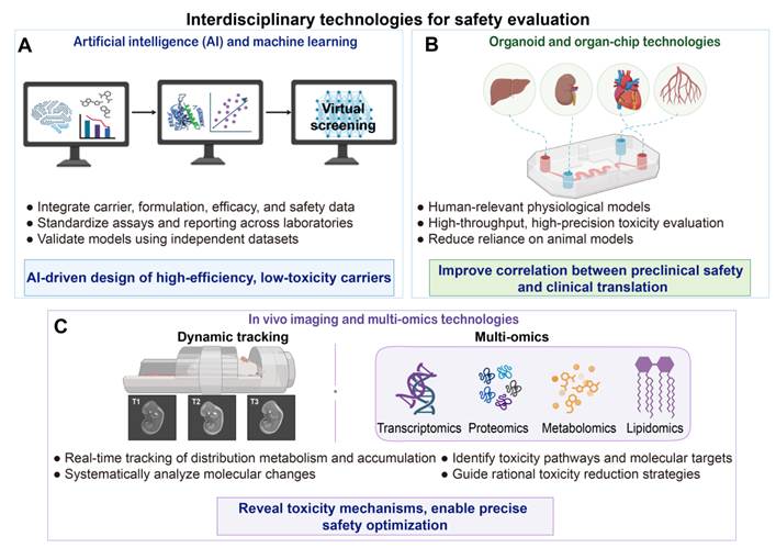
6.3 Research and development directions
We propose four research and development directions for mRNA delivery systems: stimuli-responsive design, critically evaluated biologically derived or biomimetic materials, multifunctional carriers, and precise tissue or cell targeting. Each direction should address charge exposure, membrane activity, or biopersistence of delivery systems in the tissue and intracellular compartment within the duration for the intended application.
First, intelligent and stimulus-responsive carriers are intended to improve biological selectivity by responding to endogenous stimuli, such as pH, reactive oxygen species, or enzymes [193-195]. They should remain sufficiently stable during circulation and disassemble after reaching the intended tissue or intracellular compartment. Such a design may reduce off-target exposure, but this benefit should be demonstrated with the corresponding data for biodistribution, degradation, delivery efficacy, and toxicity assessment. Externally triggered platforms, including light- and magnetic-responsive systems, may provide additional spatial and temporal control over mRNA delivery to mitigate off-target exposure [196].
Second, biologically derived and biomimetic platforms, including peptides, proteins, extracellular vesicles (EVs), and nucleic acid assemblies, may provide favorable trafficking, cellular interactions, and degradation pathways. However, the biological origin of these platforms does not imply their intrinsic safety. Peptides and proteins may be immunogenic or unstable, whereas EV heterogeneity and biomolecular cargoes within the EVs can complicate reproducibility and safety assessment. Product characterization and regulatory standards are essential for clinical development of the biologically derived and biomimetic platforms. Future development should be devoted to compositionally defined systems, controlled sourcing and manufacturing, scalable loading and purification, and consistent quality attributes. Their translational advantages should be established by assessing delivery efficiency, biodistribution, clearance, and repeated-dose immune responses from both biologically derived/biomimetic carriers and synthetic carriers under equivalent experimental conditions.
Third, multifunctional carriers can improve delivery while locally modulating inflammatory or oxidative stress [197, 198]. This can be achieved either by redesigning the carrier to reduce its intrinsic toxicity or by incorporating pharmacological groups that suppress downstream inflammatory responses. The latter may reduce cytokine production without necessarily alleviating membrane damage, material persistence, or organ exposure. The suitability of these approaches depends on the intended application. Vaccines and cancer immunotherapies may require sufficient antigen-presenting-cell activation, whereas repeated systemic protein replacement and gene editing generally require tighter control of innate and complement activation [22, 47]. Anti-inflammatory functionalization should therefore be tailored to the therapeutic context, release site, and repeated-dose requirements rather than treated as a universal safety strategy.
Finally, tissue- and cell-specific targeting is central to broadening the therapeutic window. Modification of surface ligands and tuning of formulation-level physicochemical properties can direct mRNA carriers toward intended organs or cell populations to reduce off-target exposure. Because current LNPs preferentially target the liver, delivery systems should be developed for robust extrahepatic delivery to the lung, spleen, brain, muscle, and immune-cell subsets. Extrahepatic targeting should be supported by organ-level accumulation, target-cell expression, whole-body biodistribution, and off-target toxicity (Figure 13).
Research and development directions for balancing delivery efficiency and safety of mRNA delivery systems. (A) Stimuli-responsive carriers can remain stable during circulation and release mRNA in response to endogenous or external triggers, improving spatiotemporal control. (B) Representative biologically derived mRNA delivery platforms, including peptide-, protein-, and extracellular vesicle-based carriers. (C) Multifunctional carriers can combine mRNA delivery with local anti-inflammatory, antioxidant, or immune-regulatory activity to control treatment-associated stress while preserving therapeutic function. (D) Tissue- and cell-specific targeting may enhance delivery to extrahepatic organs and defined cell populations. Created in https://BioRender.com.
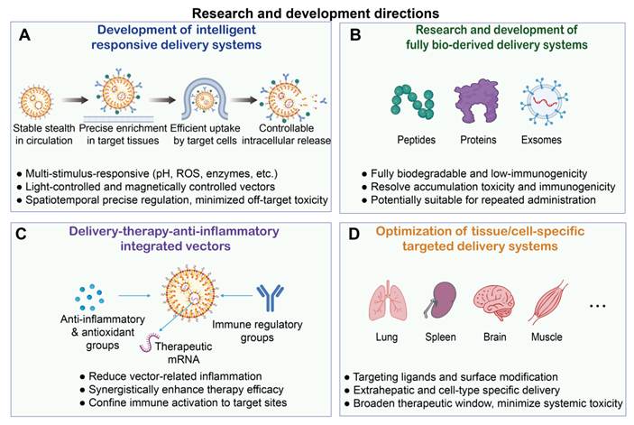
7. Conclusion
Effective mRNA delivery is achieved through protection of the mRNA cargo, efficient delivery into target cells, and mRNA release into the cytosol by the mRNA carrier. The effectiveness is correlated with material properties including charge exposure, membrane activity, and material persistence. However, these properties may also contribute to membrane damage, immune and complement activation, off-target exposure, and tissue accumulation. Moreover, mRNA vaccines may benefit from controlled immune activation, whereas systemic exposure and immune responses should be strictly controlled for repeated protein replacement and gene editing. Therefore, carrier design should strike a balance between improving delivery efficiency and mitigating carrier-associated risks. Based on the available evidence for LNPs and polymeric carriers, we propose five practical design principles for their clinical translation. First, the charge and membrane activity should be sufficient for mRNA loading, cellular uptake, and endosomal escape, but they should be controllable at the required site within a specific timeframe. Second, carriers should remain stable during transport and undergo predictable degradation and clearance after cargo release at the target site. Third, delivery efficiency should be evaluated alongside carrier dose, biodistribution, and the magnitude and duration of protein expression in target cells. Fourth, safety evaluation should cover blood compatibility, inflammatory and complement responses, organ injury, clearance, and cumulative toxicity after repeated administration. Fifth, the acceptable balance between delivery efficiency and safety should be tailored to the therapeutic application. Together, these principles provide a shared design framework for developing LNPs and polymeric carriers with effective delivery, acceptable safety, and sustained therapeutic benefits.
Abbreviations
ABC: accelerated blood clearance
ALT: alanine aminotransferase
AST: aspartate aminotransferase
BUN: blood urea nitrogen
CARPA: complement activation-related pseudoallergy
CRISPR: clustered regularly interspaced short palindromic repeats
Dex: dexamethasone
FPEI: fluorinated polyethyleneimine
GSDMD: gasdermin D
HCQ: hydroxychloroquine
IAA: imidazole-acetic acid
LNP: lipid nanoparticle
MHC: major histocompatibility complex
MPS: mononuclear phagocyte system
mRNA: messenger RNA
NF-κB: nuclear factor kappa B
NLRP3: NLR family pyrin domain-containing 3
OVA: ovalbumin
PACE: poly(amine-co-ester)
PAMAM: poly(amidoamine) dendrimer
PBAE: poly(β-amino ester)
PBMC: peripheral blood mononuclear cell
PCB: poly(carboxybetaine)
PEG: polyethylene glycol
PEI: polyethyleneimine
pSar: polysarcosine
ROS: reactive oxygen species
TLR: Toll-like receptor
Acknowledgements
This work was financially supported by the National Natural Science Foundation of China (32271445, 51903173, 82202227), Department of Science and Technology of Sichuan Province (2024NSFJQ0050, 2025ZNSFSC1417, 2025NSFSCR0142), Post-Doctor Research Project, West China Hospital, Sichuan University (2024HXBH036). All diagrams, including the graphical abstract image and figures, were created with BioRender.com (https://www.biorender.com/). The funders had no role in the conception or writing of this review, interpretation of the literature, or decision to submit the manuscript.
Competing Interests
The authors have declared that no competing interest exists.
References
1. Parhiz H, Atochina-Vasserman EN, Weissman D. mRNA-based therapeutics: looking beyond COVID-19 vaccines. Lancet. 2024;403:1192-204
2. Petsch B, Schnee M, Vogel AB, Lange E, Hoffmann B, Voss D. et al. Protective efficacy of in vitro synthesized, specific mRNA vaccines against influenza A virus infection. Nat Biotechnol. 2012;30:1210-6
3. Qin S, Tang X, Chen Y, Chen K, Fan N, Xiao W. et al. mRNA-based therapeutics: powerful and versatile tools to combat diseases. Signal Transduct Target Ther. 2022;7:166
4. Huang X, Kong N, Zhang X, Cao Y, Langer R, Tao W. The landscape of mRNA nanomedicine. Nat Med. 2022;28:2273-87
5. Barbier AJ, Jiang AY, Zhang P, Wooster R, Anderson DG. The clinical progress of mRNA vaccines and immunotherapies. Nat Biotechnol. 2022;40:840-54
6. Sahin U, Kariko K, Tureci O. mRNA-based therapeutics-developing a new class of drugs. Nat Rev Drug Discov. 2014;13:759-80
7. Bai C, Wang C, Lu Y. Novel vectors and administrations for mRNA delivery. Small. 2023;19:e2303713
8. Su K, Qiu J, Xu T, Liu S. Artificial intelligence-guided design of lipid nanoparticles for mRNA delivery. Acta Pharm Sin B. 2026;16:709-27 doi: 10.1016/j.apsb.2025.11.029
9. Xiong Q, Lee GY, Ding J, Li W, Shi J. Biomedical applications of mRNA nanomedicine. Nano Res. 2018;11:5281-309
10. Dong S, Tsai SN, Xu Y, Gong F, Young TL, Solek NC. et al. A modular mRNA platform for programmable induction of tumour-specific immunogenic cell death. Nat Nanotechnol. 2025;20:1856-70 doi: 10.1038/s41565-025-02045-5
11. Rojas LA, Sethna Z, Soares KC, Olcese C, Pang N, Patterson E. et al. Personalized RNA neoantigen vaccines stimulate T cells in pancreatic cancer. Nature. 2023;618:144-50
12. Erasmus JH, Archer J, Fuerte-Stone J, Khandhar AP, Voigt E, Granger B. et al. Intramuscular delivery of replicon RNA encoding ZIKV-117 human monoclonal antibody protects against Zika virus infection. Mol Ther Methods Clin Dev. 2020;18:402-14
13. Dagan N, Barda N, Kepten E, Miron O, Perchik S, Katz MA. et al. BNT162b2 mRNA COVID-19 vaccine in a nationwide mass vaccination setting. N Engl J Med. 2021;384:1412-23
14. Pardi N, Hogan MJ, Porter FW, Weissman D. mRNA vaccines-A new era in vaccinology. Nat Rev Drug Discov. 2018;17:261-79
15. Maruggi G, Zhang C, Li J, Ulmer JB, Yu D. mRNA as a transformative technology for vaccine development to control infectious diseases. Mol Ther. 2019;27:757-72
16. Chaudhary N, Weissman D, Whitehead KA. mRNA vaccines for infectious diseases: principles, delivery and clinical translation. Nat Rev Drug Discov. 2021;20:817-38
17. Miao L, Zhang Y, Huang L. mRNA vaccine for cancer immunotherapy. Mol Cancer. 2021;20:41
18. Liu C, Shi Q, Huang X, Koo S, Kong N, Tao W. mRNA-based cancer therapeutics. Nat Rev Cancer. 2023;23:526-43
19. You Y, Tian Y, Yang Z, Shi J, Kwak KJ, Tong Y. et al. Intradermally delivered mRNA-encapsulating extracellular vesicles for collagen-replacement therapy. Nat Biomed Eng. 2023;7:887-900
20. Popovitz J, Sharma R, Hoshyar R, Soo Kim B, Murthy N, Lee K. Gene editing therapeutics based on mRNA delivery. Adv Drug Deliv Rev. 2023;200:115026
21. Cheng Q, Wei T, Farbiak L, Johnson LT, Dilliard SA, Siegwart DJ. Selective organ targeting (SORT) nanoparticles for tissue-specific mRNA delivery and CRISPR-Cas gene editing. Nat Nanotechnol. 2020;15:313-20
22. Bitounis D, Jacquinet E, Rogers MA, Amiji MM. Strategies to reduce the risks of mRNA drug and vaccine toxicity. Nat Rev Drug Discov. 2024;23:281-300
23. Paunovska K, Loughrey D, Dahlman JE. Drug delivery systems for RNA therapeutics. Nat Rev Genet. 2022;23:265-80
24. Nzila M-M, Koehl U, Ivics Z. Non-viral vectors as beacons of hope for reducing genotoxic risks of gene therapy. Nat Biomed Eng. 2026;10:1045-50 doi: 10.1038/s41551-025-01581-8
25. Blanco E, Shen H, Ferrari M. Principles of nanoparticle design for overcoming biological barriers to drug delivery. Nat Biotechnol. 2015;33:941-51
26. Gupta SK, Haigh BJ, Griffin FJ, Wheeler TT. The mammalian secreted RNases: mechanisms of action in host defence. Innate Immun. 2013;19:86-97
27. Malone RW, Felgner PL, Verma IM. Cationic liposome-mediated RNA transfection. Proc Natl Acad Sci U S A. 1989;86:6077-81
28. Boussif O, Lezoualch F, Zanta MA, Mergny MD, Scherman D, Demeneix B. et al. A versatile vector for gene and oligonucleotide transfer into cells in culture and in vivo-polyethylenimine. Proc Natl Acad Sci U S A. 1995;92:7297-301
29. Semple SC, Akinc A, Chen JX, Sandhu AP, Mui BL, Cho CK. et al. Rational design of cationic lipids for siRNA delivery. Nat Biotechnol. 2010;28:172-6
30. Wu S, Zhang Z, Zhao Z, Cui C, Tan W. Navigating the next frontier in biomedicine: breakthroughs and insights in nucleic acid therapeutics. Chem Sci. 2026;17:3377-409 doi: 10.1039/d5sc06966a
31. Zhong Y, Du S, Dong Y. mRNA delivery in cancer immunotherapy. Acta Pharm Sin B. 2023;13:1348-57
32. Rampado R, Peer D. Design of experiments in the optimization of nanoparticle-based drug delivery systems. J Control Release. 2023;358:398-419
33. Witten J, Raji I, Manan RS, Beyer E, Bartlett S, Tang Y. et al. Artificial intelligence-guided design of lipid nanoparticles for pulmonary gene therapy. Nat Biotechnol. 2025;43:1790-9 doi: 10.1038/s41587-024-02490-y
34. Hou X, Zaks T, Langer R, Dong Y. Lipid nanoparticles for mRNA delivery. Nat Rev Mater. 2021;6:1078-94
35. Wang H, Cheng Y. Polymers for mRNA delivery. Wiley Interdiscip Rev Nanomed Nanobiotechnol. 2025;17:e70002 doi: 10.1002/wnan.70002
36. Jacob S, Varkey NR, Boddu SHS, Gorain B, Rao R, Nair AB. Advances in lipid-polymer hybrid nanoparticles: design strategies, functionalization, oncological and non-oncological clinical prospects. Pharmaceuticals (Basel). 2025;18:1772 doi: 10.3390/ph18121772
37. Ndeupen S, Qin Z, Jacobsen S, Bouteau A, Estanbouli H, Igyarto BZ. The mRNA-LNP platform's lipid nanoparticle component used in preclinical vaccine studies is highly inflammatory. iScience. 2021;24:103479
38. Sharma P, Hoorn D, Aitha A, Breier D, Peer D. The immunostimulatory nature of mRNA lipid nanoparticles. Adv Drug Deliv Rev. 2024;205:115175
39. Zelkoski AE, Lu Z, Sukumar G, Dalgard C, Said H, Alameh MG. et al. Ionizable lipid nanoparticles of mRNA vaccines elicit NF-κB and IRF responses through Toll-like receptor 4. NPJ Vaccines. 2025;10:73 doi: 10.1038/s41541-025-01124-x
40. Hall A, Lachelt U, Bartek J, Wagner E, Moghimi SM. Polyplex evolution: understanding biology, optimizing performance. Mol Ther. 2017;25:1476-90
41. Wang H, Chang H, Zhang Q, Cheng Y. Fabrication of low-generation dendrimers into nanostructures for efficient and nontoxic gene delivery. Top Curr Chem (Cham). 2017;375:62
42. Ahn JH, Lee J, Roh G, Lee NY, Bae HJ, Kwon E. et al. Impact of administration routes and dose frequency on the toxicology of SARS-CoV-2 mRNA vaccines in mice model. Arch Toxicol. 2025;99:755-73 doi: 10.1007/s00204-024-03912-1
43. Xu Y, Gong F, Golubovic A, Strilchuk A, Chen J, Zhou M. et al. Rational design and modular synthesis of biodegradable ionizable lipids via the Passerini reaction for mRNA delivery. Proc Natl Acad Sci U S A. 2025;122:e2409572122 doi: 10.1073/pnas.2409572122
44. Kenjo E, Hozumi H, Makita Y, Iwabuchi KA, Fujimoto N, Matsumoto S. et al. Low immunogenicity of LNP allows repeated administrations of CRISPR-Cas9 mRNA into skeletal muscle in mice. Nat Commun. 2021;12:7101
45. Arral ML, Whitehead KA. Design principles of lipid nanoparticles for RNA delivery. Nat Rev Bioeng. 2026;4:417-34 doi: 10.1038/s44222-026-00401-1
46. Wang X, Xu LT, Lan ZW, Hui Y, Liu Y, Yang GZ. et al. Advances in the development of lipid nanoparticles for mRNA delivery. Small Struct. 2026;7:e202500568 doi: 10.1002/sstr.202500568
47. Deng HZ, Li L, Zhao CY, Wang ZL, Xiao YF, Huang P. et al. Nanotechnology-mediated precision delivery of mRNA. Nat Mater. 2026 doi: 10.1038/s41563-026-02623-5
48. Zak MM, Zangi L. Clinical development of therapeutic mRNA applications. Mol Ther. 2025;33:2583-609 doi: 10.1016/j.ymthe.2025.03.034
49. Mendes BB, Conniot J, Avital A, Yao DB, Jiang XY, Zhou X. et al. Nanodelivery of nucleic acids. Nat Rev Methods Primers. 2022;2:24
50. Fraiman J, Erviti J, Jones M, Greenland S, Whelan P, Kaplan RM. et al. Serious adverse events of special interest following mRNA COVID-19 vaccination in randomized trials in adults. Vaccine. 2022;40:5798-805
51. Giannotta G, Murrone A, Giannotta N. COVID-19 mRNA vaccines: the molecular basis of some adverse events. Vaccines (Basel). 2023;11:747
52. Lonez C, Vandenbranden M, Ruysschaert JM. Cationic lipids activate intracellular signaling pathways. Adv Drug Deliv Rev. 2012;64:1749-58
53. Kedmi R, Ben-Arie N, Peer D. The systemic toxicity of positively charged lipid nanoparticles and the role of Toll-like receptor 4 in immune activation. Biomaterials. 2010;31:6867-75
54. Cui S, Wang Y, Gong Y, Lin X, Zhao Y, Zhi D. et al. Correlation of the cytotoxic effects of cationic lipids with their headgroups. Toxicol Res (Camb). 2018;7:473-9
55. Omo-Lamai S, Wang Y, Patel MN, Milosavljevic A, Zuschlag D, Poddar S. et al. Limiting endosomal damage sensing reduces inflammation triggered by lipid nanoparticle endosomal escape. Nat Nanotechnol. 2025;20:1285-97 doi: 10.1038/s41565-025-01974-5
56. Bates SM, Munson MJ, Trovisco V, Pereira S, Miller SR, Sabirsh A. et al. The kinetics of endosomal disruption reveal differences in lipid nanoparticle induced cellular toxicity. J Control Release. 2025;386:114047 doi: 10.1016/j.jconrel.2025.114047
57. Wu J, Zhao Z, Wu H, Lin S, Huang L, Chen G. et al. Novel swelling-lytic cell death triggered by cargo-free ionizable lipid nanoparticles. Adv Sci (Weinh). 2025;12:e09208 doi: 10.1002/advs.202509208
58. Chaudhary N, Kasiewicz LN, Newby AN, Arral ML, Yerneni SS, Melamed JR. et al. Amine headgroups in ionizable lipids drive immune responses to lipid nanoparticles by binding to the receptors TLR4 and CD1d. Nat Biomed Eng. 2024;8:1483-98
59. Li HL, Monslow MA, Freed DC, Chang D, Li FS, Gindy M. et al. Novel adjuvants enhance immune responses elicited by a replication-defective human cytomegalovirus vaccine in nonhuman primates. Vaccine. 2021;39:7446-56
60. Debnath M, Forster J, Ramesh A, Kulkarni A. Protein corona formation on lipid nanoparticles negatively affects the NLRP3 inflammasome activation. Bioconjug Chem. 2023;34:1766-79
61. Liu T, Zhang L, Joo D, Sun S-C. NF-κB signaling in inflammation. Signal Transduct Target Ther. 2017;2:17023
62. Korzun T, Moses AS, Jozic A, Grigoriev V, Newton S, Kim J. et al. Lipid nanoparticles elicit reactogenicity and sickness behavior in mice via Toll-like receptor 4 and myeloid differentiation protein 88 axis. ACS Nano. 2024;18:24842-59
63. Tahtinen S, Tong AJ, Himmels P, Oh J, Paler-Martinez A, Kim L. et al. IL-1 and IL-1ra are key regulators of the inflammatory response to RNA vaccines. Nat Immunol. 2022;23:532-42
64. Magtanong L, Ko PJ, To M, Cao JY, Forcina GC, Tarangelo A. et al. Exogenous monounsaturated fatty acids promote a ferroptosis-resistant cell state. Cell Chem Biol. 2019;26:420-32.e9
65. Noguchi N, Saito Y, Niki E. Lipid peroxidation, ferroptosis, and antioxidants. Free Radic Biol Med. 2025;237:228-38 doi: 10.1016/j.freeradbiomed.2025.05.393
66. Spickett CM. The lipid peroxidation product 4-hydroxy-2-nonenal: advances in chemistry and analysis. Redox Biol. 2013;1:145-52
67. Ioannou MS, Jackson J, Sheu SH, Chang CL, Weigel AV, Liu H. et al. Neuron-astrocyte metabolic coupling protects against activity-induced fatty acid toxicity. Cell. 2019;177:1522-35.e14
68. Sato Y, Matsui H, Yamamoto N, Sato R, Munakata T, Kohara M. et al. Highly specific delivery of siRNA to hepatocytes circumvents endothelial cell-mediated lipid nanoparticle-associated toxicity leading to the safe and efficacious decrease in the hepatitis B virus. J Control Release. 2017;266:216-25
69. Ibrahim M, Ramadan E, Elsadek NE, Emam SE, Shimizu T, Ando H. et al. Polyethylene glycol (PEG): the nature, immunogenicity, and role in the hypersensitivity of PEGylated products. J Control Release. 2022;351:215-30
70. Tenchov R, Sasso JM, Zhou QA. PEGylated lipid nanoparticle formulations: immunological safety and efficiency perspective. Bioconjug Chem. 2023;34:941-60
71. Liu YL, Liao TY, Ho KW, Liu ES, Huang BC, Hong ST. et al. Impact of pre-existing anti-polyethylene glycol antibodies on the pharmacokinetics and efficacy of a COVID-19 mRNA vaccine (Comirnaty) in vivo. Biomater Res. 2024;28:0112
72. Barta BA, Radovits T, Dobos AB, Tibor Kozma G, Meszaros T, Berenyi P. et al. Comirnaty-induced cardiopulmonary distress and other symptoms of complement-mediated pseudo-anaphylaxis in a hyperimmune pig model: causal role of anti-PEG antibodies. Vaccine X. 2024;19:100497
73. Ju Y, Lee WS, Pilkington EH, Kelly HG, Li S, Selva KJ. et al. Anti-PEG antibodies boosted in humans by SARS-CoV-2 lipid nanoparticle mRNA vaccine. ACS Nano. 2022;16:11769-80
74. Ju Y, Carreno JM, Simon V, Dawson K, Krammer F, Kent SJ. Impact of anti-PEG antibodies induced by SARS-CoV-2 mRNA vaccines. Nat Rev Immunol. 2023;23:135-6
75. Szebeni J, Storm G, Ljubimova JY, Castells M, Phillips EJ, Turjeman K. et al. Applying lessons learned from nanomedicines to understand rare hypersensitivity reactions to mRNA-based SARS-CoV-2 vaccines. Nat Nanotechnol. 2022;17:337-46
76. Kozma GT, Shimizu T, Ishida T, Szebeni J. Anti-PEG antibodies: properties, formation, testing and role in adverse immune reactions to PEGylated nano-biopharmaceuticals. Adv Drug Deliv Rev. 2020;154-155:163-75
77. Hamad I, Hunter AC, Szebeni J, Moghimi SM. Poly(ethylene glycol)s generate complement activation products in human serum through increased alternative pathway turnover and a MASP-2-dependent process. Mol Immunol. 2008;46:225-32
78. Klimek L, Novak N, Cabanillas B, Jutel M, Bousquet J, Akdis CA. Allergenic components of the mRNA-1273 vaccine for COVID-19: possible involvement of polyethylene glycol and IgG-mediated complement activation. Allergy. 2021;76:3307-13
79. Kozma GT, Meszaros T, Vashegyi I, Fulop T, Orfi E, Dezsi L. et al. Pseudo-anaphylaxis to polyethylene glycol (PEG)-coated liposomes: roles of anti-PEG IgM and complement activation in a porcine model of human infusion reactions. ACS Nano. 2019;13:9315-24
80. Mihindukulasooriya SP, Wang ZR, Li SY, Saini A, Wilson K, Plebanski M. et al. Anti-PEG antibodies in nanomedicine: mechanisms, risks, and opportunities. Adv Drug Deliv Rev. 2026;236:115925 doi: 10.1016/j.addr.2026.115925
81. Saini A, Li SY, Wang ZR, Plebanski M, Ju Y. Navigating the biomolecular corona: biological interactions and therapeutic implications of RNA-lipid nanoparticles. ACS Nano. 2026;20:15835-74 doi: 10.1021/acsnano.6c00334
82. Mazrad ZAI, Ju Y, Kent SJ, Pouton CW, Kempe K. Beyond PEG: emerging polymer lipid alternatives for lipid nanoparticle (LNP) formulations. Adv Sci (Weinh). 2026;13:e77145 doi: 10.1002/advs.77145
83. Patel S, Ashwanikumar N, Robinson E, Xia Y, Mihai C, Griffith JP 3rd. et al. Naturally-occurring cholesterol analogues in lipid nanoparticles induce polymorphic shape and enhance intracellular delivery of mRNA. Nat Commun. 2020;11:983
84. Back PI, Yu M, Modaresahmadi S, Hajimirzaei S, Zhang Q, Islam MR. et al. Immune implications of cholesterol-containing lipid nanoparticles. ACS Nano. 2024;18:28480-501
85. Shi L, Su K, Lin L, Yan X, Zhang X, He S. et al. Phospholipids tailor mRNA lipid nanoparticle delivery efficacy and immunogenicity. Mater Today. 2026;92:490-8 doi: 10.1016/j.mattod.2026.01.004
86. Iwakawa K, Sato R, Konaka M, Yamada Y, Harashima H, Sato Y. Cubic phase-inducible zwitterionic phospholipids improve the functional delivery of mRNA. Adv Sci (Weinh). 2025;12:e2413016 doi: 10.1002/advs.202413016
87. Guerrero ED, Vaidya A, Santelli J, Tian Z, Pazzi GA, Siegwart DJ. Investigating the functional contributions of phospholipids in selective organ targeting lipid nanoparticles. Biomaterials. 2026;326:123671 doi: 10.1016/j.biomaterials.2025.123671
88. Kesharwani P, Jain K, Jain NK. Dendrimer as nanocarrier for drug delivery. Prog Polym Sci. 2014;39:268-307
89. Yan Y, Liu XY, Lu A, Wang XY, Jiang LX, Wang JC. Non-viral vectors for RNA delivery. J Control Release. 2022;342:241-79
90. Yadava SK, Tripathi A, Kabilan M, Champion JA, Cicerone MT. Surface-engineered robust hybrid lipid nanocapsules: a next-generation nanoplatform for mRNA delivery. J Control Release. 2025;388:114312 doi: 10.1016/j.jconrel.2025.114312
91. Monnery BD, Wright M, Cavill R, Hoogenboom R, Shaunak S, Steinke JHG. et al. Cytotoxicity of polycations: relationship of molecular weight and the hydrolytic theory of the mechanism of toxicity. Int J Pharm. 2017;521:249-58
92. Breunig M, Lungwitz U, Liebl R, Goepferich A. Breaking up the correlation between efficacy and toxicity for nonviral gene delivery. Proc Natl Acad Sci U S A. 2007;104:14454-9
93. Xue L, Yan Y, Kos P, Chen X, Siegwart DJ. PEI fluorination reduces toxicity and promotes liver-targeted siRNA delivery. Drug Deliv Transl Res. 2021;11:255-60
94. Parhamifar L, Andersen H, Wu L, Hall A, Hudzech D, Moghimi SM. Polycation-mediated integrated cell death processes. Adv Genet. 2014;88:353-98
95. Grandinetti G, Ingle NP, Reineke TM. Interaction of poly(ethylenimine)-DNA polyplexes with mitochondria: implications for a mechanism of cytotoxicity. Mol Pharm. 2011;8:1709-19
96. Uchida S, Lau CYJ, Oba M, Miyata K. Polyplex designs for improving the stability and safety of RNA therapeutics. Adv Drug Deliv Rev. 2023;199:114972
97. Casper J, Schenk SH, Parhizkar E, Detampel P, Dehshahri A, Huwyler J. Polyethylenimine (PEI) in gene therapy: current status and clinical applications. J Control Release. 2023;362:667-91
98. Dabbaghi M, Kazemi Oskuee R, Hashemi K, Afkhami Goli A. Evaluating polyethyleneimine/DNA nanoparticles-mediated damage to cellular organelles using endoplasmic reticulum stress profile. Artif Cells Nanomed Biotechnol. 2018;46:192-9
99. Zhong D, Jiao Y, Zhang Y, Zhang W, Li N, Zuo Q. et al. Effects of the gene carrier polyethyleneimines on structure and function of blood components. Biomaterials. 2013;34:294-305
100. Piotrowski-Daspit AS, Kauffman AC, Bracaglia LG, Saltzman WM. Polymeric vehicles for nucleic acid delivery. Adv Drug Deliv Rev. 2020;156:119-32
101. Toy R, Pradhan P, Ramesh V, Di Paolo NC, Lash B, Liu J. et al. Modification of primary amines to higher order amines reduces in vivo hematological and immunotoxicity of cationic nanocarriers through TLR4 and complement pathways. Biomaterials. 2019;225:119512
102. Kopiasz RJ, Sallah HJ, Cavanagh RJ, Jones S, Moloney C, O'Shaughnessy L. et al. Modular supramolecular polycations enable efficient delivery of diverse RNA therapeutics and vaccines. Adv Mater. 2026;38:e13315 doi: 10.1002/adma.202513315
103. Zhou L, Emenuga M, Kumar S, Lamantia Z, Figueiredo M, Emrick T. Designing synthetic polymers for nucleic acid complexation and delivery: from polyplexes to micelleplexes to triggered degradation. Biomacromolecules. 2022;23:4029-40
104. Zuckerman JE, Gritli I, Tolcher A, Heidel JD, Lim D, Morgan R. et al. Correlating animal and human phase Ia/Ib clinical data with CALAA-01, a targeted, polymer-based nanoparticle containing siRNA. Proc Natl Acad Sci U S A. 2014;111:11449-54
105. Kumar R, Santa Chalarca CF, Bockman MR, Bruggen CV, Grimme CJ, Dalal RJ. et al. Polymeric delivery of therapeutic nucleic acids. Chem Rev. 2021;121:11527-652
106. Gao X, Jin Z, Tan X, Zhang C, Zou C, Zhang W. et al. Hyperbranched poly(beta-amino ester)-based polyplex nanoparticles for delivery of the CRISPR/Cas9 system and treatment of HPV infection-associated cervical cancer. J Control Release. 2020;321:654-68
107. Nelson J, Sorensen EW, Mintri S, Rabideau AE, Zheng W, Besin G. et al. Impact of mRNA chemistry and manufacturing process on innate immune activation. Sci Adv. 2020;6:eaaz6893
108. Wu WH, Wang ZW. Adverse effects of intravenous LNP-mediated mRNA therapy in clinical trials: a systematic review and meta-analysis. Expert Opin Drug Deliv. 2025;22:1395-403 doi: 10.1080/17425247.2025.2517363
109. Chen Y, De Lombaerde E, Bugler-Lamb A, Zhong Z, Schuijs MJ, Gomez CMB. et al. Thioamides adjacent to the ionizable amine headgroup in ionizable lipids reduce the pKa of lipid nanoparticles and enhance mRNA transfection efficiency in vitro and in vivo. Angew Chem Int Ed Engl. 2025;64:e202506954 doi: 10.1002/anie.202506954
110. Zhao Y, Li R, Liu P, Wang J, Cui Y, Ma Y. et al. Low reactogenicity and high tumour antigen expression from mRNA-LNPs with membrane-destabilizing zwitterionic lipids. Nat Biomed Eng. 2025 doi: 10.1038/s41551-025-01577-4
111. Liu J, Xiao B, Yang Y, Jiang Y, Wang R, Wei Q. et al. Low-dose mildronate-derived lipidoids for efficient mRNA vaccine delivery with minimal inflammation side effects. ACS Nano. 2024;18:23289-300
112. Lin Y, Li M, Luo Z, Meng Y, Zong Y, Ren H. et al. Tissue-specific mRNA delivery and prime editing with peptide-ionizable lipid nanoparticles. Nat Mater. 2026;25:133-45 doi: 10.1038/s41563-025-02320-9
113. Wang Q, Lin Y, Xiao J, Xu K, Luo Z, Ren H. et al. Optimizing peptide ionizable lipids enables efficient and low-toxicity mRNA delivery for in vivo prime editing and protein replacement therapy. Adv Mater. 2026;38:e22552 doi: 10.1002/adma.202522552
114. Men Y, Popoola DO, Cao Z, Li Y, Wilkens S, Teng Y. et al. Sulfonium lipid nanoparticles for intranasal mRNA delivery to lung epithelial and immune cells. Acta Biomater. 2025;205:616-33 doi: 10.1016/j.actbio.2025.08.032
115. Popoola DO, Cao Z, Men Y, Li X, Viapiano M, Wilkens S. et al. Lung-specific mRNA delivery enabled by sulfonium lipid nanoparticles. Nano Lett. 2024;24:8080-8
116. Li Y, Yuan W, Tian X, Zhu C, Li X, Chen R. et al. A facile method to prepare non-cationic mRNA lipid nanoparticles based on a frame-guided assembly strategy. Nano Today. 2023;52:101991
117. Wang C, Zhao C, Wang W, Liu X, Deng H. Biomimetic noncationic lipid nanoparticles for mRNA delivery. Proc Natl Acad Sci U S A. 2023;120:e2311276120
118. Liu Z, Wu JC, Wang N, Lin YQ, Song RT, Zhang M. et al. Structure-guided design of endosomolytic chloroquine-like lipid nanoparticles for mRNA delivery and genome editing. Nat Commun. 2025;16:4241 doi: 10.1038/s41467-025-59501-y
119. Chen K, Li X, Feng S, Li Y, Jiang T, Liu Y. et al. Hydroxychloroquine-functionalized ionizable lipids mitigate inflammatory responses in mRNA therapeutics. J Control Release. 2025;387:114267 doi: 10.1016/j.jconrel.2025.114267
120. Jozić A, Le Roux C, Kim J, Berchel M, Sahel DK, Bodi EK. et al. In vivo endosomal escape assay identifies mechanisms for efficient hepatic LNP delivery. Nat Biotechnol. 2026 doi: 10.1038/s41587-026-03022-6
121. Wu S, Shi L, Su K, Lin L, Yan X, Zhang Y. et al. Carbonate-bearing ionizable lipids for mRNA delivery to splenic NK cells. J Am Chem Soc. 2025;147:28665-73 doi: 10.1021/jacs.5c01389
122. Wang Q, Lin Y, Luo Z, Xiao J, Xu K, Yu X. et al. Hydrogen bond-assisted two-component lipid nanoparticles for spleen-specific mRNA delivery. J Am Chem Soc. 2025;147:42711-23 doi: 10.1021/jacs.5c14194
123. Kawai A, Shimizu T, Tanaka H, Shichinohe S, Anindita J, Hirose M. et al. Low-inflammatory lipid nanoparticle-based mRNA vaccine elicits protective immunity against H5N1 influenza virus with reduced adverse reactions. Mol Ther. 2025;33:529-47 doi: 10.1016/j.ymthe.2024.12.032
124. Wang S, Zhang Y, Zhong Y, Xue Y, Liu Z, Wang C. et al. Accelerating diabetic wound healing by ROS-scavenging lipid nanoparticle-mRNA formulation. Proc Natl Acad Sci U S A. 2024;121:e2322935121
125. Lv K, Yu Z, Wang J, Li N, Wang A, Xue T. et al. Discovery of ketal-ester ionizable lipid nanoparticle with reduced hepatotoxicity, enhanced spleen tropism for mRNA vaccine delivery. Adv Sci (Weinh). 2024;11:e2404684
126. Li YC, Cao Y, Bai H, Zhang LW, Chen BQ, Wu CF. et al. Incorporation of disulfide bonds into ionizable lipids enables efficient splenic mRNA delivery and potent immunotherapy in mice. Chem Eng J. 2025;516:164285 doi: 10.1016/j.cej.2025.164285
127. Ding H, Tan P, Fu S, Tian X, Zhang H, Ma X. et al. Preparation and application of pH-responsive drug delivery systems. J Control Release. 2022;348:206-38
128. Oyama R, Ishigame H, Tanaka H, Tateshita N, Itazawa M, Imai R. et al. An ionizable lipid material with a vitamin E scaffold as an mRNA vaccine platform for efficient cytotoxic T cell responses. ACS Nano. 2023;17:18758-74
129. Tanaka H, Takahashi T, Konishi M, Takata N, Gomi M, Shirane D. et al. Self-degradable lipid-like materials based on “hydrolysis accelerated by the intra-particle enrichment of reactant (HyPER)” for messenger RNA delivery. Adv Funct Mater. 2020;30:1910575
130. Li S, Zheng L, Zhang Z, Zhang J, Liu X, Xu J. et al. Glutarimide-derived ionizable lipid enables mRNA lipid nanoparticle vaccines with enhanced safety and immunogenicity. J Control Release. 2026;389:114482 doi: 10.1016/j.jconrel.2025.114482
131. Ge X, He Z, Yang H, Pan X, Yan L, Shi Y. et al. Impact of tail unsaturation in ionizable lipids on mRNA delivery efficiency and immunogenicity of lipid nanoparticles. J Control Release. 2025;384:113906 doi: 10.1016/j.jconrel.2025.113906
132. Yu X, Qi S, Zhang W, Li H, Cao F, Wang Y. et al. Lipid nanoparticle chaperone with redox-adaptive intracellular microenvironment modulation potentiates the potency of mRNA vaccines. J Am Chem Soc. 2025;147:44072-86 doi: 10.1021/jacs.5c10972
133. Gong N, Kim D, Alameh M-G, El-Mayta R, Han EL, Dwivedi G. et al. Mannich reaction-based combinatorial libraries identify antioxidant ionizable lipids for mRNA delivery with reduced immunogenicity. Nat Biomed Eng. 2025;9:2181-95 doi: 10.1038/s41551-025-01422-8
134. Zong Y, Zhong H, Wang Y, Lin Y, Zhao L, Wang X. et al. Safe and highly efficient lipid-pro-dexamethasone nanoparticles for mRNA delivery and base editing. J Am Chem Soc. 2026;148:10225-40 doi: 10.1021/jacs.6c01708
135. Liu M, Li J, Zhao D, Yan N, Zhang H, Liu M. et al. Branched PEG-modification: a new strategy for nanocarriers to evade of the accelerated blood clearance phenomenon and enhance anti-tumor efficacy. Biomaterials. 2022;283:121415
136. Pozzi D, Colapicchioni V, Caracciolo G, Piovesana S, Capriotti AL, Palchetti S. et al. Effect of polyethyleneglycol (PEG) chain length on the bio-nano-interactions between PEGylated lipid nanoparticles and biological fluids: from nanostructure to uptake in cancer cells. Nanoscale. 2014;6:2782-92
137. Shi D, Beasock D, Fessler A, Szebeni J, Ljubimova JY, Afonin KA. et al. To PEGylate or not to PEGylate: immunological properties of nanomedicine's most popular component, polyethylene glycol and its alternatives. Adv Drug Deliv Rev. 2022;180:114079
138. Sherman MR, Williams LD, Sobczyk MA, Michaels SJ, Saifer MG. Role of the methoxy group in immune responses to mPEG-protein conjugates. Bioconjug Chem. 2012;23:485-99
139. Sanchez A, Loughrey D, Echeverri ES, Huayamares SG, Radmand A, Paunovska K. et al. Substituting poly(ethylene glycol) lipids with poly(2-ethyl-2-oxazoline) lipids improves lipid nanoparticle repeat dosing. Adv Healthc Mater. 2024;13:e2304033
140. Yao X, Qi C, Sun C, Huo F, Jiang X. Poly(ethylene glycol) alternatives in biomedical applications. Nano Today. 2023;48:101738
141. Fu S, Zhu X, Huang F, Chen X. Anti-PEG antibodies and their biological impact on PEGylated drugs: challenges and strategies for optimization. Pharmaceutics. 2025;17:1074 doi: 10.3390/pharmaceutics17081074
142. Kang DD, Hou X, Wang L, Xue Y, Li H, Zhong Y. et al. Engineering LNPs with polysarcosine lipids for mRNA delivery. Bioact Mater. 2024;37:86-93
143. Xiao Y, Lian X, Sun Y, Sung YC, Vaidya A, Chen Z. et al. High-density brush-shaped polymer lipids reduce anti-PEG antibody binding for repeated administration of mRNA therapeutics. Nat Mater. 2025;24:1840-51 doi: 10.1038/s41563-024-02116-3
144. Qian H, Wang K, Lv M, Zhao C, Wang H, Wen S. et al. Recent advances on next generation of polyzwitterion-based nano-vectors for targeted drug delivery. J Control Release. 2022;343:492-505
145. Overby C, Park S, Summers A, Benoit DSW. Zwitterionic peptides: tunable next-generation stealth nanoparticle modifications. Bioact Mater. 2023;27:113-24
146. Luozhong S, Liu P, Li R, Yuan Z, Debley E, Chen Y. et al. Poly(carboxybetaine) lipids enhance mRNA therapeutics efficacy and reduce their immunogenicity. Nat Mater. 2025;24:1852-61 doi: 10.1038/s41563-025-02240-8
147. Kent SJ, Li SY, Amarasena TH, Reynaldi A, Lee WS, Leeming MG. et al. Blood distribution of SARS-CoV-2 lipid nanoparticle mRNA vaccine in humans. ACS Nano. 2024;18:27077-89
148. Kent SJ, Li S, Amarasena TH, Reynaldi A, Leeming MG, Juno JA. et al. In vivo blood kinetics and transcript integrity of three mRNA-lipid nanoparticle vaccines in humans. medRxiv. 2026 doi: 10.64898/2026.03.13.26348310
149. Ju Y, Li SY, Tan AEQ, Pilkington EH, Brannon PT, Plebanski M. et al. Patient-specific nanoparticle targeting in human leukemia blood. ACS Nano. 2024;18:29021-35
150. Li SY, Cortez-Jugo C, Ju Y, Caruso F. Approaching two decades: biomolecular coronas and bio-nano interactions. ACS Nano. 2024;18:33257-63
151. Ju Y, Kelly HG, Dagley LF, Reynaldi A, Schlub TE, Spall SK. et al. Person-specific biomolecular coronas modulate nanoparticle interactions with immune cells in human blood. ACS Nano. 2020;14:15723-37
152. Zhang H, Han X, Alameh MG, Shepherd SJ, Padilla MS, Xue L. et al. Rational design of anti-inflammatory lipid nanoparticles for mRNA delivery. J Biomed Mater Res A. 2022;110:1101-8
153. Safford HC, Shuler CF, Geisler HC, Thatte AS, Swingle KL, Han EL. et al. Probing the role of lipid nanoparticle elasticity on mRNA delivery to the placenta. Nano Lett. 2025;25:4800-8 doi: 10.1021/acs.nanolett.4c06241
154. Wang X, Shi X, Wang R. Regulating mRNA endosomal escape through lipid rafts: a review. Int J Pharm. 2025;675:125571 doi: 10.1016/j.ijpharm.2025.125571
155. Cheng MHY, Zhang Y, Fox K, Leung J, Strong C, Kang E. et al. Liposomal lipid nanoparticles for extrahepatic delivery of mRNA. Nat Commun. 2025;16:4135 doi: 10.1038/s41467-025-58523-w
156. Ling DD, Li N, Pan XB, Gao WX, Li H, Tian Q. et al. A new type of lipid nanoparticles with sphingomyelin-induced active lipid raft structures for improved mRNA cellular uptake. J Control Release. 2026;391:114640 doi: 10.1016/j.jconrel.2026.114640
157. Yang X, Zhou S, Zeng J, Zhang S, Li M, Yue F. et al. A biodegradable lipid nanoparticle delivers a Cas9 ribonucleoprotein for efficient and safe in situ genome editing in melanoma. Acta Biomater. 2024;190:531-47
158. Lin L, Su K, Zhang X, Shi L, Yan X, Fu Q. et al. A versatile strategy to transform cationic polymers for efficient and organ-selective mRNA delivery. Angew Chem Int Ed Engl. 2025;64:e202500306 doi: 10.1002/anie.202500306
159. Long J, Ma S, Wan R, Sun A, Zhu Z, Wang R. et al. Thiourea-modified poly(l-lysine) carriers with multiple hydrogen bonding interactions for safe and robust mRNA vaccine therapy. J Control Release. 2025;388:114371 doi: 10.1016/j.jconrel.2025.114371
160. Bayraktutan H, Kopiasz RJ, Elsherbeny A, Martinez Espuga M, Gumus N, Oz UC. et al. Polysarcosine functionalised cationic polyesters efficiently deliver self-amplifying mRNA. Polym Chem. 2024;15:1862-76
161. Li M, Zhao M, Fu Y, Li Y, Gong T, Zhang Z. et al. Enhanced intranasal delivery of mRNA vaccine by overcoming the nasal epithelial barrier via intra- and paracellular pathways. J Control Release. 2016;228:9-19
162. Tan L, Zheng T, Li M, Zhong X, Tang Y, Qin M. et al. Optimization of an mRNA vaccine assisted with cyclodextrin-polyethyleneimine conjugates. Drug Deliv Transl Res. 2020;10:678-89
163. Vlasova KY, Kerr A, Pennock ND, Jozic A, Sahel DK, Gautam M. et al. Synthesis of ionizable lipopolymers using split-Ugi reaction for pulmonary delivery of various size RNAs and gene editing. Nat Commun. 2025;16:4021 doi: 10.1038/s41467-025-59136-z
164. Li J, Wu Y, Xiang J, Wang H, Zhuang Q, Wei T. et al. Fluoroalkane modified cationic polymers for personalized mRNA cancer vaccines. Chem Eng J. 2023;456:140930
165. Li S, Lv M, Mei W, Yu X. Fluorinated polyethylenimine and fluorinated choline phosphate lipids complex system for efficient mRNA delivery to deep-seated tumor tissues. Biomacromolecules. 2024;25:5251-9
166. Lv J, Fan Q, Zhang Y, Zhou X, Yu P, Yu X. et al. A serum resistant polymer with exceptional endosomal escape and mRNA delivery efficacy for CRISPR gene therapy. Adv Sci (Weinh). 2025;12:e2413006 doi: 10.1002/advs.202413006
167. Li X, Qi J, Wang J, Hu W, Zhou W, Wang Y. et al. Nanoparticle technology for mRNA: delivery strategy, clinical application and developmental landscape. Theranostics. 2024;14:738-60
168. Suberi A, Grun MK, Mao T, Israelow B, Reschke M, Grundler J. et al. Polymer nanoparticles deliver mRNA to the lung for mucosal vaccination. Sci Transl Med. 2023;15:eabq0603
169. Kavanagh EW, Tzeng SY, Sharma N, Cutting GR, Green JJ. Ligand-free biodegradable poly(beta-amino ester) nanoparticles for targeted systemic delivery of mRNA to the lungs. Biomaterials. 2025;313:122753 doi: 10.1016/j.biomaterials.2024.122753
170. Gurnani P, Blakney AK, Terracciano R, Petch JE, Blok AJ, Bouton CR. et al. The in vitro, ex vivo, and in vivo effect of polymer hydrophobicity on charge-reversible vectors for self-amplifying RNA. Biomacromolecules. 2020;21:3242-53
171. Zhou H, Liao YQ, Han XF, Chen DS, Hong XC, Zhou K. et al. ROS-responsive nanoparticle delivery of mRNA and photosensitizer for combinatorial cancer therapy. Nano Lett. 2023;23:3661-8
172. Yang WQ, Chen PW, Boonstra E, Hong TH, Cabral H. Polymeric micelles with pH-responsive cross-linked core enhance in vivo mRNA delivery. Pharmaceutics. 2022;14:1205
173. Xiao XH, Gao Y, Li AY, Jia YY, Li JJ, Zhang MJ. et al. Smart dendrimer nanogels boost mRNA-based cancer therapy via synergistic glycolysis inhibition and immune activation. Bioact Mater. 2026;56:3-14 doi: 10.1016/j.bioactmat.2025.10.008
174. Li Z, Amaya L, Pi R, Wang SK, Ranjan A, Waymouth RM. et al. Charge-altering releasable transporters enhance mRNA delivery in vitro and exhibit in vivo tropism. Nat Commun. 2023;14:6983
175. AbdElwakil MM, Ni J, Ramsay-Burrough S, Hurst PJ, McClellan RL, Khasnavis SR. et al. In vivo mRNA delivery to the lung vascular endothelium by dicationic charge-altering releasable transporters. J Am Chem Soc. 2025;147:40146-57 doi: 10.1021/jacs.5c06654
176. Lee S, Nasr S, Rasheed S, Liu Y, Hartwig O, Kaya C. et al. Proteoid biodynamers for safe mRNA transfection via pH-responsive nanorods enabling endosomal escape. J Control Release. 2023;353:915-29
177. Ren S, Lin X, Xie Q, Yu S, Zheng X, Pan H. et al. Redox-responsive peptide coacervates for enhanced mRNA delivery and intracellular release. ACS Nano. 2026;20:459-74 doi: 10.1021/acsnano.5c13501
178. Ben-Akiva E, Karlsson J, Hemmati S, Yu H, Tzeng SY, Pardoll DM. et al. Biodegradable lipophilic polymeric mRNA nanoparticles for ligand-free targeting of splenic dendritic cells for cancer vaccination. Proc Natl Acad Sci U S A. 2023;120:e2301606120
179. Vlatkovic I. Non-immunotherapy application of LNP-mRNA: maximizing efficacy and safety. Biomedicines. 2021;9:530
180. Kon E, Ad-El N, Hazan-Halevy I, Stotsky-Oterin L, Peer D. Targeting cancer with mRNA-lipid nanoparticles: key considerations and future prospects. Nat Rev Clin Oncol. 2023;20:739-54
181. Tregoning JS, Stirling DC, Wang Z, Flight KE, Brown JC, Blakney AK. et al. Formulation, inflammation, and RNA sensing impact the immunogenicity of self-amplifying RNA vaccines. Mol Ther Nucleic Acids. 2023;31:29-42
182. Sahin U, Muik A, Derhovanessian E, Vogler I, Kranz LM, Vormehr M. et al. COVID-19 vaccine BNT162b1 elicits human antibody and TH1 T cell responses. Nature. 2020;586:594-9
183. Zhang Z, Fan YN, Jiang SQ, Ma YJ, Yu YR, Qing YX. et al. Recent advances in mRNA delivery systems for cancer therapy. Adv Sci (Weinh). 2025;12:e17571 doi: 10.1002/advs.202417571
184. Chen WF, Zhu YN, He JH, Sun X. Path towards mRNA delivery for cancer immunotherapy from bench to bedside. Theranostics. 2024;14:96-115
185. Qin Y, Zeng W-F, Liang W. Development of therapeutic cancer vaccines using nanomicellar preparations. Oncol Transl Med. 2023;9:265-8
186. Cao QN, Yao YL, Zheng WM, Liu HQ, Jiang MX, Xie DY. et al. An inherent T cell-activating mRNA delivery carrier for in vivo CAR T generation. Nat Mater. 2026 doi: 10.1038/s41563-026-02675-7
187. Vavilis T, Stamoula E, Ainatzoglou A, Sachinidis A, Lamprinou M, Dardalas I. et al. mRNA in the context of protein replacement therapy. Pharmaceutics. 2023;15:166
188. Du X, Yada E, Terai Y, Takahashi T, Nakanishi H, Tanaka H. et al. Comprehensive evaluation of lipid nanoparticles and polyplex nanomicelles for muscle-targeted mRNA delivery. Pharmaceutics. 2023;15:2291
189. Dawidczyk CM, Russell LM, Searson PC. Recommendations for benchmarking preclinical studies of nanomedicines. Cancer Res. 2015;75:4016-20
190. Faria M, Björnmalm M, Thurecht KJ, Kent SJ, Parton RG, Kavallaris M. et al. Minimum information reporting in bio-nano experimental literature. Nat Nanotechnol. 2018;13:777-85
191. Ma YT, VanKeulen-Miller R, Fenton OS. mRNA lipid nanoparticle formulation, characterization and evaluation. Nat Protoc. 2025;20:2618-51 doi: 10.1038/s41596-024-01134-4
192. Hu X, Lin H, Ma L, Guo Z, Zhang M, Zhu Y. et al. Engineering the future of cancer nanomedicine: from rational design to clinical translation. Oncol Transl Med. 2026 doi: 10.1097/OT9.0000000000000152
193. Zhou H, Chen DS, Hu CJ, Hong X, Shi J, Xiao Y. Stimuli-Responsive Nanotechnology for RNA Delivery. Adv Sci. 2023;10:2303597
194. Zhao S, Gao K, Han H, Stenzel M, Yin B, Song H. et al. Acid-degradable lipid nanoparticles enhance the delivery of mRNA. Nat Nanotechnol. 2024;19:1702-1711
195. Li B, Zhao M, Lai W, Zhang X, Yang B, Chen X. et al. Activatable NIR-II photothermal lipid nanoparticles for improved messenger RNA delivery. Angew Chem Int Ed Engl. 2023;62:e202302676
196. Yang Z, Shi J, Xie J, Wang Y, Sun J, Liu T. et al. Large-scale generation of functional mRNA-encapsulating exosomes via cellular nanoporation. Nat Biomed Eng. 2020;4:69-83
197. Kirian RD, Steinman D, Jewell CM, Zierden HC. Extracellular vesicles as carriers of mRNA: Opportunities and challenges in diagnosis and treatment. Theranostics. 2024;14:2265-89
198. Zhang H, Han X, Alameh M-G, Shepherd SJ, Padilla MS, Xue L. et al. Rational design of anti-inflammatory lipid nanoparticles for mRNA delivery. J Biomed Mater Res A. 2022;110:1101-1108
Author contact
Corresponding author: wangbedu.cn; qiyonggongorg.cn; luokuiedu.cn.