Impact Factor




Theranostics 2027; 17(1):177-198. doi:10.7150/thno.135840 This issue Cite
Review
Next-generation intelligent microneedle systems for precision tumor immunotherapy
Hong-Xiang Zhao1, Yanhui Shi2, Hao Zhang2, Wanchuan Ding4,  , Ming Xia3, , Miaoqing Zhao1,2,
1. Cancer Center, Shandong University, Jinan 250117, China.
2. Department of Pathology and Laboratory Medicine, Shandong Cancer Hospital and Institute, Shandong First Medical University and Shandong Academy of Medical Sciences, Jinan 250117, China.
3. Department of Otolaryngology, Shandong Provincial Hospital Affiliated to Shandong First Medical University, Key Laboratory for Hearing Reconstruction of Shandong Province, Jinan 250021, China.
4. Department of Rheumatology and Immunology, Nanjing Drum Tower Hospital, Nanjing 210008, China.
Received 2026-4-8; Accepted 2026-9-21; Published 2027-1-1
Abstract
Microneedles have emerged as a promising platform for cancer immunotherapy, offering targeted approaches to both drug delivery and diagnosis. Owing to their minimally invasive nature and precise control over drug administration, microneedles have the potential to improve therapeutic outcomes for cancer patients. This review provides an overview of recent advances in the use of microneedles for tumor immunotherapy. We first introduce their fundamental mechanisms and various classifications, followed by an examination of their roles in therapeutic intervention and diagnostic monitoring within the context of immunotherapy. The distinctive design of microneedles facilitates efficient therapeutic delivery and supports synergistic integration of immunotherapy with other treatment modalities, including chemotherapy, radiotherapy, and photothermal therapy. Finally, we explore future directions for microneedle-based systems and their potential to further improve the efficacy of cancer immunotherapy.
Keywords: microneedle technology, tumor immunotherapy, immune checkpoint inhibitors, combination therapy, tumor diagnosis and monitoring
Introduction
Cancer remains the second leading cause of mortality worldwide, with its high incidence and mortality rates posing a major challenge to global public health [1-10]. Conventional therapeutic approaches, such as chemotherapy, radiotherapy, and surgery, remain widely employed; however, they are frequently accompanied by serious adverse effects. In particular, systemic toxicity and drug resistance can significantly compromise patients’ quality of life [11-16]. These treatment-related burdens may also have a substantial impact on patients’ overall well-being [17-22].Immunotherapy has emerged as a promising alternative; however, its effectiveness is still limited by two principal barriers. The tumor microenvironment (TME) with immunosuppressive properties can inhibit the activity of effector cells. Meanwhile, the dense extracellular matrix of solid tumors limits drug penetration and distribution [23,24].
Transdermal drug delivery systems offer a complementary therapeutic approach [25-27]. These systems deliver drugs directly to local targets, which reduces the liver's metabolic clearance of the drugs and also helps lower systemic exposure and toxicity. Many compounds have been previously studied that support transdermal drug delivery [28-31]. However, there are a number of significant issues. One is the characteristic of numerous anti-cancer drugs. The molecules are large or very polar. Other medicines are prone to crystallizing. These features make it hard for them to pass through the skin’s stratum corneum [32,33]. Another issue is a lack of specificity for tumor tissue. This can harm the surrounding normal tissues. Moreover, maintaining the effective drug release continuously is challenging [34-36]. All of these problems restrain the use of transdermal systems in oncology. Therefore, there is a need for improved drug delivery methods. An important objective for further development of localized cancer therapy is the precise and controllable release of drugs.
Microneedles have the potential to enhance the transdermal delivery of drugs. These devices consist of small projections. Their size can be as small as the micrometer range [37-44], forming microchannels in the deep layers of the skin. Drugs can then be delivered to specific local areas [45-51]. Compared to traditional transdermal methods, microneedles are painless. These are minimally invasive treatments that can deliver medication efficiently and precisely. Drug release can also be controlled. Microneedles can bypass the liver and intestines, allowing the drug to enter the bloodstream directly. This raises drug concentration and bioavailability [52,53]. The technology also enables sustained release, thereby maintaining stable drug levels in the body over an extended period. The minimal pain associated with microneedles may improve patient adherence while reducing fear of needles [54,55]. Given their minimally invasive nature and efficient delivery capabilities, microneedles are emerging as a promising option for cancer therapy. They can also facilitate the combination of immunotherapy with other treatments, potentially achieving improved antitumor effects [56]. Furthermore, microneedles are considered safe, well tolerated, and cost-effective, and have shown substantial progress in tumor immunotherapy. Therefore, a comprehensive review of their current applications and recent developments is warranted.
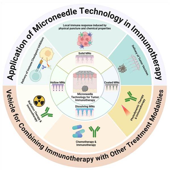
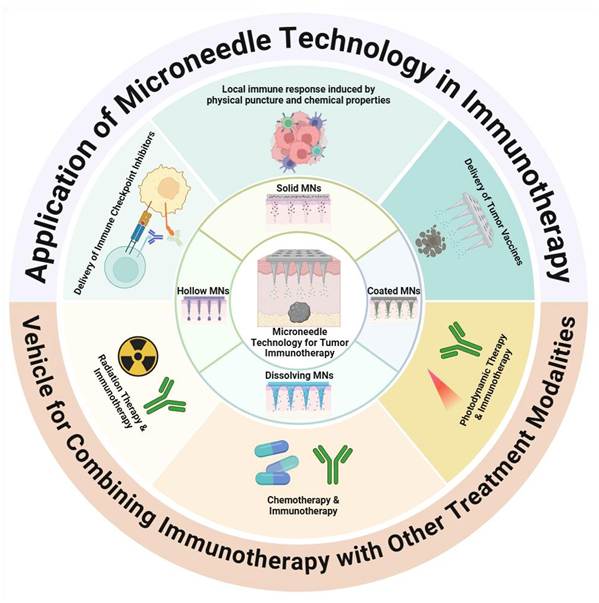
This review discusses microneedle technology for cancer immunotherapy. We summarize recent progress in this field (Figure 1). We first explain how microneedle systems work, and sort them into categories. Then we talk about their current applications in immunotherapy and diagnostic monitoring. We analyze the potential of combining microneedles with other therapies. We also discuss existing problems and future research directions. Our goal is to clarify how microneedles can be translated into clinical use for cancer immunotherapy.
An Overview of Microneedle Technology in Cancer Immunotherapy. Microneedles bypass the stratum corneum and deliver immunotherapeutics directly into the skin with minimal invasion. Solid microneedles create microchannels while providing physical and material cues that stimulate local immunity; hollow microneedles enable controlled infusion of checkpoint inhibitors or vaccine components; coated microneedles release surface-loaded drugs or adjuvants rapidly; and dissolving microneedles embed payloads in biodegradable matrices for release as the needles dissolve. These platforms support transdermal tumor vaccination, cutaneous checkpoint blockade, and combination regimens with photodynamic therapy, chemotherapy, or radiotherapy.
Fundamental principles and classification of microneedle technology
Microneedles are miniature needle-like devices ranging in size from tens to hundreds of micrometers [57,58]. They are capable of penetrating the stratum corneum without irritating the nerve endings beneath it. The main applications of such microscale systems are transdermal drug delivery, vaccine delivery, and collection of biological samples [56,59-68]. The microneedle structure is typically composed of three basic components. It contains a sharp tip for breaking through the skin barrier, a needle shaft that transports the active substance to be administered, and a support base for stability and easy handling. The special design produces temporary microchannels on the surface of the skin [69]. Microneedles have several clinical advantages. They skip the first-pass elimination, and they enhance patient compliance with drug therapy. Meanwhile, they reduce the number of specialist clinical staff. The major benefit of their use is the capacity to exert localized therapeutic activity, but with reduced systemic toxicity [70].
Classification of microneedle systems
Depending on their structural design and drug delivery mechanisms, commonly used microneedles can be divided into four major categories. All categories have their own specialties and drawbacks [71-73]. Generally, skin pretreatment is performed by using solid microneedles. Microneedles form tiny holes in the skin. This facilitates the absorption of other medications. The needles are devoid of any ingredients. Hollow microneedles are microneedles that contain a channel inside the needles. They can be used for sustained drug delivery. They also have the ability to harvest biological fluids [74-76]. The needles are applied in vaccination and biosensing [77]. Soluble microneedles are made of biodegradable polymers [61]. They are absorbed by the body slowly when in the skin. Their drug release profile is sustained-release [78]. This approach is useful in tumor therapy and immunotherapies [79-81]. Coated microneedles are needles coated with a layer of drug. They eject the drug rapidly following penetration of the skin. This is helpful in a treatment that requires quick action [79, 82].
Solid and hollow microneedles offer high mechanical integrity and precise volumetric delivery. However, they do need to be used singly at any given time, or they are difficult to make. This makes it hard to use the product on oneself. The coated type of microneedles carries less drug. They can also come out with their coating during insertion. A soluble microneedle is easy to use and enables controlled release. However, it is not very potent. Residual polymer in the skin could lead to safety concerns. Soluble microneedles are considered to be the most promising for clinical applications (assessed for vaccines and immunotherapies). However, the challenges of either large-scale manufacture at room temperature or long-term storage stability remain significant challenges.
Materials determine performance and biocompatibility
Microneedle performance is determined by the materials used. It has an influence on mechanical strength, drug release, and safety. Metal needles are tough but may not be very biocompatible [23]. Polymer systems are better tolerated by the tissues. They can also be degraded at a variable rate [60,83-85]. Depending on composition and structure, various functional microneedles are available. Solid ones are those that are made of non-degradable materials such as silicon or metal. They are structurally good but may raise questions about complex handling and dosage accuracy. The main application of these is skin pretreatment and low-level delivery [86]. The microneedles coated with the drug are also comprised of high-strength materials like silicon or metal. They suffer the drawbacks of potential instability of coatings and limited capability for drug loading. Vaccines [87-89] are typical highly active drugs for which these microneedles are used. Compared with solid and coated microneedles, the hollow microneedles have greater drug loading capabilities and can be used to develop sustained and controlled releases of drugs [90-92]. However, they are prone to channel blockage due to their complex design, and their manufacturing process is relatively complex. They can be employed for the delivery of sensitive biological compounds and can also be applied to diagnostic methods like interstitial fluid sampling [93]. Dissolvable microneedles are manufactured from water-soluble polysaccharide materials or biodegradable polymer materials [94]. Once injected, the needle-like matrix dissolves in tissue, and the therapeutic drug contained within the matrix is released from it. They are biocompatible, user-friendly, and cheap to make. They may be able to transport several medications, and the release can be timed or controlled. Their disadvantages, however, are that the drug tends to diffuse during their manufacture and that cross-linking agents leave behind residues in the skin [61,95-98]. Hydrogel-based microneedles exhibit considerable swelling capacity. The needle tip absorbs the tissue interstitial fluid, leading to controlled drug elution [99-104]. Typically constructed from hydratable polymer matrices, they provide excellent tissue compatibility without leaving residues, feature straightforward fabrication and application, and allow regulated drug release [105-108].
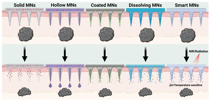
The clinical applicability of each type of microneedle depends on addressing type-specific limitations. Solid and hollow microneedles carry the risk of needle breakage. They also require trained personnel to perform multistep procedures. These requirements limit their use in large-scale vaccination campaigns. Coated microneedles have a low drug-loading capacity. They are therefore suitable only for the delivery of highly potent drugs. Dissolvable microneedles offer a good balance between safety, convenience, and controlled drug release. However, they have low mechanical strength. Batch-to-batch variation also requires engineering solutions. Smart microneedles are stimulus-responsive devices. Unlike conventional systems, they enable dynamic control (Figure 2).
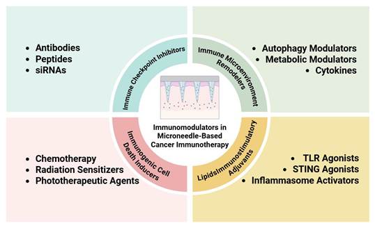
Classification of immunomodulators in microneedle-based cancer immunotherapy. Microneedle-based cancer immunotherapy leverages four classes of immunomodulators. Immune checkpoint inhibitors, including antibodies, peptides, and siRNAs, restore T cell function by blocking coinhibitory pathways. Microenvironment remodellers, such as autophagy modulators, metabolic agents, and cytokines, dismantle the immunosuppressive barriers in the tumor stroma. Cell death inducers, including chemotherapeutics, radiation sensitisers, and phototherapeutic agents, release damage-associated molecular patterns and expose tumor antigens to initiate adaptive immunity. Lipid-based adjuvants, such as Toll-like receptor agonists, Stimulator of Interferon Genes (STING) agonists, and inflammasome activators, trigger innate sensors to drive dendritic cell maturation and antigen presentation. Encapsulated in dissolving, hydrogel, or stimuli-responsive microneedles, these agents achieve localised transcutaneous delivery with limited systemic exposure and improved efficacy against primary and metastatic tumors.
Next-generation microneedle systems
There is a continuous development of materials science and flexible electronics. The applications of microneedles continue to grow. They are now not limited to the delivery of drugs transdermally. They are evolving into an intelligent diagnostic and therapeutic platform. Smart microneedles are based on stimulus-responsive materials and sensors. They sense abnormal signals from the tumor microenvironment (TME). Then they take drugs to certain areas on certain dates [109,110]. This is in contrast to traditional passive diffusion microneedles. In the field of cancer immunotherapy, tumor sites tend to be more acidic, have greater temperature changes, have more abnormally active enzymes, and have more redox imbalance. These features help trigger smart microneedles precisely. pH-responsive polymers are polymers that degrade or change structure in the acidic TME. This allows for the local release of immunostimulants, chemotherapeutics, or immune checkpoint inhibitors. It also has the advantage of minimizing exposure to normal tissues. The temperature-responsive hydrogels can undergo phase transitions in response to external temperature stimuli or temperature changes in inflammation. Enzyme-responsive microneedles rely on the overexpression of the enzyme matrix metalloproteinases in tumors. These enzymes are able to degrade the needle matrix and lead to drug release. Redox-responsive nanocarriers make use of high levels of glutathione in tumor cells, which break disulfide bonds. This will permit the selective release of the drug within cells [111-116]. Other than single-stimulus systems, multi-response microneedles enhance spatiotemporal control in tumor immunomodulation. They deliver several drugs successively as per the requirements of the treatment. They, for instance, first secrete chemotherapeutics to induce immunogenic cell death (ICD). Then they set free dendritic cell stimulants or immunostimulants. This enhances the presentation of antigens and adaptive immunity. This is similar to the way the natural immune response takes place, albeit to some extent. It helps to boost immunity in the area where the tumor is present. Smart microneedles coupled with nanocarriers increase drug retention time. They maintain the levels of local drugs throughout the treatment. This makes drug administration less frequent. It also plays a role in immune memory [117-123]. Recently, microneedles with embedded sensors are of increasing interest. These systems are based on arrays of microneedles containing microelectrodes, electrochemical sensors, or optical probes. They track in real time the pH, inflammatory factors, and tumor markers in interstitial fluid [124-126]. In this mode, the sensor is used to identify abnormal biological signals. This in turn leads to the liberation of the drug or to an external physical stimulus [115]. Most of the current studies are still at concept validation or animal testing. Power sources, wireless transmission, and intelligent algorithms are still improving to make them biocompatible. Smart microneedles are expected to progress further (Figure 3).
Microneedle (MN) designs before and after skin insertion. Solid MNs pierce the stratum corneum and are withdrawn, leaving microchannels for permeation and local immune cues. Hollow MNs use a central lumen/reservoir to infuse liquid cargo after insertion. Coated MNs carry drug or adjuvant on the needle surface, which hydrates and detaches in skin. Dissolving MNs are the dosage form: the loaded matrix swells/erodes and releases cargo as the needles dissolve. Smart MNs remain intact but release in response to NIR/radiation, pH, or temperature cues.
Application of microneedles in tumor immunotherapy
Microneedle-induced local immune responses
The use of microneedles allows for minimally invasive drug delivery. They also regulate the tumor microenvironment by their physical and chemical characteristics. This enhances anti-tumor immune responses.
Mechanical penetration triggers innate immune activation
Microneedles initially penetrate the epidermis and dermis. Temporary microchannels form. There is minor damage to the tissue. DAMPs are released. Components of the extracellular matrix are exposed. The stiffness and shape of microneedles will affect penetration depth. The needles are stiff, are of the right size, and penetrate the stratum corneum. This allows antigens to be more easily delivered to immune cells [127]. These signals are detected by skin-resident immune cells, particularly epidermal Langerhans cells and dermal dendritic cells and macrophages. They detect damage. An innate immune response starts. The activation of TLR4 leads to the activation of downstream signaling. This is based on MyD88 (myeloid differentiation primary response 88). The NF-κB pathway within these sentinel cells gets activated [128-132]. Keratinocytes and fibroblasts react. They produce IL-1β, IL-6, and TNF-α [133-135]. Chemokine expression increases. More immune cells are recruited to the area.
Material engineering strategies for immune modulation
Material choice involves considerations that extend beyond physical properties. The skin contains numerous immune cells [136,137], providing a natural environment in which microneedles can directly regulate immunity. The chemical properties of the matrix, together with its degradation properties and surface characteristics, play important roles in controlling the release of antigens and immunomodulators. These factors can subsequently influence the extent of dendritic cell maturation and the level of T-cell activation [138-140].
Hyaluronic acid is a naturally occurring glycosaminoglycan. The release of drugs can be controlled by the extent of cross-linking and molecular weight. Low-molecular-weight hyaluronic acid (< 50 kDa) is easily dissolved and soon provokes an inflammatory reaction and attraction of antigen-presenting cells. High-molecular-weight hyaluronic acid (> 500 kDa), in contrast, is a sustained-release system, which extends immune stimulation and memory T cells. The extent of immunomodulation by this polysaccharide is affected by changes in molecular weight differential binding to CD44 and Toll-like receptors (TLR2/4). Specifically, high-molecular-weight hyaluronic acid (> 500 kDa) binds to CD44 receptors to exert immunomodulatory effects, while low-molecular-weight fragments (< 50 kDa) preferentially interact with TLR2 and TLR4 to trigger pro-inflammatory signaling. It is also not restricted to a physical barrier. Degradation products also bind to CD44 receptors on macrophages and promote the polarisation of M2-like immunosuppressive macrophages into M1-like immune-activating macrophages [141,142]. Co-delivered medications have the ability to suppress regulatory T cells and myeloid-derived suppressive cells. They can increase the tumour's blood supply and thus promote the infiltration of immune cells into the tumour [143].
The proportion of lactic acid to glycolic acid in polylactic-co-glycolic acid (PLGA) has a great influence on immunological results. A high lactic acid ratio raises the level of hydrophobicity, thereby leading to aggregation of proteins and development of antibodies. Increasing the glycolic acid ratio accelerates degradation and reduces local acidification. This may mitigate the immunosuppressive environment caused by tissue damage. Besides, the lactic acid produced by PLGA degradation can be directly utilised by regulatory T cells. This promotes the expansion and functional maintenance of regulatory T cells. Therefore, when the aim is to overcome immune tolerance, materials with high lactic acid release should be avoided, or lactate dehydrogenase inhibitors should be considered.
Chitosan and its derivatives have unique advantages due to their cationic characteristics. These characteristics confer excellent mucosal adhesion and membrane permeability [138-140]. However, charge density must be carefully balanced with immunogenicity. When chitosan is highly deacetylated, it has a greater number of free amine groups. This can improve the delivery of intracellular DNA vaccines but can at the same time trigger the complement system and induce acute inflammation. Moderate acetylation leads to a decrease in the cytotoxicity, as does PEGylation. Good transfection efficiencies are still obtained for intracellular delivery. These modifications will also decrease clearance, which will limit the rapid clearance through the reticuloendothelial system.
Immune regulation and spatiotemporal precision are controlled by stimulus-responsive materials. Antigen and adjuvants can be released in specific microenvironments from pH-sensitive and enzyme-responsive materials. This facilitates efficient uptake of antigens and accelerated maturation of dendritic cells. Adaptive immune responses kick in more strongly. Temperature-responsive materials also exist. A representative example is poly(N-isopropylacrylamide), which exhibits a phase transition at 32–34 °C. These materials can pair with local hyperthermia or photothermal nanoparticles. Together they trigger release by heat. Drug delivery timing can be adjusted. Real-time conditions in the tumor immune microenvironment guide this.
An alternative strategy exists in which the material itself functions as an immune modulator. For example, CpG oligonucleotides attach to microneedle surfaces. When the microneedles puncture the skin, they activate plasmacytoid dendritic cells. This happens directly in the dermis. Manganese dioxide nanoparticles can also be used. These nanoparticles create reactive oxygen species. They do this through a Fenton-like reaction. The redox balance at the tumor site gets disrupted. ICD is then induced. These integrated designs blur the distinction between carriers and active ingredients. Thus, immunological effects should be considered at the earliest stages of material synthesis.
The topography of the material surface is equally important. Nanoscale roughness can mimic the patterns found in pathogens. This activates phagocytosis in the macrophages. Conversely, overly smooth surfaces may trigger a foreign body reaction and lead to the formation of fibrous capsules, which block contact between the microneedles and immune cells. Constructing biomimetic microgrooves or arrays of nanocolumns on the surface of microneedles using soft lithography or electrospinning has become a common approach to actively guide immune cell behaviour.
From a clinical translation perspective, the immunological validation of material design requires broader assessment than current standards provide. Traditional standards primarily focus on acute inflammation and sensitisation, and may not capture changes in the chronic immune microenvironment. Establishing a graded evaluation system for assessing immunocompatibility is therefore warranted. Initially, innate immune activation should be assessed, including complement activation and the cytokine profile. Next, adaptive immune biases, including the T helper 1/T helper 2/regulatory T cell balance, should be examined. Finally, the establishment and maintenance of immune memory should be monitored. Material properties must be considered. The release kinetics and the immunological outcome are also significant. For microneedle technology to evolve beyond merely a delivery system to an immunotherapy platform, these three elements must be carefully considered and incorporated.
Adjuvant co-delivery and ICD amplify antitumor immunity
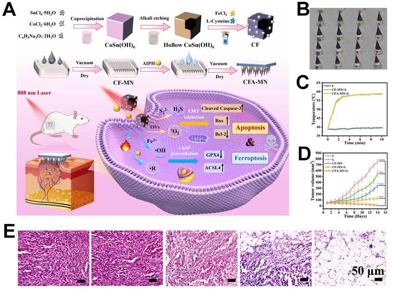
The platform is able to deliver agents directly to the target site, where ICD is triggered. The DAMPs released by the malignant cells activate dendritic cells. Antigen presentation improves; more antigen-presenting cells enter the tumor; and immune signals activate tumor-specific T cells. Immune memory can be established, leading to a lower risk of relapse (Figure 4) [144,145].
(A) Preparation workflow of the CF-CFA-MN dual-layer microneedle system for synergistic induction of cell death and ferroptosis for tumor treatment. (B) Representative micrograph of CFA-MN. (C) Body temperature of 4T1 tumor-bearing mice. (D) Relative tumor volume among the different groups of treatment. (E) Haematoxylin and eosin-stained representative tumor sections from mice of each treatment group. Adapted with permission from [145]. Copyright © 2025 Elsevier Inc.
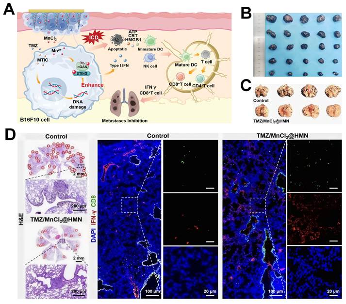
Microneedles can also carry immune-boosting agents. This is on top of the ICD immune cascade and enhances the antitumor response of the body (Figure 5) [146]. As an example, microneedles have the ability to deliver granulocyte-macrophage colony-stimulating factor (GM-CSF). Its release fosters the recruitment of the dendritic cell and augmentation of the antigen uptake. This enhances the Langerhans-cell density and T-cell clonal diversity in tumor-draining lymph nodes [68]. The platform has the potential to present immune modulators like CpG oligonucleotides, which elicit antitumor effects and suppress tumor growth. Diagnosis is done with the use of microneedles to remove skin interstitial fluid, and in this way longitudinal analysis of immune parameters, such as macrophage and effector T-cell responses, is monitored [142]. To conclude, material-based immune modulation, ICD induction, adjuvant delivery, and diagnostic monitoring allow a strong long-term antitumor immune response [147].
Microneedle-induced local immune response. (A) The composite microneedle system demonstrates synergistic antitumor efficacy through activation of the Stimulator of Interferon Genes (STING) pathway and induction of immunogenic cell death. (B) tumor growth curves across experimental groups. (C) Representative images of lung metastatic foci on day 14 post-inoculation, with quantitative analysis of metastatic nodules and lung weight ratio. (D) Haematoxylin and eosin-stained lung tissue sections (with insets showing metastatic lesions) and immunofluorescence staining for CD8⁺ T cells and interferon-γ. Adapted with permission from [146]. Copyright © 2023 Wiley.
Delivery of immune checkpoint inhibitors
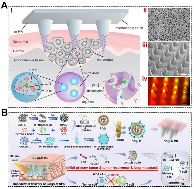
Microneedle technology offers a novel and highly efficient approach for the administration of immune checkpoint inhibitors. Conventional intravenous delivery is associated with the risk of systemic toxicity and an increased potential for drug resistance. In contrast, microneedles facilitate precise localised drug delivery with on-demand release capabilities. These devices can breach the stratum corneum to administer immune checkpoint inhibitors directly into the TME. This method substantially increases drug bioavailability and concurrently minimises off-target adverse effects. Microneedle systems may therefore improve the effectiveness and safety of immunotherapeutic agents. For instance, biodegradable patches loaded with antibodies against programmed cell death protein 1 (PD-1) have demonstrated superior antitumor efficacy compared with traditional intravenous injections. These platforms also show marked synergistic outcomes when administered with therapy targeting cytotoxic T-lymphocyte-associated protein 4 (CTLA-4) [148]. Advancements in this field have led to the development of core-shell microneedle designs capable of co-delivering anti-PD-L1 antibodies and the indoleamine 2,3-dioxygenase inhibitor 1-methyltryptophan, thereby augmenting T cell recruitment and improving melanoma growth suppression [149,150]. Gilardi et al. devised a localised delivery strategy for immune checkpoint inhibitors using dissolvable microneedles. This study has targeted and administered an anti-CTLA-4 antibody via microneedles to tumors of the head and neck. This preserved the antitumor activity of CD8+ T cells and conventional type 1 dendritic cells (cDC1s) [151] and may limit the systemic distribution of the antibodies and the resulting side effects, minimizing unwanted systemic exposure. Drugs can be released from stimulus-responsive materials under specific conditions. Some systems are degraded in the acidic tumor environment and release drugs locally and are more effective for treatment and also for local control. Healthy tissue stays untouched. This method sharpens immunotherapy. It also fights immunosuppressive environments. The same technology stops cancer from coming back after surgery. Yin et al. used soluble microneedles. They delivered photoimmunotherapy nanoparticles to the tumor. These nanoparticles were coated with M1 macrophage membranes. They also carried black phosphorus quantum dots and doxorubicin. This approach induced ICD through chemophototherapy, substantially enhancing the effectiveness of anti-PD-L1 immunotherapy. This strategy inhibited postoperative recurrence and metastasis in breast cancer and melanoma models, accompanied by observable T cell activation and the establishment of long-term immunological memory (Figure 6) [152].
Research on the application of microneedle technology in immune checkpoint inhibitor delivery systems. (A) (i) Schematic of the anti-PD-1 delivery system based on microneedle patches applied for melanoma treatment. (ii) Scanning electron microscopy (SEM) image of the nanoparticles. (iii) SEM image of the microneedle patch. (iv) Fluorescence imaging of fluorescein isothiocyanate-labelled antibody-loaded nanoparticles in a microneedle patch. The scale bars are 100 nm in (ii), 200 μm in (iii), and 200 μm in (iv). Adapted with permission from [148]. Copyright © 2016, American Chemical Society. (B) Technical scheme for the combined use of the BD@LM microneedle patch with anti-PD-L1 immunotherapy. Adapted with permission from [152]. Copyright © 2025 Elsevier B.V.
Xiang et al. engineered a dissolvable microneedle patch for the transdermal co-delivery of the TLR7/8 agonist R848 and an anti-PD-1 inhibitor. This combination stimulates dendritic cell maturation directly within the tumor, enhances CD8+ T cell infiltration, and disrupts the PD-1/programmed death ligand 1 (PD-L1) pathway to alleviate immunosuppression. It inhibited both tumor recurrence and pulmonary metastasis in a postoperative model of triple-negative breast cancer [153]. Zhang et al. developed a biodegradable, implantable microneedle device designed to slowly release factors that activate glioblastoma-associated macrophages, along with a TLR9 agonist, into the tumor cavity following resection. This method reprograms remaining macrophages. They become a proinflammatory type. It also helps recruit T cells [45]. This lowers the chance of tumor regrowth. These studies show microneedles can deliver immune checkpoint inhibitors locally. This boosts cancer immunotherapy effectiveness. It also cuts down on systemic side effects (Table 1).
Application of dissolving microneedles for localised delivery of immune checkpoint inhibitors in cancer treatment
| Composition | Diseases | Treatment | Mechanism | Key Advantages | Limitations | Ref. |
|---|---|---|---|---|---|---|
| Anti-PD-1 antibodies | Melanoma | Immunotherapy | Localised delivery of ICI, enhanced antitumor immunity | Superior efficacy vs. IV injection, synergistic with anti-CTLA-4 | Systemic toxicity and drug resistance risks of conventional IV delivery | [148] |
| Anti-PD-L1 antibodies + IDO inhibitor 1-MT | Melanoma | Immunotherapy | Co-delivery augments T cell recruitment, suppresses tumor growth | Enhanced T cell recruitment, improved melanoma suppression | Insufficient inherent drug-loading capacity of microneedles, anti-PD-L1 loading dependent on electrostatic interactions, risking drug migration or inactivation during storage, potential immunogenicity of chitosan | [149,150] |
| Anti-CTLA-4 antibody | Head and Neck Squamous Cell Carcinoma | Immunotherapy | Sustained CD8+ T cell/cDC1-dependent antitumor activity via intratumoral delivery | Considerably reduced irAEs by limiting systemic antibody exposure | Requires intratumoral administration, accessibility limitation | [151] |
| M1 macrophage membrane-modified photoimmunological NPs, black phosphorus quantum dots, doxorubicin | Breast cancer, Melanoma | Immunotherapy and Chemo-phototherapy | ICD via chemophototherapy potentiates anti-PD-L1 immunotherapy | Prevented postoperative recurrence and metastasis, T cell activation, long-term immunological memory | Multi-component complexity, potential phototoxicity | [152] |
| TLR7/8 agonist R848 + anti-PD-1 inhibitor | Triple-negative breast cancer | Immunotherapy | Stimulates DC maturation, enhances CD8+ T infiltration, disrupts PD-1/PD-L1 axis | Inhibited postoperative recurrence and pulmonary metastasis | Potential TLR agonist-related inflammation, transdermal depth limitation | [153] |
| Glioblastoma-associated macrophage-activating factors + TLR9 agonist | Glioblastoma | Immunotherapy | Reprograms residual macrophages to proinflammatory phenotype, promotes T cell recruitment | Sustained intracavitary release post-resection, reduced tumor regrowth | Invasive implantation required, applicable only to resectable tumors | [45] |
CTLA-4: cytotoxic T-lymphocyte-associated protein 4, IDO: indoleamine-pyrrole 2,3-dioxygenase, irAEs: immune-related adverse events, IV: intravenous, 1-MT: 1-methyltryptophan, PD-1: programmed cell death 1, PD-L1: programmed death ligand 1, TLR: Toll-like receptor
Delivery of tumor vaccines
Cancer vaccines are designed to trigger targeted immune responses against specific cancers. They are often grouped by how they are made. Microneedles help deliver tumor vaccines. They improve immunogenicity and targeting. Traditional delivery routes often fail for tumor vaccines. They cannot take up lymph efficiently. This stops antigen presentation and immune activation. Microneedles break through the skin's outer layer. They deliver vaccines into the dermis. This area is rich in immune cells. Antigen presentation then becomes more effective. The studies on melanoma have demonstrated that microneedle vaccines can deliver a potent antitumor response with acceptable overall safety and hence have potential applications in personalized and targeted cancer immunotherapy [154].
These vaccines are split up into groups based on their composition. Nucleic acid-based vaccines are eye-catching due to their high immunogenicity, low cost, and flexibility of design. DNA vaccines are stable during storage and transport, and mRNA vaccines do not require going into the cell nucleus. Both typically, however, need delivery systems to gain access to cells. During microneedle formulation, interactions between the microneedle matrix materials and nucleic acid carriers need to be controlled to maintain overall transfection efficiency. Microneedle delivery results in higher expression of genes and higher immune responses such as antibody production and T cell activity when compared with standard needle injections [155].
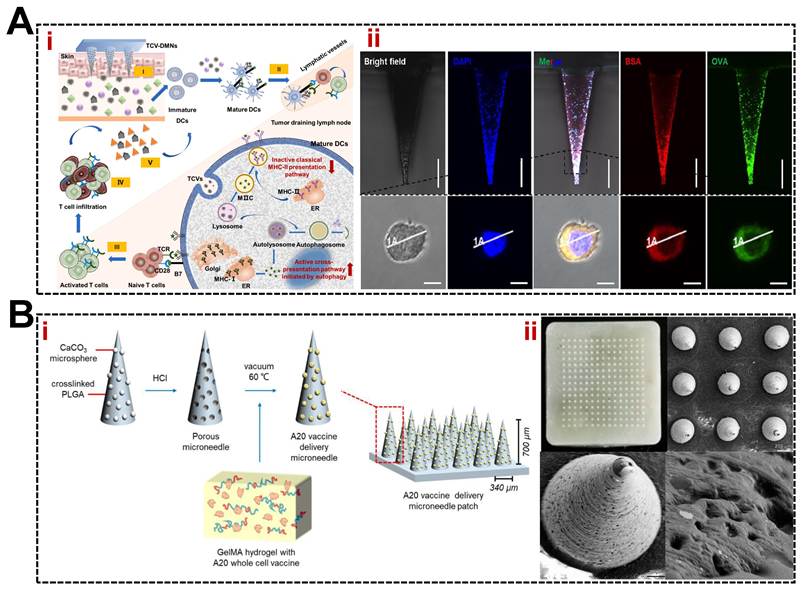
This technology is confirmed in real-world tests. The delivery of RALA/pDNA nanoparticles was done using freeze-dried dissolvable microneedles (MNs) made of polyvinyl alcohol (PVA). Approx. 50 µg was obtained from each array, which is in the effective range of DNA vaccines. This approach has been shown to cause greater cytotoxic and immune responses, delay the onset of tumors, and slow the growth of tumors in a cervical cancer model, compared with intramuscular injections [156]. A second study employed cationic RALA for the condensation of DNA to form nanoparticles that were administered via a polyvinylpyrrolidone microneedle patch. This induced a long-term cellular immune response to prostate stem cell antigens in skin immunization in both preventive and treatment settings. It inhibited growth and decreased the tumor burden [157]. There has been extensive research studying dendritic cell and whole tumor cell vaccines. Whole-cell vaccines are effective in exposing a variety of tumor-associated antigens and counteracting the immunosuppressive effects of the tumor. In a study, the researchers used Prussian blue nanoparticles to induce ICD in A20 lymphoma cells. The whole-cell vaccine was able to activate dendritic cell responses. Microneedle patches in porous gel of GelMA and PLGA showed enhanced lymphatic uptake and comfort [158]. GM-CSF and Tat-beclin 1 were delivered in another whole-cell system, TCV-DMNs. These combined to increase antigen uptake, autophagy, dendritic cell maturation, and presentation, leading to antitumor immunity. The system resulted in the prevention of melanoma, and in mice with established tumors, it shrank the tumors, leaving the mice free of tumors for more than 40 days [159].
Viral vectors are employed in third-generation vaccines. They are similar to viruses but do not contain any viral genome and are created by the interaction of viral structural proteins. This involves cloning of the genes in an expression cassette [160]. In this regard, Boone et al. created a self-propelled microneedle system based on reaction-driven thrust. PvBNP adjuvant nanoparticles are pushed by the action of magnesium microparticles into the tissue fluids in the TME, where they can penetrate deep into the tumor [161]. This was an active microneedle that performed better than passive microneedles and conventional injections. It inhibited the growth of tumors, prolonged survival, and triggered local innate and systemic adaptive anti-tumor immunity in melanoma models.
The fourth category covers protein or peptide antigen vaccines. Ovalbumin is a classic model antigen. It is often used to test microneedle immunogenicity because it targets lymph nodes well. One study showed that adding particulate adjuvants, loaded onto polylactic-polylactic copolymer nanoparticles, into dissolving microneedles triggered a stronger immune response than standard intradermal injection [162]. Later work created a hyaluronic acid-based dissolving microneedle system. It delivered ovalbumin antigen and poly(lactic-co-glycolic acid) nanoparticles together through the skin. Those nanoparticles carried the polyinosinic-polycytidylic acid adjuvant [163]. In summary, microneedle technology presents a promising avenue for advancing tumor vaccine development towards highly effective, low-toxicity, and personalised immunotherapeutic strategies (Figure 7).
Application of microneedle technology in tumor vaccine delivery systems. (A) (i) Schematic representation of the TCV-DMN system for tumor immunotherapy. (ii) Imaging analysis of samples using confocal laser scanning microscopy. The top-row images show the lateral morphology of a single microneedle. Scale bar = 200 μm. The bottom row shows the B16 cells in the TCV-DMN. Scale bar = 10 μm. These cells underwent multicolour fluorescent labelling prior to imaging: OVA-FITC (green fluorescence), BSA-Cy5 (red fluorescence), and DAPI (blue fluorescence). Adapted with permission from [159]. Copyright © 2023 Elsevier B.V. (B) (i) Fabrication process of poly(lactic-co-glycolic acid) (PLGA) porous microneedle patches. (ii) Macro top view of PLGA porous microneedle patches, scanning electron microscopy (SEM) morphology of surface microneedle arrays, and SEM images of microneedle bodies and surface pore structures. Adapted with permission from [158]. Copyright © 2024 by the authors.
Microneedles as delivery vehicles for combining cancer immunotherapy and other treatments
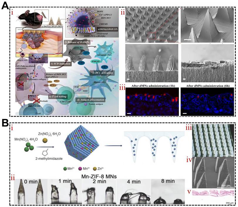
Microneedle technology offers a highly efficient platform for the co-delivery of cancer immunotherapy and other treatment modalities. In the field of combined chemo-immunotherapy, early studies developed nanoparticles incorporating both anti-PD-1 antibodies and cisplatin for administration via microneedles. This method had synergistic effects on melanoma models and was able to overcome drug resistance [83,164-167]. Progress has seen a programmable delivery system created by Tian et al., based on dissolvable microneedles (si/DOX@LRGD dMNs). It uses pH-sensitive, melanoma-targeting liposomes to co-deliver doxorubicin and an anti-PD-L1 agent. It also potently suppressed tumor growth in mouse models using the same doses. This was the result of a T-cell response: active T cells were CD8+ T cells, which produced IFN-γ [168]. Lan et al. have created a patch of microneedle patch based on pH-sensitive liposomal nanoparticles to achieve the delivery of anti-PD-1 and cisplatin to the tumor. The nanoformulations stimulated immunity and retarded tumor development. This local therapy was more effective with animals that were resistant to systemic anti-PD-1 therapy [164]. Combining radiotherapy with immunotherapy can be achieved using microneedles. Hu et al. developed a manganese X-ray responsive, microneedle system that is soluble and loaded with manganese ions. It enhances the melanoma radiosensitivity and activates ICD, due to the stimulation of the STING pathway. These microneedles used together with radiotherapy and immune checkpoint blockade stimulates the maturation of dendritic-cells and enhances T-cell-mediated immune response, which greatly enhances systemic antitumor effects (Figure 8) [169].
Microneedle technology is used as a delivery system of tumor immunotherapy that is synergistically combined with radio- and chemotherapy. (A) si/DOX@LRGD dMN system: (i) Mechanism of melanoma chemoimmunotherapy, (ii) Scanning electron microscopy (SEM). Scale bar = 100 μm and (iii) Rhodamine B-labelled dMN penetration in mouse skin. Scale bar = 100 μm. Based on [168] with modification. Copyright © 2023 Elsevier B.V. (B) Polylactic-co-glycolic acid (PLGA) and Mn (20%)-ZIF-8 MNs: (i) PLGA porous MN fabrication, (ii) Mn-ZIF-8 MN solubility, (iii) MN array image, (iv) SEM image of MNs, and (v) haematoxylin and eosin-stained image of rat skin post-puncture. With permission, adapted in [169]. Copyright © 2025 The Author(s).
Microneedle platforms have the potential to combine immunotherapy with phototherapeutic strategies to allow photothermal agents to be targeted at tumor locations, enhancing bioavailability and reducing off-target toxicity. The hyaluronic acid or polyvinyl alcohol matrix can incorporate gold nanoparticles, polydopamine, and indocyanine green [170]. Microinjected microneedles degrade in tissue fluid and discharge their cargo once inside the skin. Localised hyperthermia is induced by follow-up near-infrared irradiation that kills cancerous cells. This approach uses two mechanisms, which are microneedle penetration of the tumor stromal barrier to enhance access to drugs and photothermal effects of inducing apoptotic or necrotic cell-death. A single system was loaded with anti-PD-1 peptides and thiol-modified gold nanorods. On insertion, and near-infrared exposure, the gold nanorods produce ablative heat, and emitted peptides boost T-lymphocyte cytotoxicity [171]. Equally, Chen et al. created a self-degrading hyaluronic acid microneedle platform comprising of pH-sensitive glucan nanoparticles to be co-introduced with hydrophobic zinc phthalocyanine and hydrophilic anti-CTLA-4 antibody (Figure 9) [172].
Microneedle technology serves as a synergistic delivery platform for combined tumor immunotherapy and phototherapy. (A) Schematic illustration of the mechanism by which microneedle (MN)-assisted photodynamic therapy (PDT) combined with immune checkpoint blockade activates latent antitumor immune responses through MN-mediated co-delivery systems. (B) Scanning electron microscopy image of a hyaluronic acid (HA)-based microneedle patch. Scale bar = 500 μm. (C) Haematoxylin and eosin-stained cross-section of mouse skin after microneedle puncture. Scale bar = 200 μm. (D) Photographs of mice captured on days 1, 5, and 20 following treatment. (E) Normalised tumor volume. Adapted with permission from [172]. Copyright © 2020 Elsevier B.V.
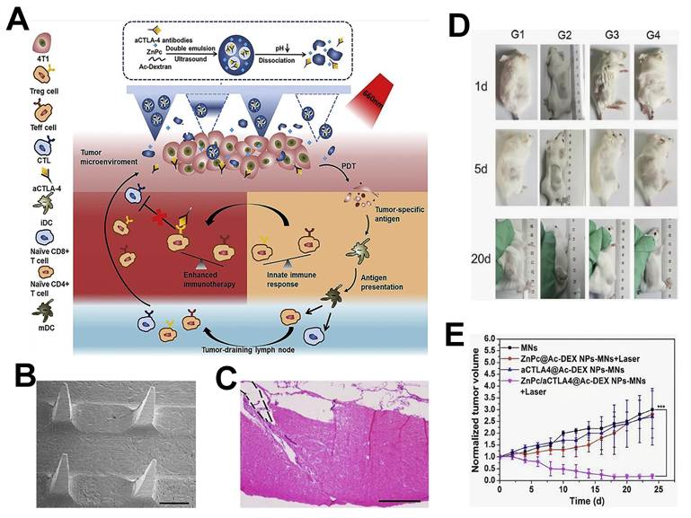
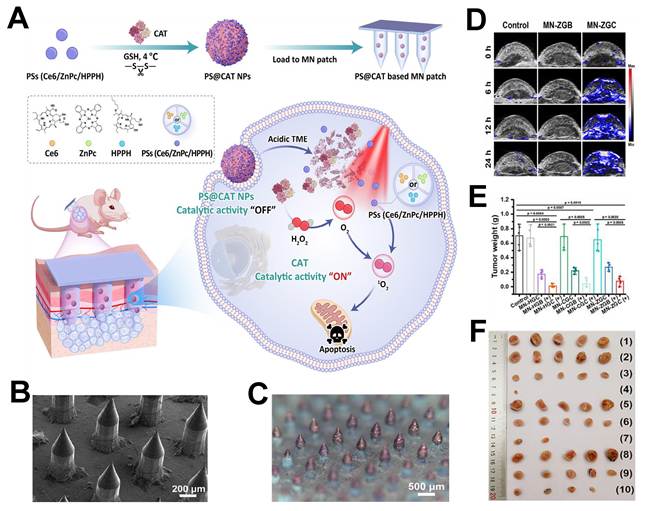
Other approaches use composite microneedles loaded with both photothermal agents and chemotherapeutic drugs, which have shown favourable results in melanoma models. The immunotherapeutic or chemotherapeutic synergy achieved through microneedle-assisted photothermal treatment arises from thermally induced increases in tumor vascular permeability, which enhances subsequent drug penetration and uptake [173-175]. This form of synergistic approach can help to surpass the drawbacks of traditional monotherapies. These mechanisms boost therapy and synchronize immune response and tumor destruction. The use of microneedles can support transdermal co-delivery of agents with different solubility profiles such as anti-CTLA-4 antibodies and zinc phthalocyanine [172]. Coupling of photothermal agents and chemotherapeutic drugs into composite microneedles has also produced encouraging results in melanoma models. The other method involves carrier-free nanoplatform that employs catalase as a scaffold. It stands in a state of equilibrium until it comes in contact with the acidic TME where it restores enzyme activity, degrades hydrogen peroxide, produces oxygen, and decreases tumor hypoxia, thus enhancing photodynamic therapy [176]. The team of Chen et al. designed self-assembling microneedles that, when used in vivo, self-assemble and deliver micelles containing ICD inducers and autophagy inhibitors to tumors. The complex delivered after the insertion self-assembles with a size dependent on the photothermal conditions to enable deep penetration of tumors. The platform inhibits autophagy and increases the release of DAMP in tumors. Moreover, chloroquine inactivates NF-κB that leads to M1 polarisation of tumor-associated macrophages [177]. Overall, microneedle-based platforms may enable the precise spatiotemporal co-administration of multiple therapeutics. This approach enhances antitumor immunity while minimising systemic exposure. Future work should evaluate integrating smart stimuli-responsive materials with patient-specific treatment regimens to advance tumor immunotherapy towards minimally invasive, highly efficient, and personalised therapeutic solutions (Table 2, Figure 10).
Application of dissolving microneedles in combined cancer treatment modalities
| MNs Matrix Material | Composition | Diseases | Treatment | Mechanism | Key Advantages | Limitations | Ref. |
|---|---|---|---|---|---|---|---|
| Amylopectin, Phosphate | Liposomes capable of effectively loading DOX and siPD-L1 | Melanoma | Immunotherapy and Chemotherapy | si/DOX@LRGD suppresses PD-L1-mediated immune evasion while inducing immunogenic cell death and tumor apoptosis via doxorubicin. Simultaneously, it stimulates CD8+ T cells to secrete interferon-γ and elicits potent immune responses | Dual-response precise controlled release reduces systemic toxicity; siRNA silencing is more efficient than antibody blockade and simultaneously enhances chemotherapy sensitivity and reshapes the immune microenvironment | siRNA has poor stability in vivo and requires liposomal protection; the durability of PD-L1 silencing is limited | [168] |
| Polyvinylpyrrolidone (PVP) | Lipid nanoparticles loaded with anti-PD-1 and cisplatin | Melanoma | Immunotherapy and Chemotherapy | By inducing T cell immune responses, blocking the PD-1 signalling pathway, and enhancing the direct cytotoxic effects of CDDP on tumor cells, synergistic antitumor effects were jointly activated | Combined chemotherapy and immunotherapy delivery via a single platform, lipid nanoparticles enhance the solubility of hydrophobic drugs, pH-responsive tumor-targeted release | Concerns remain regarding the nephrotoxicity and neurotoxicity of cisplatin | [164] |
| Tips: hyaluronic acid and Mn-ZIF-8 Base layer: polyvinylpyrrolidone (PVP K90) | Zeolite imidazolate framework-8 (ZIF-8) capable of effectively loading manganese ions | Melanoma | Immunotherapy and Radiation Therapy | Following X-ray irradiation, Mn-ZIF-8 microneedles continuously release Mn²⁺ to activate the cGAS-STING pathway. This enhances antigen presentation by dendritic cells and further amplifies T cell-dominant immune responses | High stability of inorganic ions, dual functions of radiotherapy sensitisation and immune activation, spatiotemporal precision control via X-ray response | Mn²⁺ has a narrow therapeutic window and is neurotoxic at high doses; the temporal control of the X-ray response is complex | [169] |
| PMVE-MA (Gantrez S-97) | AuNRs and anti-PD-1 peptides | Breast cancer | Immunotherapy and Photothermal Therapy | Enhances the ability of cytotoxic T lymphocytes to destroy tumor cells. The heat generated upon exposure to near-infrared light aids tumor elimination | Precise and controllable photothermal effects, easy functionalisation of AuNRs, low systemic toxicity, simultaneous imaging-guided therapy | Limited light penetration depth, the long-term in vivo metabolism of AuNRs is unknown | [171] |
| Hyaluronic acid | Photosensitiser and anti-CTLA-4 antibody (aCTLA-4) | Breast cancer | Immunotherapy and Photodynamic Therapy | PDT first exerts its tumor-killing effect and triggers an immune response, subsequently promoting aCTLA-4-based immunotherapy | Dual photodynamic-immunological mechanism, pH-sensitive, enhanced tumor targeting, hyaluronic acid offers good biocompatibility | Photodynamic therapy has limited light penetration depth; aCTLA-4 is associated with significant systemic toxicity (colitis, hypophysitis); and there is a risk of cutaneous phototoxicity with photosensitisers | [172] |
| Hyaluronic acid | Immunogenic Cell Death Inducer (IR780) and Autophagy Inhibitor (Chloroquine, CQ) | Melanoma | Immunotherapy and Phototherapy | Upon entering the skin, C/I-Mil undergoes self-assembly and is extensively internalised into cells. It inhibits autophagy and promotes the release of DAMPs. Concurrently, chloroquine activates the NF-κB pathway, reprogramming tumor-associated macrophages to the M1 phenotype | Diagnosis and Treatment Integration: Near-infrared fluorescence imaging guidance, in situ self-assembly-enhanced tumor targeting, reversal of the immunosuppressive microenvironment | The efficiency of immunogenic cell death is influenced by tumor heterogeneity; chloroquine may interfere with autophagy in normal cells; near-infrared light has limited tissue penetration depth | [177] |
CTLA-4: cytotoxic T-lymphocyte-associated protein 4, DAMP: damage-associated molecular patterns, NF-κB: nuclear factor kappa B, PD-1: programmed cell death 1, PD-L1: programmed death ligand 1, PDT: photodynamic therapy, STING: Stimulator of interferon genes.
(A) Schematic of dissolvable MNs with in situ self-assembled nanospheres for photoimmunotherapy. (B, C) Scanning electron microscopy and stereomicroscopy images of the MN-ZGC patch. (D) Photoacoustic imaging of tumor sO₂ in 4T1-bearing mice after MN-ZGB or MN-ZGC treatment. (E, F) tumor weights and images of excised tumors from each group. Copyright © 2021 American Chemical Society [177].
Applications of microneedles in tumor diagnosis and monitoring
The functional characteristics of microneedle technology support its use for both tumor biomarker detection and monitoring of the TME. Dermal interstitial fluid contains various cancer-related biomarkers, including proteins, lipids, nucleic acids, and extracellular vesicles. Together, these components form tumor-specific molecular signatures. Microneedles can penetrate the stratum corneum to directly obtain samples from the dermis or interstitial fluid and collect biological fluids near the tumor in real time. This enables continuous monitoring of important indicators [178]. Circulating tumor DNA is clinically relevant, and so are some patterns of microRNAs and tumor-derived exosomes. These support less invasive liquid biopsy platforms. Specific molecules, such as metabolites and cytokines, can be captured on the surface of microneedles with attached antibodies or nanomaterials. They can identify early recurrence or metastasis and help direct modifications in tumor immunotherapy treatment [58]. These tools, in combination, enable early cancer screening. They help check treatment outcomes. They also aid personalized treatment design [179-182].
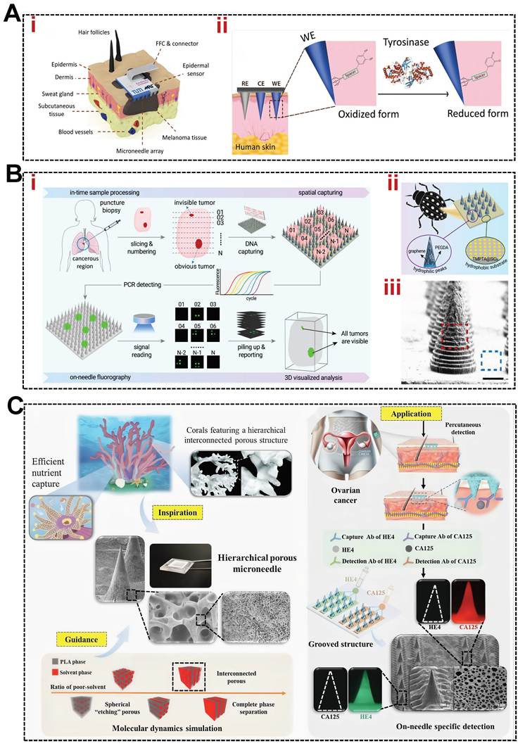
Related studies have supported this approach. For example, Tran et al. used a microneedle-assisted method to analyse interstitial fluid in the skin and found that the levels of proteins such as albumin and α-1 antitrypsin were 40% to 60% higher than those in serum. This may reflect tumor-associated inflammatory responses or changes in the microenvironment. Analysing interstitial fluid biomarkers has the potential to improve the accuracy of predicting cancer recurrence or metastasis by approximately 30%, while continuous monitoring of changes in its composition can aid in assessing treatment response [183]. Based on this foundation, He et al. developed a biomimetic three-dimensional porous microneedle system. The system has a strong liquid absorption capacity and high-density antibody immobilisation capability, enabling the high-density binding of human epididymis protein 4 and cancer antigen 125 on the microneedle surface. Its unique groove design effectively prevents cross-reactions between different detection components, achieving nanomolar-level detection sensitivity [184]. Poursharifi et al. developed a microneedle-based wearable biosensor patch fabricated using a polyurethane-PEDOT:PSS composite via a single-step drop-casting process. Mechanical simulations confirmed that the microneedles exhibit good penetration and structural stability while maintaining electrical conductivity. Validation using a human skin model demonstrated that the device specifically responds to tyrosinase at physiological concentrations and did not cause substantial skin irritation or allergic reactions. These findings support its use for the non-invasive detection of melanoma [185]. Another study developed a microneedle array platform based on wettability control. Hydrogel scaffolds at the tips facilitate tissue penetration and three-dimensional sampling. The immobilised graphene aerogel efficiently concentrates nucleic acid biomarkers, while the hydrophobic substrate forms isolated compartments for tip-based polymerase chain reaction amplification and reduces fluorescence interference. This platform enabled the spatial localisation of lung cancer nucleic acid biomarkers and highly sensitive detection of occult lesions in animal tissues and clinical samples (Figure 11) [178].
Application value of microneedle technology in tumor diagnosis. (A) Surface-immobilised smart probe for epidermal sensing. (i) Operational mechanism of the probe on microneedles and its specific chemical reaction with tyrosinase (Tyr). (ii) Representative square-wave voltammetry signal illustrating the signal-on detection principle. Adapted with permission from [185]. Copyright © 2025 Wiley. (B) F-PCR-MNA for spatial tumor tissue biopsy. (i) Workflow schematic. (ii) Structural composition of the MNA and its biomimetic narrow-mite morphology. (iii) Scanning electron microscopy image of the MNA. Copyright © 2023 The Author(s) [178]. (C) Core workflow for ovarian cancer diagnosis via multi-biomarker detection using immune-specifically functionalised hp-MN patches. Adapted with permission from [184]. Copyright © 2025 Wiley.
Building on this foundation, the dynamic sampling of these biomarkers via the therapeutic microneedle platform can provide real-time insights into the intensity of local immune responses and the efficacy of immunotherapy, thereby establishing a monitoring system that is highly aligned with tumor immunotherapy centres. In a related study, Dosta et al. designed a hydrogel-based microneedle platform. The platform combines drug delivery and fluid sampling. This platform delivers immunostimulatory agents directly into the tumor core. It also collects interstitial fluid from the local lesion. The platform was tested on mice with melanoma and colorectal cancer. The fluid was analyzed, and its immune cell profile was very similar to the solid tumor tissue. Following treatment, there was a significant increase in the number of macrophages and in the influx of effector T cells [142]. The combination of treatment and monitoring opens a novel approach to investigate antitumor immune responses in situ. The majority of studies address static biomarkers, and a limited number have analyzed immune gene expression before and after therapy. But such monitoring of several TME parameters in vivo at high spatiotemporal resolution is still scarce. This gap could be bridged by therapeutic microneedle platforms, which simultaneously deliver drugs and monitor real-time responses. They are able to gather marker information on TME along with physiological or drug measurements in real time [186-188]. In the case of cancer patients suffering from diabetes, this technology can monitor their blood glucose [189]. Live data can help to quickly adjust treatment and identify side effects.
Ways to deal with microneedle cancer diagnostics are not perfect. The sample volumes are small, and there is a danger of dilution of samples due to extraction pressure, and it is difficult to obtain intact specimens. Minor proteins and important biomarkers in interstitial fluid from skin are difficult to detect [190,191]. Antibody binding efficiency may be affected by tissue conditions, and biomarker transport from blood to interstitial fluid may contribute to a delay in diagnosis. Current systems might not be sensitive and specific enough. Since the devices cannot be integrated, electrochemical biosensors have limited lifespans and can only be used for short-term monitoring. There is a need for better in vitro skin models to validate the use of these, as well as improvements in biocompatibility, device durability, and the ability to scale up manufacture. These issues will lead to greater use and clinical uptake.
Summary and outlook
The field of microneedle technology has made great advances and may prove beneficial for cancer immunotherapy. It delivers drugs to specific locations, is less invasive, and may overcome the limitations of drugs in relation to targeting. This review highlights the basics and the types of microneedles, therapeutic and diagnostic applications, and the use of microneedles with other therapies. It provides an analytical approach that allows us to compare different microneedle designs and their impact on the tumor microenvironment and immune response. It provides an overview of the relationship between delivery accuracy and speed of release and the need for closed-loop systems. These systems enable real-time monitoring, provide feedback, and integrate multiple treatment modalities. Besides, this review proposes a new classification method for combination therapy strategies. This is based on coordination in time and space, changing the surroundings, and making more antigens. These ideas might be useful for future work.
Despite rapid progress, several issues still need to be addressed before these methods can be applied to patients. Major challenges include mass production, long-term safety testing, and patient-specific customization [34,61,63,123,186-189]. A major issue is how the physical insertion of microneedles alters the local tumor immune microenvironment [71]. Our current understanding of the molecular cascades triggered by tissue injury remains limited [131]. The dense extracellular matrix impedes the penetration of substances. Abnormal blood vessels present an additional barrier [72]. These factors may attenuate pro-inflammatory signaling and limit drug penetration. Therefore, there is a need to develop advanced in vivo monitoring tools with high resolution and real-time operational capabilities [58,178]. Another objective is to develop smart materials sensitive to and responsive to specific conditions in the tumor microenvironment, such as certain enzymes, abnormal pH changes, or abnormal levels of matrix metalloproteinases [115,126]. Drug carriers should avoid generating immunosuppressive signals and be able to penetrate thick tissue matrices [138,147]. Biodegradable microneedle materials like hyaluronic acid, collagen, gelatin, and polydopamine [64,80] degrade slowly and produce fragments and intermediate compounds [64]. How these fragments bind to immune cell receptors is not well understood. The signaling mechanisms after binding and the resulting immune response remain unclear. Whether these interactions result in inflammation, prevention of inflammation, or exacerbation of immunosuppression is unclear. One of the biggest challenges in clinical practice is the difference between the prototype that is tested in the laboratory and the prototype tested in patients. A key obstacle to clinical translation is that, to date, most microneedle studies have been conducted on rodents. However, their skin differs from human skin. These differences directly affect drug delivery. The human stratum corneum is much thicker, consisting of 5–10 layers of keratinocytes. In contrast, the stratum corneum of mice contains only 2–3 layers of keratinocytes. The density of hair follicles differs by more than an order of magnitude between the two species. Furthermore, the mouse epidermis is densely populated with γδ T cells, whereas the human epidermis contains only sparse populations of γδ T cells [192,193]. Furthermore, the microbial environment of laboratory mice is far cleaner than that of human skin; consequently, the development of the resident immune cell populations in their skin is constrained by a microbiome that is far less diverse than that of humans [194]. Therefore, the antitumor immune responses induced in patients may differ from those observed in mice, both in intensity and quality. What’s more, Laboratory-scale microneedle geometry, drug loading, and release profile may not scale up to large-scale production [34,61]. Quality control, long-term storage, and biosafety should be prioritized for future studies [128]. The next stage is to create systems that are capable of being mass-produced, conform to the regulatory requirements, and can be adapted to patients. There are challenges in cancer immunotherapy for microneedle technology, but it has tremendous potential for translation. This review helps to deepen understanding and further innovations.
Abbreviations
CLSM: confocal laser scanning microscope; CTLA-4: cytotoxic T-lymphocyte associated protein 4; DAMPs: damage-associated molecular patterns; DC: dendritic cell; FITC: fluorescein isothiocyanate; GM-CSF: granulocyte-macrophage colony-stimulating factor; ICD: immunogenic cell death; ICI: immune checkpoint inhibitor; IDO: indoleamine-pyrrole 2,3-dioxygenase; irAE: immune-related adverse event; MMP: matrix metalloproteinase; MN: microneedle; mRNA: messenger RNA; NF-κB: nuclear factor kappa B; NP: nanoparticle; PD-1: programmed cell death protein 1; PD-L1: programmed death-ligand 1; PDT: photodynamic therapy; PLGA: polylactic-co-glycolic acid; SEM: scanning electron microscopy; siRNA: small interfering RNA; STING: stimulator of interferon genes; TLR: Toll-like receptor; TME: tumor microenvironment.
Acknowledgements
This work was supported by the National Natural Science Foundation of China (No. 82071035, 82371165), Natural Science Foundation of Shandong Province (No. ZR2022LZL001), Noncommunicable Chronic Diseases-National Science and Technology Major Project (2024ZD0533203), Collaborative Academic Innovation Project of Shandong Cancer Hospital (FC002), and Jiangsu Funding Program for Excellent Postdoctoral Talent (2024ZB003). We thank the Shandong Provincial Key Medical and Health Discipline of Clinical Laboratory Diagnostics (Cancer Hospital of Shandong First Medical University) for its support and academic resources. Parts of the figures were drawn with the aid of BioRender (http://www.biorender.com), licenced under a Creative Commons Attribution 3.0 Unported Licence.
Author contributions
H.X.Z., M.Q.Z., W.C.D., and M.X. designed the review. H.X.Z., Y.H.S., and H.Z. drafted the manuscript and prepared the figures. H.X.Z., M.Q.Z., W.C.D., and M.X. revised the manuscript. All authors have read and approved the final manuscript.
Competing Interests
The authors have declared that no competing interest exists.
References
1. Passaro A, Al Bakir M, Hamilton EG, Diehn M, André F, Roy-Chowdhuri S. et al. Cancer biomarkers: emerging trends and clinical implications for personalized treatment. Cell. 2024;187:1617-35
2. Wahida A, Buschhorn L, Fröhling S, Jost PJ, Schneeweiss A, Lichter P. et al. The coming decade in precision oncology: six riddles. Nat Rev Cancer. 2023;23:43-54
3. Khamsi R. Computing cancer’s weak spots. Science. 2020;368:1174-7
4. Mullard A. Addressing cancer’s grand challenges. Nat Rev Drug Discov. 2020;19:825-6
5. Hegde PS, Chen DS. Top 10 challenges in cancer immunotherapy. Immunity. 2020;52:17-35
6. Lu Z, Chen Y, Liu D, Jiao X, Liu C, Wang Y. et al. The landscape of cancer research and cancer care in China. Nat Med. 2023;29:3022-32
7. Tan AC, Bagley SJ, Wen PY, Lim M, Platten M, Colman H. et al. Systematic review of combinations of targeted or immunotherapy in advanced solid tumors. J Immunother Cancer. 2021;9:e002459
8. Ding H, Xue L, Zhu S, Wang Z, Gui H, Zhang G. et al. Polyamine metabolism promotes the progression of thyroid carcinoma by regulating the immune phenotype of tumor associated macrophages. Smart Med. 2025;4:e70009
9. Zhang J, Liu Z, Zhao W, Li C, Liu F, Wang J. Targeting KRAS sensitizes ferroptosis by coordinately regulating the TCA cycle and Nrf2-SLC7A11-GPX4 signaling in hepatocellular carcinoma. Smart Med. 2025;4:e70005
10. Mandal R, Chan TA. Personalized oncology meets immunology: the path toward precision immunotherapy. Cancer Discov. 2016;6:703-13
11. Herrmann J. Vascular toxic effects of cancer therapies. Nat Rev Cardiol. 2020;17:503-22
12. Verginadis II, Citrin DE, Ky B, Feigenberg SJ, Georgakilas AG, Hill-Kayser CE. et al. Radiotherapy toxicities: mechanisms, management, and future directions. Lancet. 2025;405:338-52
13. Schuurhuizen CSEW, Braamse AMJ, Konings IRHM, Sprangers MAG, Ket JCF, Dekker J. et al. Does severe toxicity affect global quality of life in patients with metastatic colorectal cancer during palliative systemic treatment? A systematic review. Ann Oncol. 2017;28:478-86
14. Du XL, Osborne C, Goodwin JS. Population-based assessment of hospitalizations for toxicity from chemotherapy in older women with breast cancer. J Clin Oncol. 2002;20:4636-42
15. Stone JB, DeAngelis LM. Cancer-treatment-induced neurotoxicity-focus on newer treatments. Nat Rev Clin Oncol. 2016;13:92-105
16. Tallet AV, Dhermain F, Le Rhun E, Noël G, Kirova YM. Combined irradiation and targeted therapy or immune checkpoint blockade in brain metastases: toxicities and efficacy. Ann Oncol. 2017;28:2962-76
17. Passaro A, Brahmer J, Antonia S, Mok T, Peters S. Managing resistance to immune checkpoint inhibitors in lung cancer: treatment and novel strategies. J Clin Oncol. 2022;40:598-610
18. Vasseur D, Bigot L, Beshiri K, Flórez-Arango J, Facchinetti F, Hollebecque A. et al. Deciphering resistance mechanisms in cancer: final report of MATCH-R study with a focus on molecular drivers and PDX development. Mol Cancer. 2024;23:221
19. Hangauer MJ, Viswanathan VS, Ryan MJ, Bole D, Eaton JK, Matov A. et al. Drug-tolerant persister cancer cells are vulnerable to GPX4 inhibition. Nature. 2017;551:247-50
20. O’Donnell JS, Teng MWL, Smyth MJ. Cancer immunoediting and resistance to T cell-based immunotherapy. Nat Rev Clin Oncol. 2019;16:151-67
21. Ge M, Chen XY, Huang P, Fleishman JS, Yang D-H, Wu Z-X. et al. Understanding and overcoming multidrug resistance in cancer. Nat Rev Clin Oncol. 2025;22:760-80
22. Burgers FH, van der Mijn JCK, Seijkens TTP, Jedema I, Bex A, Haanen JBAG. Immunological features of clear-cell renal-cell carcinoma and resistance to immune checkpoint inhibitors. Nat Rev Nephrol. 2025;21:687-701
23. Riley RS, June CH, Langer R, Mitchell MJ. Delivery technologies for cancer immunotherapy. Nat Rev Drug Discov. 2019;18:175-96
24. Roerden M, Spranger S. Cancer immune evasion, immunoediting and intratumor heterogeneity. Nat Rev Immunol. 2025;25(5):353-369
25. Phatale V, Vaiphei KK, Jha S, Patil D, Agrawal M, Alexander A. Overcoming skin barriers through advanced transdermal drug delivery approaches. J Control Release. 2022;351:361-80
26. Sabbagh F, Kim BS. Recent advances in polymeric transdermal drug delivery systems. J Control Release. 2022;341:132-46
27. Qu F, Geng R, Liu Y, Zhu J. Advanced nanocarrier- and microneedle-based transdermal drug delivery strategies for skin diseases treatment. Theranostics. 2022;12:3372-406
28. Mantia C, Choudhury AD. Transdermal oestrogen for advanced prostate cancer. Lancet. 2021;397:556-7
29. Moses MA, Brem H, Langer R. Advancing the field of drug delivery: taking aim at cancer. Cancer Cell. 2003;4:337-41
30. Olson AC, Link JS, Waisman JR, Kupiec TC. Breast cancer patients unknowingly dosing themselves with estrogen by using topical moisturizers. J Clin Oncol. 2009;27:e103-4
31. Verges B, Cercueil JP, Pfitzenmeyer P, Pascuad F, Vaillant G, Brun JM. et al. Percutaneous ethanol injection of parathyroid adenomas in primary hyperparathyroidism. Lancet. 1991;337:1421-2
32. Thakor AS, Gambhir SS. Nanooncology: the future of cancer diagnosis and therapy. CA Cancer J Clin. 2013;63:395-418
33. Ma H, Liu J, Ali MM, Mahmood MAI, Labanieh L, Lu M. et al. Nucleic acid aptamers in cancer research, diagnosis and therapy. Chem Soc Rev. 2015;44:1240-56
34. Moawad F, Pouliot R, Brambilla D. Dissolving microneedles in transdermal drug delivery: a critical analysis of limitations and translation challenges. J Control Release. 2025;383:113794
35. Blagus T, Markelc B, Cemazar M, Kosjek T, Preat V, Miklavcic D. et al. In vivo real-time monitoring system of electroporation mediated control of transdermal and topical drug delivery. J Control Release. 2013;172:862-71
36. Jiang Y, Zhang W, Liu L, Wu Y, Li W, Liang J. et al. Gelatin methacryloyl xerogel puncture implants loaded with Cu0.5Mn2.5O4 nanoparticles synergizes cuproptosis and STING activation for enhanced breast cancer immunotherapy. ACS Nano. 2025;19:27902-18
37. Coulborn RM, Danet C, Alsalhani A. Measles and rubella vaccine microneedle patch: new hope to reach the unreached children. Lancet. 2024;403:1825-7
38. Han J, Kanelli M, Liu Y, Daristotle JL, Pardeshi A, Forster TA. et al. On-patient medical record and mRNA therapeutics using intradermal microneedles. Nat Mater. 2025;24:794-803
39. Harris E. Microneedle vaccine patches generated immune response in children. JAMA. 2024;331:1982
40. Moreira AF, Rodrigues CF, Jacinto TA, Miguel SP, Costa EC, Correia IJ. Microneedle-based delivery devices for cancer therapy: a review. Pharmacol Res. 2019;148:104438
41. Fang A, Wang Y, Guan N, Zuo Y, Lin L, Guo B. et al. Porous microneedle patch with sustained delivery of extracellular vesicles mitigates severe spinal cord injury. Nat Commun. 2023;14:4011
42. Xu R, Guo H, Chen X, Xu J, Gong Y, Cao P. et al. Smart hydrothermally responsive microneedle for topical tumor treatment. J Control Release. 2023;358:566-78
43. Xue H, Jin J, Huang X, Tan Z, Zeng Y, Lu G. et al. Wearable flexible ultrasound microneedle patch for cancer immunotherapy. Nat Commun. 2025;16:2650
44. Song M, Ha M, Shin S, Kim M, Son S, Lee J. et al. A hierarchical short microneedle-cupping dual-amplified patch enables accelerated, uniform, pain-free transdermal delivery of extracellular vesicles. Nanomicro Lett. 2025;18:11
45. Zhang Y, Fang Z, Liu Z, Xi K, Zhang Y, Zhao D. et al. Implantable microneedle-mediated eradication of postoperative tumor foci mitigates glioblastoma relapse. Adv Mater. 2024;36:e2409857
46. Li N, Mu W, Xia Z, Ma Q, Feng R, Gu P. et al. Soluble-microneedle enhance three T-cell activation signals as efficient tumor vaccines for melanoma prevention and treatment. J Control Release. 2025;383:113726
47. Zhang J, Shi Y, Zhang Y, Fang Z, Zhou Y, Bian F. et al. Antibacterial and proangiogenic hydrogel microneedle patches for wound healing. Smart Med. 2025;4:e70014
48. Song C, Lu M, Li N, Gu H, Li M, Lu L. et al. MXene-integrated responsive hydrogel microneedles for oral ulcers healing. Smart Med. 2025;4:e135
49. Fan L, Wang L, Wang X, Li M, Gu H, Zhang H. Multifunctional silk and gelatin composed microneedle patches for enhanced wound healing. Smart Med. 2025;4:e137
50. Zhang W, Cai L, Gan J, Zhao Y. Photothermal responsive porous hollow microneedles as Chinese medicine versatile delivery system for wound healing. Smart Med. 2024;3:e20240007
51. Zhang X, Lu M, Cao X, Zhao Y. Functional microneedles for wearable electronics. Smart Med. 2023;2:e20220023
52. Wang M, Li X, Du W, Sun M, Ling G, Zhang P. Microneedle-mediated treatment for superficial tumors by combining multiple strategies. Drug Deliv Transl Res. 2023;13:1600-20
53. Zhang W, Jiang Y, Liu L, Shen H, Huang X, Zheng W. et al. Implantable microneedles loaded with nanoparticles surface engineered Escherichia coli for efficient eradication of triple-negative breast cancer stem cells. Nano Lett. 2025;25:2041-51
54. Desai VM, Priya S, Gorantla S, Singhvi G. Revolutionizing therapeutic delivery with microneedle technology for tumor treatment. Pharmaceutics. 2022;15:14
55. Chu Z, Zheng W, Fu W, Liang J, Wang W, Xu L. et al. Implanted microneedles loaded with sparfloxacin and zinc-manganese sulfide nanoparticles activates immunity for postoperative triple-negative breast cancer to prevent recurrence and metastasis. Adv Sci (Weinh). 2025;12:e2416270
56. Mbituyimana B, Adhikari M, Qi F, Shi Z, Fu L, Yang G. Microneedle-based cell delivery and cell sampling for biomedical applications. J Control Release. 2023;362:692-714
57. Lyu S, Dong Z, Xu X, Bei H-P, Yuen H-Y, James Cheung C-W. et al. Going below and beyond the surface: microneedle structure, materials, drugs, fabrication, and applications for wound healing and tissue regeneration. Bioact Mater. 2023;27:303-26
58. Hu Y, Chatzilakou E, Pan Z, Traverso G, Yetisen AK. Microneedle sensors for point-of-care diagnostics. Adv Sci (Weinh). 2024;11:e2306560
59. Zhao P, Zhou Z, Wolter T, Womelsdorf J, Somers A, Feng Y. et al. Engineering microneedles for biosensing and drug delivery. Bioact Mater. 2025;52:36-59
60. Ahmed Saeed Al-Japairai K, Mahmood S, Hamed Almurisi S, Reddy Venugopal J, Rebhi Hilles A, Azmana M. et al. Current trends in polymer microneedle for transdermal drug delivery. Int J Pharm. 2020;587:119673
61. Sartawi Z, Blackshields C, Faisal W. Dissolving microneedles: applications and growing therapeutic potential. J Control Release. 2022;348:186-205
62. Van der Maaden K, Jiskoot W, Bouwstra J. Microneedle technologies for (trans)dermal drug and vaccine delivery. J Control Release. 2012;161:645-55
63. Ingrole RSJ, Azizoglu E, Dul M, Birchall JC, Gill HS, Prausnitz MR. Trends of microneedle technology in the scientific literature, patents, clinical trials and internet activity. Biomaterials. 2021;267:120491
64. Saha I, Rai VK. Hyaluronic acid based microneedle array: recent applications in drug delivery and cosmetology. Carbohydr Polym. 2021;267:118168
65. Glover K, Mishra D, Gade S, Vora LK, Wu Y, Paredes AJ. et al. Microneedles for advanced ocular drug delivery. Adv Drug Deliv Rev. 2023;201:115082
66. Wang G, Moriyama N, Tottori S, Nishizawa M. Recent advances in iontophoresis-assisted microneedle devices for transdermal biosensing and drug delivery. Mater Today Bio. 2025;31:101504
67. Rzhevskiy AS, Singh TRR, Donnelly RF, Anissimov YG. Microneedles as the technique of drug delivery enhancement in diverse organs and tissues. J Control Release. 2018;270:184-202
68. Wu C, Yu Q, Huang C, Li F, Zhang L, Zhu D. Microneedles as transdermal drug delivery system for enhancing skin disease treatment. Acta Pharm Sin B. 2024;14:5161-80
69. Yang Y, Sun H, Sun X, Wang Y, Xu F, Xia W. et al. From mechanism to applications: advanced microneedles for clinical medicine. Bioact Mater. 2025;51:1-45
70. Xu Z, Chi J, Qin F, Liu D, Lai Y, Bao Y. et al. Nanoparticles-incorporated hydrogel microneedle for biomedical applications: fabrication strategies, emerging trends and future prospects. Asian J Pharm Sci. 2025;20:101069
71. Wang T, Liu H, Li M, Ji Z, Zhang X, Wang N. et al. Microneedle-based nanodrugs for tumor immunotherapy. J Control Release. 2025;380:539-62
72. Shaha S, Rodrigues D, Mitragotri S. Locoregional drug delivery for cancer therapy: preclinical progress and clinical translation. J Control Release. 2024;367:737-67
73. Hussain Y, You BG, Huang L, Liu X, Dormocara A, Shah KA. et al. Dissolving microneedles for melanoma: most recent updates, challenges, and future perspectives. Int J Pharm. 2025;673:125382
74. Kim J, Jeong J, Jo JK, So H. Hollow microneedles as a flexible dosing control solution for transdermal drug delivery. Mater Today Bio. 2025;32:101754
75. Xenikakis I, Tsongas K, Tzimtzimis EK, Zacharis CK, Theodoroula N, Kalogianni EP. et al. Fabrication of hollow microneedles using liquid crystal display (LCD) vat polymerization 3D printing technology for transdermal macromolecular delivery. Int J Pharm. 2021;597:120303
76. Lhernould MS, Deleers M, Delchambre A. Hollow polymer microneedles array resistance and insertion tests. Int J Pharm. 2015;480:152-7
77. Waghule T, Singhvi G, Dubey SK, Pandey MM, Gupta G, Singh M. et al. Microneedles: a smart approach and increasing potential for transdermal drug delivery system. Biomed Pharmacother. 2019;109:1249-58
78. Wang B, Zhang S, Yang G, Zhou Z, Xing M, Liu H. et al. Dissolvable polymeric microneedles loaded with aspirin for antiplatelet aggregation. Asian J Pharm Sci. 2023;18:100776
79. Ma G, Wu C. Microneedle, bio-microneedle and bio-inspired microneedle: a review. J Control Release. 2017;251:11-23
80. Kim MJ, Seong KY, Kim DS, Jeong JS, Kim SY, Lee S. et al. Minoxidil-loaded hyaluronic acid dissolving microneedles to alleviate hair loss in an alopecia animal model. Acta Biomater. 2022;143:189-202
81. Sun J, Chen QZ, Hong AZ, Ju F, Wang HL, Zhang B. et al. Dissolvable microneedle-assisted transdermal administration of diacerein nanoparticles achieved satisfactory therapeutic effects in tendon-bone insertion repair by reducing oxidative stress and inflammation. Mater Today Bio. 2025;33:101999
82. Liu GS, Kong Y, Wang Y, Luo Y, Fan X, Xie X. et al. Microneedles for transdermal diagnostics: recent advances and new horizons. Biomaterials. 2020;232:119740
83. Chen MC, Ling MH, Lai KY, Pramudityo E. Chitosan microneedle patches for sustained transdermal delivery of macromolecules. Biomacromolecules. 2012;13:4022-31
84. Ali M, Namjoshi S, Benson HAE, Mohammed Y, Kumeria T. Dissolvable polymer microneedles for drug delivery and diagnostics. J Control Release. 2022;347:561-89
85. Li Y, Wu Z, Zhou Y, Manar S, Wang R, Jiang G. Review on engineered polymer microneedles for drug delivery and disease diagnosis. Biofabrication. 2025;17:042001
86. Tucak A, Sirbubalo M, Hindija L, Rahić O, Hadiabdić J, Muhamedagić K. et al. Microneedles: characteristics, materials, production methods and commercial development. Micromachines (Basel). 2020;11:961
87. Ajmal Mokhtar SM, Yamada M, Prow TW, Moore M, Strudwick XL, Evans DR. PEDOT coated microneedles towards electrochemically assisted skin sampling. J Mater Chem B. 2023;11:5021-31
88. Ruan W, Zhai Y, Yu K, Wu C, Xu Y. Coated microneedles mediated intradermal delivery of octaarginine/BRAF siRNA nanocomplexes for anti-melanoma treatment. Int J Pharm. 2018;553:298-309
89. Jain AK, Lee CH, Gill HS. 5-Aminolevulinic acid coated microneedles for photodynamic therapy of skin tumors. J Control Release. 2016;239:72-81
90. Gade S, Glover K, Mishra D, Sharma S, Guy O, Donnelly RF. et al. Hollow microneedles for ocular drug delivery. J Control Release. 2024;371:43-66
91. O’Mahony C, Sebastian R, Tjulkins F, Whelan D, Bocchino A, Hu Y. et al. Hollow silicon microneedles, fabricated using combined wet and dry etching techniques, for transdermal delivery and diagnostics. Int J Pharm. 2023;637:122888
92. Ghate V, Renjith A, Badnikar K, Pahal S, Jayadevi SN, Nayak MM. et al. Single step fabrication of hollow microneedles and an experimental package for controlled drug delivery. Int J Pharm. 2023;632:122546
93. Liu T, Chen M, Fu J, Sun Y, Lu C, Quan G. et al. Recent advances in microneedles-mediated transdermal delivery of protein and peptide drugs. Acta Pharm Sin B. 2021;11:2326-43
94. Li JY, Feng YH, He YT, Hu LF, Liang L, Zhao ZQ. et al. Thermosensitive hydrogel microneedles for controlled transdermal drug delivery. Acta Biomater. 2022;153:308-19
95. Leone M, Mönkäre J, Bouwstra JA, Kersten G. Dissolving microneedle patches for dermal vaccination. Pharm Res. 2017;34:2223-40
96. Park W, Seong KY, Han HH, Yang SY, Hahn SK. Dissolving microneedles delivering cancer cell membrane coated nanoparticles for cancer immunotherapy. RSC Adv. 2021;11:10393-9
97. Balmert SC, Carey CD, Falo GD, Sethi SK, Erdos G, Korkmaz E. et al. Dissolving undercut microneedle arrays for multicomponent cutaneous vaccination. J Control Release. 2020;317:336-46
98. Chen M-C, Ling M-H, Kusuma SJ. Poly-γ-glutamic acid microneedles with a supporting structure design as a potential tool for transdermal delivery of insulin. Acta Biomater. 2015;24:106-16
99. Ding YW, Li Y, Zhang ZW, Dao JW, Wei D-X. Hydrogel forming microneedles loaded with VEGF and ritlecitinib/polyhydroxyalkanoates nanoparticles for mini-invasive androgenetic alopecia treatment. Bioact Mater. 2024;38:95-108
100. Li L, Qin W, Ye T, Wang C, Qin Z, Ma Y. et al. Bioactive Zn-V-Si-Ca glass nanoparticle hydrogel microneedles with antimicrobial and antioxidant properties for bone regeneration in diabetic periodontitis. ACS Nano. 2025;19:7981-95
101. Ji M, Zhan F, Qiu X, Liu H, Liu X, Bu P. et al. Research progress of hydrogel microneedles in wound management. ACS Biomater Sci Eng. 2024;10:4771-90
102. Wu Y, Tang Z, Ma S, Du L. The promising application of hydrogel microneedles in medical application. J Pharm Pharmacol. 2023;75:1011-20
103. Martins CF, García-Astrain C, Conde J, Liz-Marzán LM. Nanocomposite hydrogel microneedles: a theranostic toolbox for personalized medicine. Drug Deliv Transl Res. 2024;14:2262-75
104. Mohite P, Puri A, Munde S, Ade N, Kumar A, Jantrawut P. et al. Hydrogel-forming microneedles in the management of dermal disorders through a non-invasive process: a review. Gels. 2024;10:719
105. Luan Q, Qiao R, Wu X, Shan J, Song C, Zhao X. et al. Plant-derived Chinese herbal hydrogel microneedle patches for wound healing. Small. 2024;20:e2404850
106. Park W, Maeng S-W, Mok JW, Choi M, Cha HJ, Joo C-K. et al. Hydrogel microneedles extracting exosomes for early detection of colorectal cancer. Biomacromolecules. 2023;24:1445-52
107. Omidian H, Dey Chowdhury S. Multifunctional hydrogel microneedles (HMNs) in drug delivery and diagnostics. Gels. 2025;11:206
108. Li H, Li J, Xu J, Li L, Wang Y, Liu C. et al. Advances in dermatological application of GelMA hydrogel microneedles. Skin Res Technol. 2023;29:e13327
109. Donnelly RF, Singh TRR, Garland MJ, Migalska K, Majithiya R, McCrudden CM. et al. Hydrogel-forming microneedle arrays for enhanced transdermal drug delivery. Adv Funct Mater. 2012;22:4879-90
110. Hardy JG, Larrañeta E, Donnelly RF, McGoldrick N, Migalska K, McCrudden MTC. et al. Hydrogel-forming microneedle arrays made from light-responsive materials for on-demand transdermal drug delivery. Mol Pharm. 2016;13:907-14
111. Zhang X, Wang Y, Chi J, Zhao Y. Smart microneedles for therapy and diagnosis. Research (Wash D C). 2020;2020:7462915
112. Qi Z, Yan Z, Tan G, Kundu SC, Lu S. Smart responsive microneedles for controlled drug delivery. Molecules. 2023;28:7411
113. Bilal M, Mehmood S, Raza A, Hayat U, Rasheed T, Iqbal HMN. Microneedles in smart drug delivery. Adv Wound Care (New Rochelle). 2021;10:204-19
114. Liu M, Jiang J, Wang Y, Liu H, Lu Y, Wang X. Smart drug delivery and responsive microneedles for wound healing. Mater Today Bio. 2024;29:101321
115. Chu H, Xue J, Yang Y, Zheng H, Luo D, Li Z. Advances of smart stimulus-responsive microneedles in cancer treatment. Small Methods. 2024;8:e2301455
116. Tavafoghi M, Nasrollahi F, Karamikamkar S, Mahmoodi M, Nadine S, Mano JF. et al. Advances and challenges in developing smart, multifunctional microneedles for biomedical applications. Biotechnol Bioeng. 2022;119:2715-30
117. Colaco V, Datta D, Kudarha R, Singh AK, Dhas N. Microneedles as gateways: smart nanoparticle delivery for enhanced breast cancer treatment. ACS Omega. 2025;10:42135-50
118. Ullah A, Choi HJ, Jang M, An S, Kim GM. Smart microneedles with porous polymer layer for glucose-responsive insulin delivery. Pharmaceutics. 2020;12:606
119. He X, Zhao W, Xu H, Li H, Chen M, Fu Y. et al. Smart core-shell microneedles for psoriasis therapy: in situ self-assembly of calcium ion-coordinated dexamethasone hydrogel. J Control Release. 2025;379:786-96
120. Abraham AM, Afolabi OE, Anjani QK, Donnelly RF. PowderInjector microneedles: smart cavity microneedles as a novel intradermal delivery system for high doses of niacinamide. Int J Pharm. 2025;683:125992
121. He X, Wang C, Wang H, Wu S, Jiang P, Chen L. et al. Smart light-activated microneedles with copper-doped Prussian blue for targeted Propionibacterium acnes eradication. ACS Appl Bio Mater. 2025;8:8146-59
122. Xu Y, Zhang Y, Tian H, Zhong Q, Yi K, Li F. et al. Smart microneedle arrays integrating cell-free therapy and nanocatalysis to treat liver fibrosis. Adv Sci (Weinh). 2024;11:e2309940
123. Ma C, Li X, Mao N, Wang M, Cui C, Jia H. et al. Semi-invasive wearable clinic: solution-processed smart microneedle electronics for next-generation integrated diagnosis and treatment. Biosens Bioelectron. 2024;260:116427
124. Pan Y, Zhang Y, Shi X, Li D, Xu X, Xiao B. et al. Electrical stimulation induces anti-tumor immunomodulation via a flexible microneedle-array-integrated interdigital electrode. Sci Bull (Beijing). 2023;68:2779-92
125. Huang X, Chen L, Sha T, Lin Y, Zeng R, Xu J. et al. In situ tyrosinase monitoring by wearable microneedle patch toward clinical melanoma screening. ACS Nano. 2023;17:20073-86
126. Ullah A, Khan H, Choi HJ, Kim GM. Smart microneedles with porous polymer coatings for pH-responsive drug delivery. Polymers (Basel). 2019;11:1834
127. Menon I, Bagwe P, Gomes KB, Bajaj L, Gala R, Uddin MN. et al. Microneedles: a new generation vaccine delivery system. Micromachines (Basel). 2021;12:435
128. Lin H, Xiong W, Fu L, Yi J, Yang J. Damage-associated molecular patterns (DAMPs) in diseases: implications for therapy. Mol Biomed. 2025;6:60
129. Chen R, Zou J, Liu J, Kang R, Tang D. DAMPs in the immunogenicity of cell death. Mol Cell. 2025;85:3874-89
130. Ma M, Jiang W, Zhou R. DAMPs and DAMP-sensing receptors in inflammation and diseases. Immunity. 2024;57:752-71
131. Schwacha MG, Rani M, Nicholson SE, Lewis AM, Holloway TL, Sordo S. et al. Dermal γδ T-cells can be activated by mitochondrial damage-associated molecular patterns. PLoS One. 2016;11:e0158993
132. Portou MJ, Baker D, Abraham D, Tsui J. The innate immune system, toll-like receptors and dermal wound healing: a review. Vascul Pharmacol. 2015;71:31-6
133. Landén NX, Li D, Ståhle M. Transition from inflammation to proliferation: a critical step during wound healing. Cell Mol Life Sci. 2016;73:3861-85
134. Kuzmich NN, Sivak KV, Chubarev VN, Porozov YB, Savateeva-Lyubimova TN, Peri F. TLR4 signaling pathway modulators as potential therapeutics in inflammation and sepsis. Vaccines (Basel). 2017;5:34
135. Roh JS, Sohn DH. Damage-associated molecular patterns in inflammatory diseases. Immune Netw. 2018;18:e27
136. Kengmo Tchoupa A, Kretschmer D, Schittek B, Peschel A. The epidermal lipid barrier in microbiome-skin interaction. Trends Microbiol. 2023;31:723-34
137. Gribonika I, Band VI, Chi L, Perez-Chaparro PJ, Link VM, Ansaldo E. et al. Skin autonomous antibody production regulates host-microbiota interactions. Nature. 2025;638:1043-53
138. Gong X, Gao Y, Shu J, Zhang C, Zhao K. Chitosan-based nanomaterial as immune adjuvant and delivery carrier for vaccines. Vaccines (Basel). 2022;10:1906
139. Lin Y, Sun B, Jin Z, Zhao K. Enhanced immune responses to mucosa by functionalized chitosan-based composite nanoparticles as a vaccine adjuvant for intranasal delivery. ACS Appl Mater Interfaces. 2022;14:52691-701
140. Edwards C, Shah SA, Gebhardt T, Jewell CM. Exploiting Unique Features of Microneedles to Modulate Immunity. Adv Mater. 2023;35(52):e2302410
141. D’Amico C, Fusciello M, Hamdan F, D'Alessio F, Bottega P, Saklauskaite M. et al. Transdermal delivery of PeptiCRAd cancer vaccine using microneedle patches. Bioact Mater. 2025;45:115-27
142. Dosta P, Puigmal N, Cryer AM, Rodríguez AL, Scott E, Weissleder R. et al. Polymeric microneedles enable simultaneous delivery of cancer immunomodulatory drugs and detection of skin biomarkers. Theranostics. 2023;13:1-15
143. Singh V, Kesharwani P. Recent advances in microneedles-based drug delivery device in the diagnosis and treatment of cancer. J Control Release. 2021;338:394-409
144. Peng S, Wu W, Feng X, Zhong Z, Wang G, Gan L. et al. Nanoigniter-integrated microneedle patches for boosted photothermal-mediated multimodal therapy: in situ tumor microenvironment igniting strategy. ACS Nano. 2025;19:22931-52
145. Zhang Q, Jiang J, Hu T, Xu M, Zhang X, Yang C. et al. Oxygen vacancy-rich CoSn(OH)6/FeS2 heterostructure-based microneedles for combinatorial cancer therapy via activation of ferroptosis and apoptosis. J Colloid Interface Sci. 2025;700:138558
146. Jiang Y, Jin Y, Feng C, Wu Y, Zhang W, Xiao L. et al. Engineering hyaluronic acid microneedles loaded with Mn2+ and temozolomide for topical precision therapy of melanoma. Adv Healthc Mater. 2024;13:e2303215
147. Ye Y, Wang C, Zhang X, Hu Q, Zhang Y, Liu Q. et al. A melanin-mediated cancer immunotherapy patch. Sci Immunol. 2017;2:eaan5692
148. Wang C, Ye Y, Hochu GM, Sadeghifar H, Gu Z. Enhanced cancer immunotherapy by microneedle patch-assisted delivery of anti-PD1 antibody. Nano Lett. 2016;16:2334-40
149. Yang P, Lu C, Qin W, Chen M, Quan G, Liu H. et al. Construction of a core-shell microneedle system to achieve targeted co-delivery of checkpoint inhibitors for melanoma immunotherapy. Acta Biomater. 2020;104:147-57
150. Ye Y, Wang J, Hu Q, Hochu GM, Xin H, Wang C. et al. Synergistic transcutaneous immunotherapy enhances antitumor immune responses through delivery of checkpoint inhibitors. ACS Nano. 2016;10:8956-63
151. Gilardi M, Saddawi-Konefka R, Wu VH, Lopez-Ramirez MA, Wang Z, Soto F. et al. Microneedle-mediated intratumoral delivery of anti-CTLA-4 promotes cDC1-dependent eradication of oral squamous cell carcinoma with limited irAEs. Mol Cancer Ther. 2022;21:616-24
152. Yin Y, Tang L, Cao Y, Liu H, Fu C, Feng J. et al. Microneedle patch-involved local therapy synergized with immune checkpoint inhibitor for pre- and post-operative cancer treatment. J Control Release. 2025;379:678-95
153. Xiang M, Yang C, Zhang L, Wang S, Ren Y, Gou M. Dissolving microneedles for transdermal drug delivery in cancer immunotherapy. J Mater Chem B. 2024;12:5812-22
154. Seetharam AA, Choudhry H, Bakhrebah MA, Abdulaal WH, Gupta MS, Rizvi SMD. et al. Microneedles drug delivery systems for treatment of cancer: a recent update. Pharmaceutics. 2020;12:1101
155. Zhang S, Zhao S, Jin X, Wang B, Zhao G. Microneedles improve the immunogenicity of DNA vaccines. Hum Gene Ther. 2018;29:1004-10
156. Cole G, Ali AA, McCrudden CM, McBride JW, McCaffrey J, Robson T. et al. DNA vaccination for cervical cancer: strategic optimisation of RALA mediated gene delivery from a biodegradable microneedle system. Eur J Pharm Biopharm. 2018;127:288-97
157. Cole G, Ali AA, McErlean E, Mulholland EJ, Short A, McCrudden CM. et al. DNA vaccination via RALA nanoparticles in a microneedle delivery system induces a potent immune response against the endogenous prostate cancer stem cell antigen. Acta Biomater. 2019;96:480-90
158. Fu W, Li Q, Sheng J, Wu H, Ma M, Zhang Y. Whole-cell vaccine preparation through Prussian blue nanoparticles-elicited immunogenic cell death and loading in gel microneedles patches. Gels. 2024;10:838
159. Yang D, Chen M, Sun Y, Shi C, Wang W, Zhao W. et al. Microneedle-assisted vaccination combined with autophagy regulation for antitumor immunotherapy. J Control Release. 2023;357:641-54
160. Lim DJ, Kim HJ. Microneedles in action: microneedling and microneedles-assisted transdermal delivery. Polymers (Basel). 2022;14:1608
161. Boone CE, Wang C, Lopez-Ramirez MA, Beiss V, Shukla S, Chariou PL. et al. Active microneedle administration of plant virus nanoparticles for cancer in situ vaccination improves immunotherapeutic efficacy. ACS Appl Nano Mater. 2020;3:8037-51
162. Lee J, Neustrup MA, Slütter B, O’Mahony C, Bouwstra JA, van der Maaden K. Intradermal vaccination with PLGA nanoparticles via dissolving microneedles and classical injection needles. Pharm Res. 2024;41:305-19
163. Mönkäre J, Pontier M, van Kampen EEM, Du G, Leone M, Romeijn S. et al. Development of PLGA nanoparticle loaded dissolving microneedles and comparison with hollow microneedles in intradermal vaccine delivery. Eur J Pharm Biopharm. 2018;129:111-21
164. Lan X, Zhu W, Huang X, Yu Y, Xiao H, Jin L. et al. Microneedles loaded with anti-PD-1-cisplatin nanoparticles for synergistic cancer immuno-chemotherapy. Nanoscale. 2020;12:18885-98
165. Zhang Q, Kuang G, Chen K, Zhao M, Shang L. Bioorthogonal Janus microparticles for photothermal and chemo-therapy. Smart Med. 2024;3:e20240038
166. Wang X, Sun Y, Wangpraseurt D. Engineered photoresponsive biohybrids for tumor therapy. Smart Med. 2023;2:e20220041
167. Liang D, Kuang G, Chen X, Lu J, Shang L, Sun W. Near-infrared light-responsive nitric oxide microcarrier for multimodal tumor therapy. Smart Med. 2023;2:e20230016
168. Tian Y, Jing H, Wang Q, Hu S, Wu Z, Duan Y. Dissolving microneedles-based programmed delivery system for enhanced chemo-immunotherapy of melanoma. J Control Release. 2023;360:630-46
169. Hu W, Hong X, Zhang X, Chen H, Wen X, Lin F. et al. X-ray-responsive dissolving microneedles mediate STING pathway activation to potentiate cutaneous melanoma radio-immunotherapy. Theranostics. 2025;15:6919-37
170. Zhou W, Ma X, Wang J, Xu X, Koivisto O, Feng J. et al. Co-delivery CPT and PTX prodrug with a photo/thermo-responsive nanoplatform for triple-negative breast cancer therapy. Smart Med. 2022;1:e20220036
171. Weng J, Zheng G, Wen J, Yang J, Yang Q, Zheng X. et al. Construction and application of microneedle-mediated photothermal therapy and immunotherapy combined anti-tumor drug delivery system. Drug Deliv. 2023;30:2232950
172. Chen SX, Ma M, Xue F, Shen S, Chen Q, Kuang Y. et al. Construction of microneedle-assisted co-delivery platform and its combining photodynamic/immunotherapy. J Control Release. 2020;324:218-27
173. Lin Z, Ling J, Omer AM, Ouyang XK, Mei L, Wang N. Flexible Microneedle Platform for Synergistic Chemo Photothermal Ferroptosis Therapy via P-Glycoprotein Inhibition in Breast Cancer. ACS Appl Mater Interfaces. 2025;17:42807-23
174. Huang Y, Lai H, Jiang J, Xu X, Zeng Z, Ren L. et al. pH-activated oxidative stress amplifying dissolving microneedles for combined chemo-photodynamic therapy of melanoma. Asian J Pharm Sci. 2022;17:679-96
175. Chen MC, Lin ZW, Ling MH. Near-infrared light-activatable microneedle system for treating superficial tumors by combination of chemotherapy and photothermal therapy. ACS Nano. 2016;10:93-101
176. Li Y, Li Y, He G, Li X, Ding R, Yan R. et al. Activatable enzymatic nanoplatform incorporated into microneedle patch for relieving tumor hypoxia augmented photodynamic therapy. Adv Mater. 2025;37:e2504258
177. Chen M, Yang D, Sun Y, Liu T, Wang W, Fu J. et al. In situ self-assembly nanomicelle microneedles for enhanced photoimmunotherapy via autophagy regulation strategy. ACS Nano. 2021;15:3387-401
178. Zhang X, Chen G, Wang Y, Zhao Y. Spatial tumor biopsy with fluorescence PCR microneedle array. Innovation (Camb). 2024;5:100538
179. Merzougui C, Yang X, Meng D, Huang Y, Zhao X. Microneedle array-based dermal interstitial fluid biopsy for cancer diagnosis: advances and challenges. Adv Healthc Mater. 2025;14:e2404420
180. Wang X, Li K, Li X, Wu S, Lv Z, Guan L. et al. Phase separation-based porous microneedle array for non-invasive point-of-care dermal interstitial fluid testing. Biosens Bioelectron. 2025;287:117701
181. Jiang X, Wilkirson EC, Bailey AO, Russell WK, Lillehoj PB. Microneedle-based sampling of dermal interstitial fluid using a vacuum-assisted skin patch. Cell Rep Phys Sci. 2024;5:101975
182. Samant PP, Prausnitz MR. Mechanisms of sampling interstitial fluid from skin using a microneedle patch. Proc Natl Acad Sci U S A. 2018;115:4583-8
183. Tran BQ, Miller PR, Taylor RM, Boyd G, Mach PM, Rosenzweig CN. et al. Proteomic characterization of dermal interstitial fluid extracted using a novel microneedle-assisted technique. J Proteome Res. 2018;17:479-85
184. He YT, Feng YH, Zhao ZQ, Geng XY, Geng H, Li FF. et al. A biomimetic hierarchical porous microneedle for transdermal ovarian cancer detection. Small. 2025;21:e03615
185. Poursharifi N, Hassanpouramiri M, Zink A, Ucuncu M, Parlak O. Transdermal sensing of enzyme biomarker enabled by chemo-responsive probe-modified epidermal microneedle patch in human skin tissue. Adv Mater. 2024;36:e2403758
186. Chen S, Guo Z, Lu B, Sun M, Wang S, Li S. et al. A wearable device for continuous immunoassay-based monitoring of C-peptide in interstitial fluid. Sci Adv. 2025;11:eadw2182
187. Lin S, Cheng X, Zhu J, Wang B, Jelinek D, Zhao Y. et al. Wearable microneedle-based electrochemical aptamer biosensing for precision dosing of drugs with narrow therapeutic windows. Sci Adv. 2022;8:eabq4539
188. Wu Y, Tehrani F, Teymourian H, Mack J, Shaver A, Reynoso M. et al. Microneedle aptamer-based sensors for continuous, real-time therapeutic drug monitoring. Anal Chem. 2022;94:8335-45
189. Parrilla M, Detamornrat U, Domínguez-Robles J, Tunca S, Donnelly RF, De Wael K. Wearable microneedle-based array patches for continuous electrochemical monitoring and drug delivery: toward a closed-loop system for methotrexate treatment. ACS Sens. 2023;8:4161-70
190. Huang S, Gao W, Shang L. Advances in microneedle-based transdermal fluorescent sensors. Chem Commun (Camb). 2025;61:11298-311
191. Patil SN, Jain SN, Patil SN, Bhise YV. Microneedle-based wearable sensors: a new frontier in real-time biomarker monitoring. Drug Dev Ind Pharm. 2025;51:1149-66
192. Medetgul-Ernar K, Davis MM. Standing on the shoulders of mice. Immunity. 2022;55(8):1343-1353
193. Bhar B, Das E, Manikumar K, Mandal BB. 3D Bioprinted Human Skin Model Recapitulating Native-Like Tissue Maturation and Immunocompetence as an Advanced Platform for Skin Sensitization Assessment. Adv Healthc Mater. 2024;13(15):e2303312
194. Liu Z, Bian X, Luo L. et al. Spatiotemporal single-cell roadmap of human skin wound healing. Cell Stem Cell. 2025;32(3):479-498.e8
Author contact
Corresponding authors: Miaoqing Zhao (zhaomqsdcom), Ming Xia (xiamingsducom), and Wanchuan Ding (antoferecom).