Impact Factor




Theranostics 2027; 17(1):132-176. doi:10.7150/thno.142652 This issue Cite
Review
Decoding and regulating hypoxic-ischemic brain injury: smart theranostic materials for precision neurointervention
Jialu Cui1, Xiaopeng Zhou1, Ya Hou2, Jierui Yan1, Siwei Hua3, Xiaobo Wang1,  , Qihang Ding3, , Xianli Meng1,2,
1. Chinese Medicine Germplasm Resources Innovation and Effective Uses Key Laboratory of Sichuan Province, School of Pharmacy, Innovative Institute of Chinese Medicine and Pharmacy/Institute of Interdisciplinary Studies, Chengdu University of Traditional Chinese Medicine, Chengdu 611137, China.
2. TCM Prevention and Treatment of Metabolic and Chronic Diseases Key Laboratory of Sichuan Province, Institute of High Altitude Multimorbidity/TCM-Integrated High-Altitude Medicine Center, Hospital of Chengdu University of Traditional Chinese Medicine, Chengdu 610075, China.
3. Department of Chemistry, Korea University, Seoul 02841, South Korea.
Received 2026-8-17; Accepted 2026-9-26; Published 2027-1-1
Abstract
Hypoxic-ischemic brain injury, such as ischemic stroke and neurological disorders due to hypoxia, is an important cause of death and disability worldwide. The pathophysiological processes of this condition involve oxidative stress, inflammation of the nervous system, and metabolic disturbance but are complicated by low permeability of the blood-brain barrier, which affects diagnosis and treatment. The progress of smart biomimetic therapeutic materials has found the basis for precise neurosurgery thanks to integrated systems that carry disease-sensing imaging and drugs that can cross the blood-brain barrier. Technologies that make use of biomimetic materials taken from cell membranes, extracellular vesicles, and bioinspired nanomaterials, enable better targeting and localization in the brain, followed by advanced imaging methods such as magnetic resonance imaging for real-time monitoring. Various multifunctional materials, including nanozymes, oxygen delivery systems and responsive nanocarriers, have been applied for modification of the pathological environment and improvement of neurovascular repair. This review summarizes the modern achievements in the use of these biomimetic theranostic materials in the case of hypoxic-ischemic brain injury, draws attention to their contribution to bypassing the blood-brain barrier and precision neurointervention and discusses various existing translation obstacles and prospects for the future.
Keywords: hypoxic-ischemic brain injury, blood-brain barrier, molecular imaging, biomimetic nanoplatforms, smart responsive materials
1. Introduction
Hypoxic-ischemic brain injury (HIBI) is regarded as a serious issue in central nervous system (CNS) disorders and establishes itself as an archetype for advanced material interventions capable of making significant changes in clinical pathways, as it encompasses ischemic stroke (IS), neonatal hypoxic-ischemic encephalopathy (HIE) and hypoxic brain injury at high altitudes [1,2], along with presenting enormous prevalence rates and negative consequences in terms of morbidity and mortality all over the globe. For example, IS (the most common subtype) affects about 70-80% of adults leading to major motor, cognitive, or speech impairments [3]. In addition, individuals who have chronic hypoxia history, like high-altitude residents and soldiers, suffer from complicated neurological syndromes due to high altitudes [4]. Neonatal hypoxic-ischemic encephalopathy still remains the leading cause of infant death and can lead to neurological damage in infants [5]. Despite recent advances, clinical management is still severely bottlenecked by the stark disconnect between the intricate pathological mechanisms of HIBI and the fundamental limitations of current clinical modalities.
The pathogenesis of HIBI involves a complex cascade of events, including inflammatory storms, BBB dysfunction, oxidative stress, neuronal apoptosis, mitochondrial dysfunction, and impaired vascular responses. Because of the connected nature of these pathological processes, conventional drugs that target a single process are largely ineffective [6]. It is impossible to precisely determine the occurrence of ischemic events before irreversible injuries occur due to the absence of reliable imaging and biomarker techniques [7]. Poor BBB permeability and low lesion targeting efficiency make standard treatment methods unsuitable [8].
In combining materials science with biomedical engineering, new ways to solve these problems was established and development has started in bringing functional materials into medical practice. In this way, innovative decisions are emerged to overcome clinical limitations of HIBI in the field of early diagnostics, near-infrared II (NIR-II) imaging, advanced magnetic resonance imaging (MRI) contrast agents, and smart probes which are capable of detecting oxygen, reactive oxygen species (ROS), and pH [9-11]. To enhance targeting, BBB-penetrating nanocarriers, oxygen-transporting nanosystems, and RBC membrane (RBCM) engineered systems have been used to successfully deal with the limited bioavailability and off-target distribution of traditional drugs [12-14]. Tissue protection involved neuroprotective hydrogels, anti-inflammatory devices, and antioxidant nanozymes that modulate the injury environment by removing excess ROS and actively promoting neuronal repair [15-17]. Finally, advancing into personalized medicine, the responsive systems designed to work under local oxygen pressure or shear stress allow for the continuous monitoring of diseases and the exact modification of the treatment plan in both space and time [18, 19].
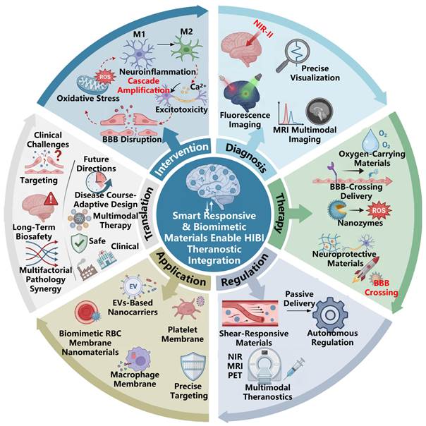
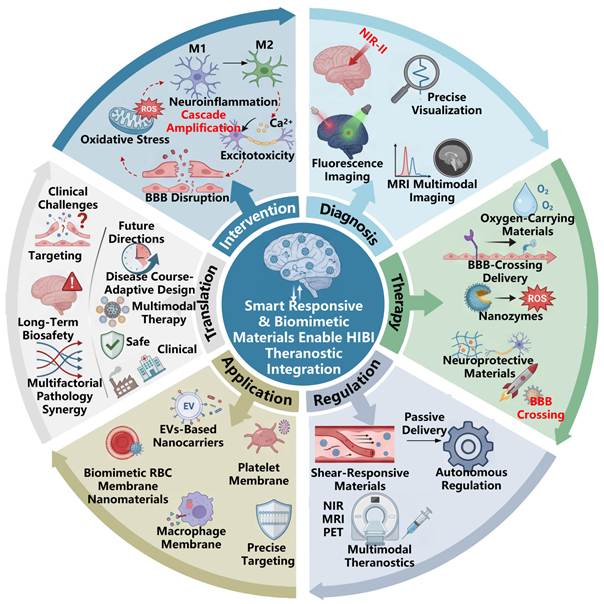
This review presents all the pathological mechanisms of HIBI and corresponding basis for material-useable interventions and constructs a three-layer conceptual framework of "designing materials based on pathological nodes and further outputting treatment diagnosis". Rather than enumerating isolated studies, we bring all known smart theranostic materials under four upending pathological cascades and provide explanation for each one based on its target. We analyze new developments in diagnostics, therapy and theranostic systems by classifying new innovations that integrate biocompatible membranes and multi-stimulus responsive elements that work in contact with various HIBI conditions. One major innovation is combining NIR-II, MRI and fluorescence imaging and therapy processes into one multimodal imaging guided biofeedback system. It is worth clarifying that although HIBI encompasses adult ischemic stroke, neonatal HIE and high-altitude hypoxic brain injury by definition, publicly-available pre-clinical studies of smart theranostic biomaterials are heavily skewed toward adult ischemic-stroke MCAO models, with limited subtype-specific material data for neonatal HIE and high-altitude-related brain injury. Nevertheless, substantial clinical-translation-related barriers persist, including BBB spatiotemporal heterogeneity, insufficient long-term biosafety data, manufacturing bottlenecks and unmet regulatory requirements. This review also discusses these unmet obstacles and outlines theoretical foundations and future research directions for developing intelligent biomimetic materials for managing HIBI (Scheme 1).
Schematic presents of smart responsive and biomimetic material-enabled integrated strategies for HIBI, highlighting the coupling of multi-mechanism pathological cascades with multimodal diagnosis, BBB-penetrating delivery, and microenvironment-regulating therapies to achieve precise and translatable theranostics. Created with Figdraw.com.
2. Pathological mechanisms
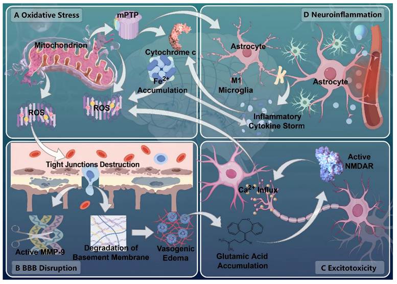
The progression of HIBI is affected by several interrelated pathways [6]. Traditional methods are incapable of stopping the cascade amplification of injury since they are restricted by their singular targets, inadequate BBB penetration and off-target distribution [20]. Targeting the right points of the pathological pathways represents the foundational concept of smart, responsive materials to allow precision neurointervention [21]. Advanced approaches allow precise regulation of the damaged CNS microenvironment by targeting the necessary nodes of the pathological cascade. [22]. This section outlines four main pathological mechanisms as well as their molecular networks, including oxidative stress and mitochondrial dysfunction, neuroinflammation, disruption of the blood-brain barrier, and excitotoxicity, and describes the principles behind material-based targeting approaches, offering a theoretical framework for the further development of theranostic systems (Figure 1) (Table 1).
Subtype-specific pathological nodes and material design priorities for HIBI.
| HIBI subtypes | Critical pathological nodes | Material design priorities | Material Properties | Ref. |
|---|---|---|---|---|
| IS | Oxidative stress-ROS Inflammation | Angiopep-2 | PH responsive drug release | [81] |
| Oxidative stress-ROS | RBCM + Mn3O4 NPs+T7 | / | [82] | |
| Oxidative stress-ROS Inflammation-M1 polarization | Ce NPs+TPP+SPIONs | PH responsive drug release | [83] | |
| Oxidative stress-ROS Inflammation-M1 polarization | RVG29 | / | [84] | |
| Hemodynamic abnormalities | Discoidal NPs | Shear gradients responsive drug release | [85] | |
| BBB penetration | MSC-EVs | / | [86] | |
| Neuronal necrosis BBB penetration | PM-coated anti-inflammatory NPs | / | [87] | |
| Oxidative stress-ROS | NPs | Neutrophil elastase responsive release | [88] | |
| Oxidative stress-ROS Inflammation | RVG29 | ROS response drug release | [89] | |
| Inflammation-M1 polarization | T7 | / | [90] | |
| Neonatal HIE | Inflammation | RVG29+NPs | / | [91] |
| Oxidative stress-ROS Inflammation | RVG29 | ROS response drug release | [92] | |
| Oxidative stress-ROS Inflammation | CeVO4 nanozyme | / | [93] | |
| Inflammation | Biomimetic RBCM nanomaterials | / | [94] | |
| Hypoxic brain injury | Inflammatory-M1 polarization Oxidative stress | PFC oxygen carrier | Oxygen carrying and release | [95] |
| Oxidative stress Inflammation | PFC NPs | Oxygen carrying and release | [96] | |
| HACE | BBB penetration Oxidative stress-ROS Inflammation | MSCs | / | [97] |
Note: SPIONs, superparamagnetic iron oxide nanoparticles.
The crosstalk network of four core interacting pathological mechanisms in HIBI. Four panels elaborate each pathological cascade and their cross-promoting effects in detail. (A) depicts oxidative stress that has been initiated by the mitochondrial permeability transition pore (mPTP) opening, which has led to cytochrome c release, Fe2+ accumulation, ROS burst, and the accumulated ROS have in turn further exacerbates mitochondrial damage through positive feedback. (B) illustrates BBB disruption, in which activated MMP-9 has disrupted tight junctions and degraded the vascular basement membrane, ultimately resulting in vasogenic edema. (C) demonstrates excitotoxicity that has been triggered by glutamate accumulation and NMDAR overactivation, which has induced detrimental Ca2+ influx into neurons. (D) presents neuroinflammation that has been driven by M1-polarized microglia and reactive astrocytes, giving rise to inflammatory cytokine storms, while the bidirectional arrows across the figure highlight the mutual positive feedback between the four pathways that have amplified HIBI pathological damage. Created with Figdraw.com.
2.1. Oxidative stress and mitochondrial dysfunction
Cerebral hypoxia damages mitochondrial energy metabolism, leading to dysfunction in electron transport and oxidative phosphorylation, causing a tremendous reduction in adenosine triphosphate (ATP) levels. The energy crisis quickly leads to termination of normal cellular functions in the brain [23]. Furthermore, at the same time, a high production of ROS occurs due to increased electron leak from the transport chain, which worsens significantly in the period of reperfusion phase after HIBI [24]. Oxidative stress occurs when the endogenous antioxidant defense system fails to cope with rising levels of reactive oxygen species. This leads to direct damage to biomacromolecules like proteins, lipids, and deoxyribonucleic acid (DNA) [25].
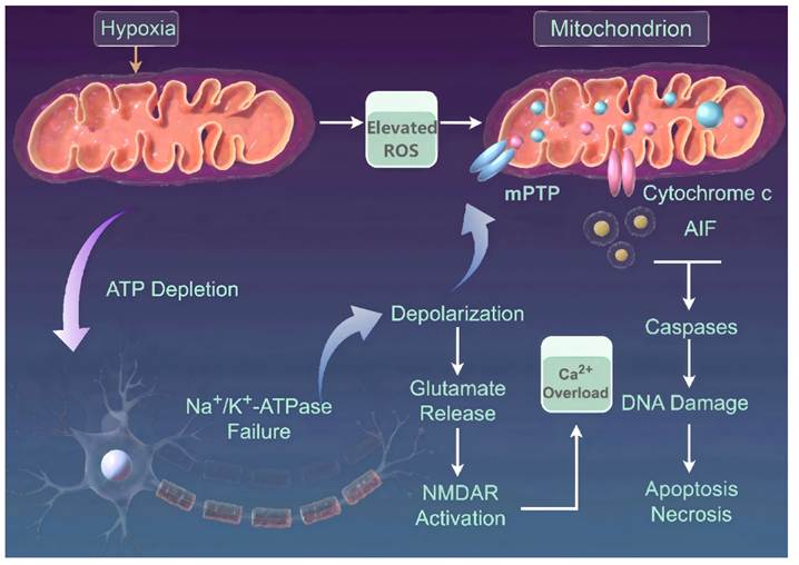
Hypoxia causes a lack of energy and impairs ion pumps using ATP. If the sodium/potassium ATPase stops functioning, the neuron is always in a depolarized state [26]. This abnormal potential not only activates the Ca2+ channels but also enhances glutamate release from presynaptic terminals. When too much glutamate accumulates in the synaptic cleft, N-methyl-D-aspartate receptor (NMDAR) on the postsynaptic membrane gets overactivated [27], leading to huge influx of extracellular Ca2+ which consequently brings about severe cytoplasmic Ca2+ overload, disturbing Ca2+ homeostasis in brain tissues. Excess glutamate further activates phospholipases stimulating catabolism and heavily inhibits glutathione peroxidase 4 (GPx4) that is the principal inhibitor of ferroptosis. Consequently, lipid peroxides fail to be eliminated, making way for iron buildup within cells and thus causing ferroptotic neuronal death [28, 29]. In the end, consistent stress and Ca2+ overload act towards mPTP opening, which causes damage to the mitochondrial membrane and releases cytochrome c, apoptosis-inducing factor, and other apoptosis-promoting molecules [30, 31], which eventually activates caspase-dependent apoptosis pathways which lead to nuclear DNA damage [32, 33]. If the pore stays open for too long, the irreversible destruction of mitochondrial structure and function becomes cemented, and a self-amplifying ROS burst is also set in motion. This vicious cycle will inevitably end in neuronal apoptosis or necrosis, which seriously worsens the progression of HIBI (Figure 2).
Mitochondrial dysfunction and neuronal cell death pathways in HIBI. Hypoxia induces mitochondrial ROS overproduction and triggers mPTP opening, which promotes the release of cytochrome c and AIF, activates caspase cascades, causes DNA damage, and finally results in neuronal apoptosis and necrosis. Meanwhile, hypoxia-caused ATP depletion leads to Na+/K+-ATPase failure, cell membrane depolarization, glutamate release and NMDAR overactivation, which further induces intracellular Ca2+ overload and in turn exacerbates mPTP opening and mitochondrial damage, forming a positive feedback loop that aggravates HIBI neuronal injury. Created with Figdraw.com.
The defining pathological hallmark of HIBI is a multistep cascading process that exacerbates injury. The activation of key components such as mitochondrial malfunction and overproduction of ROS leads to an irreversible vicious cycle causing significant neuronal death and total neurological impairment. Currently, clinical intervention is facing significant obstacles. Reperfusion techniques, including thrombolysis and thrombectomy open big vessels, but they do not restore mitochondrial functioning or remove ROS after the ischemia-reperfusion phase [34]. Moreover, their therapeutic windows are limited, thus excluding most patients from treatment. Conversely, hyperbaric oxygen therapy raises oxygen levels in the body but does not provide sufficient oxygen to the ischemic region or reverse mitochondrial damage [35]. Meanwhile, conventional antioxidants have not shown great efficacy, largely because of off-target distribution, rapid clearance in vivo, and poor BBB penetrability, all of which severely limit their accumulation at damaged mitochondria [36].
To circumvent these intrinsic limitations-namely, inadequate targeting, restricted BBB penetration, and monomodal mechanisms and to directly disrupt the oxidative stress-mitochondrial dysfunction cascade, a repertoire of multifunctional nanomaterials has been engineered. For example, the single-atom FeCo-N6 nanozyme exhibits dual catalase (CAT) and superoxide dismutase (SOD)-mimetic activities that far surpass their natural counterparts. It has successfully broken the vicious cycle of ROS-induced injury and progressive decline of mitochondria through more than 60% reduction of intracellular ROS and increased ATP generation in vitro oxygen-glucose deprivation/reperfusion (OGD/R) models [37]. Meanwhile, the cerium oxide@zeolitic imidazolate framework-8 (CeO2@ZIF-8) core-shell nanoplatform has been fabricated to address the challenge of BBB penetration and effective ROS scavenging [38]. The ZIF-8 of this platform is utilized for efficient BBB transcytosis, and the system is subjected to acid-responsive degradation to release the CeO2 core. Broad-spectrum ROS scavenging and potent anti-inflammatory effects are thus accomplished leading to combining targeted BBB penetration and thoroughly relieving oxidative stress in a synergistic manner. For example, a tissue-mimicking injectable granular hydrogel called GUD-HB@PDA was developed by embedding polydopamine-coated hemoglobin (HB@PDA) into ureidopyrimidinone/dopamine-functionalized gelatin microspheres. This innovative design accurately imitates the dynamic viscoelastic characteristics of natural brain tissue and employs HB@PDA as an oxygen source for stable oxygenation in hypoxic states. Cellular energy regulation is significantly influenced by the hydrogel's inherent viscoelastic properties, which induce transient receptor potential vanilloid 4-mediated mechanotransduction and are responsible for most of the ATP recovery. The combination of sustained oxygen delivery and mechanotransduction to relieve the conditions of hypoxia also activates the AMP-activated protein kinase/mammalian target of rapamycin (AMPK/mTOR) metabolic pathway in this synergistic mechanism, allowing an efficient neurovascular regeneration in ischemia models [39].
These newly developed materials are designed to target key nodes in the pathological cascade of disease progression. The biomaterials show superior delivery and great compatibility with the living tissues. The drawbacks of traditional therapeutic strategies have been overcome by simultaneously addressing the issues of oxidation stress, inflammation, and metabolism. Thus, these multifunctional systems offer rich routes to eliminate the clinical challenges of narrow therapeutic ranges and insufficient effectiveness, and material-based therapies have proven to be a key solution for enhancing the outcomes in HIBI patients. Nevertheless, it is worth mentioning that the use of these injectable tissue-mimicking hydrogels is confronted with significant translational obstacles when adopted for the highly sensitive CNS. While their short-term biocompatibility is promising, the long-term in vivo degradation kinetics of these complex polymeric networks, and the potential chronic foreign-body responses triggered by their degradation byproducts, remain inadequately explored. Going onwards, how to balance the duration of their mechanical properties with absorption without producing harmful substances will make the difference.
2.2. Neuroinflammation
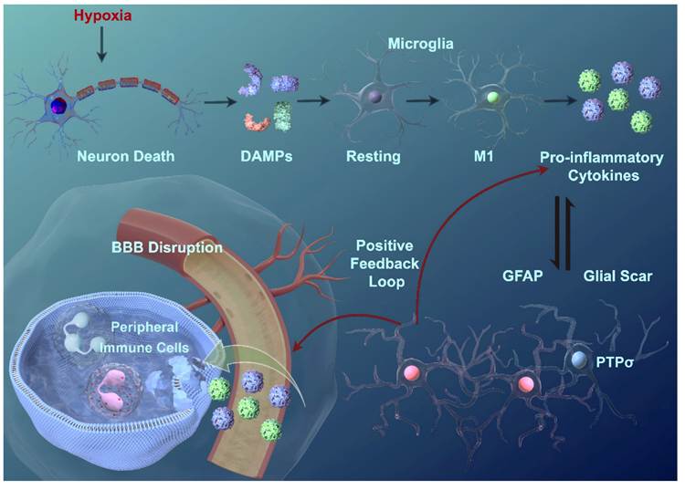
Cerebral hypoxia precipitates massive neuronal death, triggering the release of damage-associated molecular patterns (DAMPs) such as heat shock protein, ATP, and high mobility group box 1 protein [40]. These DAMPs promptly activate resident microglia, driving their transition from a ramified, surveillant resting state into a macrophage-like activated phenotype. Initially, this activation favors the M2 phenotype, characterized by anti-inflammatory and tissue repair functions; however, it rapidly shifts toward the M1 phenotype, which secretes copious pro-inflammatory cytokines including tumor necrosis factor-α, interleukins (IL-1, IL-6, IL-1β), and nitric oxide [41, 42]. Furthermore, DAMPs have been shown to stimulate various immune cells, which then secrete chemokines and adhesion molecules, and through this process, local immune homeostasis is profoundly disrupted while tissue injury gets exacerbated [43].
At the same time, BBB disruption through ischemia allows for infiltration of immune system cells from the periphery. Neutrophils attach to brain microvascular endothelial cells (BMECs) using selectins. Once they enter the brain, these immune cells form networks called neutrophil traps and start producing enzymes like elastase and matrix metallopeptidase 9 (MMP-9). These factors aggressively degrade endothelial cells and the structural components of the BBB, and barrier hyperpermeability as well as neurovascular unit (NVU) devastation are thereby exacerbated. Local immune homeostasis is further disturbed by the subsequent recruitment of monocytes [44]. Moreover, in response to inflammatory signals, astrocytes may proliferate and increase their expression of glial fibrillary acidic protein, ultimately leading to the formation of a glial scar. Although the formation of a glial scar is a temporary mechanism for restricting expansion of the ischemic penumbra, its continued presence will physically and chemically inhibit neuronal migration and functional recovery because of its interactions with protein tyrosine phosphatase σ (PTPσ) [45]. In the end, the cytokine-rich microenvironment is responsible for pushing microglia into the M1 state while stagnating their move to the M2. Inflammatory mediators secreted by activated microglia and astrocytes have created a dangerous positive feedback loop of continuous DAMP release that perpetuates the neuroinflammatory cascade and causes progressive damage to brain tissue.
The highly intertwined and self-amplifying nature of this neuroinflammatory cascade has rendered conventional pharmacological interventions largely inadequate. On one hand, restricted BBB penetrability and negligible central efficacy have fundamentally limited non-steroidal anti-inflammatory drugs. On the other hand, acute cytokine surges can be rapidly suppressed by glucocorticoids, but their prolonged administration has been associated with severe systemic risks, primarily widespread immunosuppression. Importantly, these conventional agents do not possess the spatial precision required for targeting the local neuroinflammatory microenvironment, and this has made them incapable of blocking the pathological cascade at its origin [46, 47] (Figure 3).
Hypoxia-induced neuroinflammatory cascade and glial scar formation in HIBI. Hypoxia triggers neuronal death and release of DAMPs, which activate resting microglia to polarize into the pro-inflammatory M1 phenotype and secrete large amounts of pro-inflammatory cytokines. Inflammatory mediators induce BBB disruption and subsequent infiltration of peripheral immune cells, further amplifying intracerebral inflammation. Persistent inflammatory stimulation drives astrocyte activation with elevated GFAP and PTPσ expression, leading to glial scar formation. A positive feedback loop between inflammatory factors and glial cells sustains and aggravates neuroinflammatory damage in HIBI. Created with Figdraw.com.
To counteract the neuroinflammatory cascade in HIBI, which is driven by aberrant glial activation, cytokine storms, and peripheral immune infiltration, a number of multifunctional nanomaterials have been engineered. Through targeted delivery, precise phenotype modulation, and active clearance of pathogenic mediators, the limitations of conventional treatments have been overcome by these platforms. One primary strategy has centered on reprogramming glial polarization, with the goal of stopping the inflammatory vicious cycle early on. For instance, platelet membrane (PM) camouflaged pH sensitive polymeric nanoparticles have been used to regulate the Notch1 pathway, which has promoted M1 to M2 microglial transition and helped reduce neuronal apoptosis [48]. Similarly, immunosuppressive nanoparticles coated with CXCR4-overexpressing mesenchymal stem cell (MSC) membranes block peripheral immune infiltration, and concurrently release the cGAS inhibitor A151 in a ROS-responsive manner to drive M2 polarization [49]. As CAT-loaded tannic acid (TA) self-assembled nanoparticles camouflaged with M2 microglial membranes, the biomimetic TPC@M2 nano-scavengers chelate excess ferrous ions via the TA matrix and scavenge diverse ROS including H₂O₂ via CAT, while the biomimetic coating intrinsically induces the M1 to M2 microglial shift to enhance neurological recovery [50]. Beyond single-pathway interventions, multimodal systems exert synergistic effects. A representative example is the macrophage membrane (MM) camouflaged dendrimer co-loaded with fibronectin and edaravone (M@A-F/E), in which edaravone efficiently scavenges ROS and drives M2 microglial polarization, while fibronectin promotes angiogenesis and preserves BBB integrity, ultimately achieving a potent triad of anti-inflammatory, antioxidant and pro-angiogenic effects [51].
Collectively, current nanotherapeutic strategies for neuroinflammation intervention are largely built upon the simplified paradigm of M1 to M2 phenotypic polarization. However, high-resolution analyses via single-cell RNA sequencing (scRNA-seq) and spatial transcriptomics have demonstrated that microglia and monocyte-derived macrophages in HIBI and other brain disorders do not exist as two discrete phenotypes but rather occupy a continuous, multidimensional phenotypic spectrum - a finding that challenges the oversimplified M1 to M2 switching mechanism adopted in the majority of nanomaterial studies. Multiple single-cell atlases of CNS myeloid cells have confirmed that myeloid cells under hypoxic and injurious stimuli exhibit high heterogeneity with complex transcriptional profiles, and cannot be simply categorized as purely M1 or M2 populations. For instance, a distinct subset of hypoxia-associated tumor-associated macrophages (Hypoxia-TAMs) has been identified in the perinecrotic hypoxic niche of glioblastoma. These cells mediate vascular endothelial injury via p50-dependent transcriptional regulation of adrenomedullin, and their unique glycolytic and pro-angiogenic transcriptional programs fall outside the conventional M1/M2 classification framework [52]. In ischemic brain injury, repair-associated macrophages differentiated from CCR2-positive monocytes show high expression of GPNMB and CD63. Governed by the transcription factor Mafb, they participate in myelin lipid recycling and angiogenesis, harbor mixed functional transcriptional signatures, and do not conform to the narrow definition of M2 macrophages [53]. In vitro experiments with dual inflammatory stimulation further validate that macrophages exhibit orthogonal, heterogeneous transcriptional responses under combinatorial signals. Typical M1 pro-inflammatory cytokines and M2 reparative markers are rarely co-expressed in a single cell, indicating that the binary classification inherently masks the true functional diversity of macrophages within complex microenvironments [54].
Building on the continuous, polymorphic transcriptomic features of myeloid cells, next-generation smart nanomaterials must move beyond the singular design logic of enforced M1 to M2 phenotypic switching, and shift toward precise, dynamic immunomodulation aligned with lesion heterogeneity. Multi-stimuli-responsive, region-specific delivery systems should be developed, integrating responsive moieties for microenvironmental signals such as hypoxia, ROS and lipid debris. These systems release differential regulatory molecules targeting myeloid subsets in hypoxic regions, replacing global uniform polarization induction to enable directed modulation of distinct functional subsets. Meanwhile, combinatorial loading of antioxidants and lipid metabolism regulators preserves the phagocytic function of myeloid cells for necrotic debris clearance during the acute phase, while selectively promoting the expansion and maturation of endogenous reparative subsets, rather than forcing all myeloid cells toward a single M2 phenotype.
2.3. BBB damage
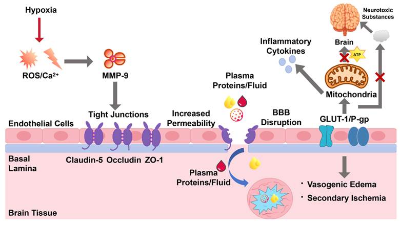
The BBB, which is composed of BMECs, tight junctions (TJs), the basement membrane, and astrocytic end-feet, serves as the main barrier for maintaining CNS homeostasis. Its integrity depends largely on core TJ proteins like Claudin-5, Occludin, and zonula occludens-1 (ZO-1). Hypoxia triggers structural and functional disruption of the BBB through multiple pathways, and this sets off a self-amplifying cycle of damage. On one hand, hypoxic stress leads to intracellular Ca2+ overload, and this activates downstream signaling that promotes the conversion of pro-MMP-9 to its active form. On the other hand, metabolic collapse and ischemia-reperfusion injury have been found to drive a massive ROS burst [55-57]. Once activated, MMP-9 cleaves critical TJ proteins and degrades key basement membrane components like laminin and collagen. This proteolytic degradation significantly widens the endothelial paracellular gaps, compromises the structural integrity of the BBB, and leads to marked hyperpermeability [58, 59]. As a result, albumin-rich plasma components and other macromolecules leak out into the brain parenchyma. The resulting increase in interstitial colloid osmotic pressure drives a large fluid influx, and this eventually causes vasogenic cerebral edema. The rise in intracranial pressure decreases cerebral perfusion and induces secondary ischemic injury [60, 61]. Furthermore, neuroinflammation initiated by hypoxia can reduce the levels of glucose transporter-1 [62] and also inhibit P-glycoprotein efflux function [63]. These changes compromise the brain's energy supply and hinder the removal of neurotoxic metabolites, further aggravating BBB dysfunction and tissue necrosis [64]. The BBB demonstrates long-lasting functional deficits and chronic leakage following HIBI, even with some endogenous repair processes. It seems that mainly, the reason behind the failure to completely recover lies in the improper assembly of TJ complexes and the abnormal interaction between Claudin-5 and ZO-1 (Figure 4).
Mechanistic pathways of BBB breakdown under hypoxic conditions. ROS accumulation and Ca2+ overload are induced by hypoxia, and these events lead to MMP-9 activation, which then degrades endothelial TJ proteins like Claudin-5, Occludin, and ZO-1, thereby increasing barrier permeability. Once the structural integrity of the BBB is compromised, plasma proteins and fluid extravasate into the brain tissue, and vasogenic edema along with secondary ischemia is brought about. At the same time, hypoxia has been found to damage BBB transporters GLUT-1 and P-gp, and mitochondrial dysfunction as well as ATP deficiency is triggered; inflammatory cytokine release and accumulation of neurotoxic substances are also promoted. These processes together establish a positive feedback loop that further aggravates BBB injury.
The development of the disease process is mainly based on the disruption of TJs and the resulting cerebral edema, but standard treatments have not provided any combined method of repairing the BBB and eliminating edema. Osmotic agents like mannitol work to reduce intracranial pressure by forming an intravascular osmotic gradient, removing extra fluid from brain tissue into the circulation. Osmotic agents cause increases in osmotic diuresis, but they do not restore damaged structures of TJs and can create electrolyte imbalance and renal intoxication in the long run [65]. Thrombolysis and thrombectomy are reperfusion techniques that open blood vessels but fail to restore TJ integrity or prevent BBB damage from ischemia-reperfusion injury [34]. Although glucocorticoids can reduce BBB permeability, the method has limited clinical value due to serious systemic side effects [46]. Conventional neuroprotective agents like butylphthalide are subject to off-target distribution, poor penetration across the compromised BBB preventing accumulation at target lesions [66].
The real value of material-based interventions comes from their ability to precisely adapt to and disrupt this interconnected pathological cascade. One example of this approach is an innovative biomimetic nanoplatform called MFP, which has been constructed by camouflaging a perfluorocarbon (PFC) and pluronic P123 core within an M2 microglial cell membrane. This platform is designed to stop the progression from hypoxia-induced TJ degradation to hyperpermeability and the resulting cerebral edema [67]. A triple-targeted therapeutic synergy has been achieved with this design. The PFC core delivers oxygen to relieve ischemic hypoxia and suppress hypoxia-driven MMP-9 release, while pluronic P123 works to inhibit any remaining MMP-9 activity and thus protect TJ integrity from inflammatory proteolytic degradation. At the same time, the M2 microglial membrane enables inflammation-targeted delivery through specific interactions between surface lymphocyte function-associated antigen 1 (LFA-1) and intercellular adhesion molecule 1 (ICAM-1), both of which are heavily upregulated in ischemic lesions. This biomimetic strategy has allowed efficient BBB penetration, with 3.47 to 4.43 times higher accumulation in the ischemic penumbra than that seen with non-targeted carriers. In vivo tests using middle cerebral artery occlusion (MCAO) rat models showed that MFP reduced BBB leakage to 38.18 percent of the model group level, and cerebral edema was markedly alleviated. The infarction area was also narrowed from 36.66 percent to 11.03 percent, and the therapeutic window was extended to 4 hours. With strong efficacy and negligible systemic toxicity, MFP has established a comprehensive approach for managing HIBI-associated edema. This advanced platform overcomes the limitations of traditional therapies and offers a critical strategy for resolving stubborn cerebral edema and saving the NVU.
Translating these BBB-targeted biomimetic platforms into clinical practice has not been straightforward, since a number of scientific and biological hurdles still stand in the way. On one hand, receptor-mediated transcytosis like the LFA-1/ICAM-1 pathway can significantly enhance brain accumulation of nanoparticles, but on the other hand a lot of the nanoparticles frequently get trapped in endolysosomal compartments of endothelial cells and do not reach deeper parenchymal targets, so new strategies will need to be employed in future designs for effective endosomal escape which would allow for successful delivery into the brain tissue. Conversely, using PFCs and man-made polymer cores has raised serious questions about their long-term removal from the CNS and whether these materials, which break down slowly, can cause prolonged neurotoxicity or sustained activation of microglia due to prolonged residence in the parenchyma. Therefore, the next generation of theranostics should be developed with the aim of ensuring the right balance between short-term barrier opening and rapid, seamless removal from the body.
2.4. Excitotoxicity
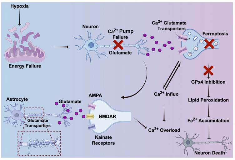
Excitotoxicity, a severe form of neurotoxicity, has been fundamentally driven by sustained and excessive glutamate release [68], and severe neuronal energy depletion is precipitated by hypoxia and glucose deprivation, which renders the maintenance of physiological ion gradients impossible and drives sustained depolarization as well as disabled Ca2+ pumps, culminating in a sharp intracellular Ca2+ influx [69]. Concurrently, astrocytic glutamate transporters are downregulated by this energy crisis, and massive glutamate efflux along with its pathological accumulation in the synaptic cleft is provoked, which effectively ignites the excitotoxic cascade and vastly amplifies the neurotoxicity of excitatory amino acids [70, 71]. NMDARs, along with AMPA and kainate receptors, are hyperactivated by synaptic glutamate overload, with NMDARs being the primary targets [72], continuous NMDAR activation, which is facilitated by the co-agonist glycine, mediates a massive Ca2+ influx, and intracellular Ca2+ overload is severely exacerbated [73, 74]. On one hand, this cytoplasmic Ca2+ overload triggers a cascade of self-destructive, Ca2+-dependent enzymatic responses, dictating widespread neuronal death and aggravating HIBI; on the other hand, synaptic plasticity, the cellular substrate for learning and cognition, is also profoundly disrupted by this aberrant Ca2+ overload. Sustained NMDAR hyperactivation disturbs the physiological balance between synaptic and extrasynaptic signaling, impairs hippocampal long-term potentiation, and facilitates pathological long-term depression, thereby abolishing the synaptic remodeling required for memory formation [75, 76], while key synaptic structural proteins like PSD-95 and cytoskeletal components are degraded by Ca2+ activated proteases such as calpains, resulting in dendritic spine loss and synaptic structure destruction [77]. These synaptic plasticity deficits directly mediate the cognitive impairment that manifests after HIBI (Figure 5).
Hypoxia-induced excitotoxicity and ferroptosis in neurons. Neuronal energy failure is caused by hypoxia, and this has led to dysfunction of Ca2+ pumps and excessive glutamate release, while the uptake of glutamate by astrocytic transporters has been impaired. Once extracellular glutamate builds up, AMPA receptors, NMDARs, and kainate receptors become activated, triggering a massive Ca2+ influx and intracellular Ca2+ overload, which then creates a positive feedback loop that makes excitotoxicity even worse. On one hand, sustained Ca2+ overload and excitotoxic stress have further inhibited GPx4 activity; on the other hand, lipid peroxidation and Fe2+ accumulation have been promoted, and these events eventually drive neuronal ferroptosis and cell death. Created with Figdraw.com.
Conventional treatment methods have their limitations which have ignited the need for effectual material-based treatment options, particularly because these conventional treatment methods do not have proper targeting and can not achieve multipathway synergistic blockade. On one side of the argument, these commonly used anti-excitotoxic drugs like NMDAR antagonists are known to interfere with proper neural signaling and cause serious side effects [78]. Conversely, single-target drugs, including Ca2+ channel blockers, have failed to interrupt interconnected pathological processes and their effectiveness compromised by the inability to penetrate the BBB and rapid clearance from the body, meaning they cannot prevent diseases from advancing [79]. Hence, there is a need to create multipurpose platforms that can be directed precisely to the targets, penetrate well the BBB, and exert synergistic therapeutic effects to solve the problem of treatment.
HIr-PS, a theranostic agent for treating ROS overproduction and Ca2+ overload, was designed with core-shell structure including very small iridium-based catalyst core and biopolymer coatings of polydopamine and sodium alginate, which gives it an ability to perform triple-targeting. The core features iridium (Ir) in mixed state (Ir0/Ir4+) that gives it strong abilities in SOD, CAT, and peroxidase-like actions, thus leading to high efficiency in scavenging superoxide and ROS, surpassing efficacy regular small molecules like N-acetylcysteine. In addition, SA coating acts like a biological sponge, specifically binding to extra Ca2+ in both the cytoplasm and the mitochondria. Within one minute, the coating chelates 80% of the free Ca2+ and is effective at that level as some of the classic chelators like ethylene glycol tetraacetic acid without the off-target toxicity of those types of compounds. Moreover, the nano-sized dimensions size of 28 nm, along with its biopolymer cover, help to achieve fast transcytosis of the BBB via caveolae and macropinocytosis and, eventually, the desired accumulation in the ischemic penumbra tissue [80]. This high degree of targeting along with multidimensional synergy make this option impossible for any classical monotherapy. Instead of just providing symptomatic treatment, these nanoplatforms break the cycle of damage in the brain.
Inorganic nanozymes, like Ir or Ce-based catalytic platforms, have high catalytic efficiency. The application of these metallic structures in clinical practice, however, encounters regulatory and biological challenges. Firstly, metals cause the problem of their clearance from the CNS, since metal-based cores can cause neurotoxic effects when they remain in the body for a long period of time. Secondly, broad-spectrum ROS scavenging, though effective against oxidative damage, might also disrupt normal redox-dependent signaling if it eliminates ROS too indiscriminately. In upcoming designs, we must aim to use biodegradable or rapidly removable metal clusters to neutralize only the pathological ROS and maintain redox equilibrium.
Four interrelated pathological cascades together form the self amplifying injury loop of HIBI. The starting point of corresponding material design is these key pathological nodes, such as cross BBB, ROS, pH stimulation response, M1 polarization, etc. Based on these pathological derived design standards, it is expected to improve the efficacy of intelligent therapeutic diagnostic materials.
3. HIBI material-assisted diagnosis platform
Diagnosis is a prerequisite for material drug intervention. Under the guidance of HIBI pathological features, advanced auxiliary diagnostic platforms synergistically monitor factors such as brain oxygenation, BBB integrity, ROS and pH levels, as well as metabolic markers such as lactate and ATP levels, overcoming the limitations of traditional clinical imaging in capturing dynamic molecular level pathological changes [98]. Traditional medical imaging techniques often are not effective in monitoring these specific biological events, as the methods are limited by limited penetration, low sensitivity, or the inability to multiplex [99]. Advanced material-based techniques overcome these limitations in an efficient manner. Smart probes help to establish a bridge between conventional diagnosis and theranostics by providing quantitative information about the microenvironment of the disease [21].
3.1. NIR-II imaging materials - achieving visualization of pathology in deep brain regions
Optical imaging in the NIR-II range of 1000-1700 nm results in reduced photon scattering from tissues and their autofluorescence, leading to improved spatial and temporal resolution and better penetration depth [100]. The developed NIR-II nanomaterials further allow for visualization of disease evolution in the brain at both macro- and microscopic levels, as compared to conventional optical probes.
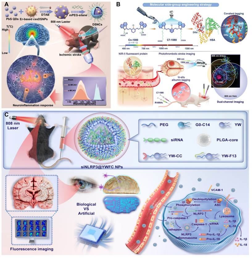
Localizing the ischemic penumbra and measuring the hemodynamic parameters is key for early HIBI detection [101]. The use of functionalized NIR-II probes can selectively locate areas of hypoxia and assess cerebral blood flow or collateral circulation [100]. Although early nanothermometers based on lanthanide-quantum dots were successful in measuring local temperature changes in IS models, they were not able to provide therapeutic tracking [102]. To gain the synergy from diagnostic and therapeutic methods, researchers succeeded in creating rare earth-element-doped NPs that produce stable NIR-II light to locate ischemia and a mesoporous silica coating with phase change materials inside [103]. Under NIR-II imaging assistance, the novel system serves to produce localized cooling effects that prevent infarct development and inflammatory response (Figure 6A). The viability of the ischemic penumbra is largely reliant on collateral flow [104], therefore, it is required to address the issue regarding non-specific binding of albumin by the currently available NIR-II contrast agents. To overcome the limitations from the unnecessary background signal and unpredictable pharmacokinetics, the albumin-evading NIR-II probe (e.g. IR-808AC) has been developed for rapid elimination. This permits short-interval repeated injections to visually map cerebral vasculature and perfusion status [105]. In addition to this, composite semiconducting organic nanoprobes (4T-BSA) have been formulated based on small-molecule dye along with BSA. These nanoprobes hence act not only as a visualization tool for collateral perfusion network but also as a sensitive medium for detecting minute abnormalities in BBB permeability, thus providing an excellent dual diagnostic system [106].
Nanoprobe and nanotherapeutic strategies for imaging and intervention in HIBI. (A) Schematic of DSNP nanoprobe synthesis and application for NIR-II thermometric imaging of neuroinflammation in IS model. The probe enables real-time temperature mapping of the ischemic brain, providing a direct basis for studying the neuroinflammatory response. Adapted with permission from [102], Copyright 2025 American Chemical Society. (B) Molecular side-group engineering strategy for constructing NIR-II fluorescent proteins (C7-1080) and their application in photothrombotic stroke imaging. This approach allows in-situ dual-channel imaging of arterial and venous vasculature via albumin tagging, enabling precise visualization of stroke-related hemodynamic changes. Adapted with permission from [112], Copyright 2025 John Wiley and Sons. (C) Theranostic application of the siNLRP3@YWFC NPs. By integrating nod-like receptor family pyrin domain-containing 3 (NLRP3)-targeted siRNA within a multifunctional carrier featuring a NIR-II fluorophore, fluorination, and targeting peptide conjugation, the developed siNLRP3@YWFC NPs effectively mitigates neuroinflammation via gene silencing, while its intrinsic fluorescence enables the non-invasive monitoring of therapeutic efficacy. Adapted with permission from [115], Copyright 2025 American Chemical Society.
Conventional imaging techniques are unable to monitor quick changes that occur in oxidative stress. In this case, stimuli-responsive NIR-II probes can convert oxidative stress events to fluorescent events [107]. A specific example is a ratiometric nanoprobe that uses a ROS-responsive IR-783 iodide dye with lanthanide NPs, to measure the amount of local ROS present in the tested area [108]. To advance this concept, scientists have created theranostic nanoplatforms which incorporate catalytic CeO2 NPs as well as three terminal thioether (3TT) cores which emit NIR-II radiation, and rabies virus glycoprotein (RVG) recognition peptides. With this combination, multiple functions are accomplished including the elimination of excessive ROS molecules from cells of the nervous system and actively traverses the BBB [109]. Another development has also been reported in order to measure the increase of myeloperoxidase (MPO) activity in HIBI. The new NIR-II probe activated by hypochlorous acid adopts a unique mechanism of operation whereby its fluorescence spectrum responds to any MPO activity through redox reactions, enabling the qualitative and quantitative tracking of local MPO activity [110].
The BBB breakdown is crucial in starting cerebral edema and secondary injury to neurovascular tissue. NIR-II imaging materials allow detection of the BBB breakdown by utilizing its discrete permeability and size-dependent aggregation [111]. For instance, albumin-based NIR-II fluorescent proteins show substantial accumulation only in the areas affected by BBB breakdown. Since the fluorescence intensity of locally accumulated NIR-II imaging material reflects the BBB permeability directly, this gives a good technique to assess the structure integrity [112] (Figure 6B). These findings are validated by dual-modal imaging techniques. Indocyanine green-doped porous poly(lactic-co-glycolic acid) (PLGA) microspheres are used to integrate high resolution ultrasound imaging with NIR-II fluorescence imaging for differentiating mild leaks from severe barrier damage in a porcine HIBI model [113]. At the capillary level, novel NIR-II segmentation allows for quantification of microvascular dysfunction through calculation of the vascular rupture rate and the perfusion unevenness index. Microscopic mapping in murine stroke models provides clear visualization of the limited capillary network in the ischemic penumbra, which supports the idea of BBB damage. In order to achieve a more practical application of these developments, several new systems have been created to integrate NIR-II imaging and small interfering RNA (siRNA) delivery [114]. This combination allows for targeted delivery and tracking of the carriers as well as assessment of the treatment outcome through NIR-II signal attenuation [115] (Figure 6C).
Hence, the NIR-II imaging agents not only allow complete visual mapping of the deep-brain pathological microenvironment but also facilitate simultaneous monitoring of brain oxygenation, oxidative stress, and BBB permeability. Despite the fact that it helps in diagnosis, it also has an important characteristic of providing real-time physiological feedback. In addition to that, through the constant evaluation of treatment effectiveness and adaptation to it, this kind of diagnostic tool contributes to bringing HIBI treatment from empirical to personalized medicine.
3.2. Advanced MRI platforms: multidimensional mapping of HIBI
Owing to its unparalleled soft-tissue contrast and non-ionizing nature, MRI remains a cornerstone modality for evaluating the structural and functional sequelae of HIBI [116]. In the acute phase of hypoxia, there is immediate disruption of the metabolic system of the brain; it is associated with an increase in lactose concentration in the brain tissue and drop in N-acetylaspartate [117]. However, classical magnetic resonance spectroscopic imaging (MRSI) does not possess appropriate resolution, speed of examination, and spatial coverage required for collecting high-quality data [118], recent technological leaps have birthed advanced three-dimensional (3D) MRSI platforms. Through improvements in pulse sequences and acquisition of signals, these novel systems make quick and complete mapping of volumes of the brain for N-acetylaspartate and lactate distribution possible. In particular in acute IS, using Diffusion Weighted Imaging (DWI) together with 3D MRSI helps clinicians to precisely delineate the boundaries between the viable ischemic penumbra (reversible ischemia) and the ischemic core (irreversible infarction) based on quantifiable lactate thresholds [119].
This diagnostic precision emphatically extends to high-altitude-induced hypoxic injuries, such as high-altitude cerebral edema (HACE). In these scenarios, DWI exquisitely captures the hyperintense signals of nascent white matter edema long before standard computed tomography (CT) can detect macroscopic structural anomalies. When coupled with apparent diffusion coefficient mapping, these advanced modalities provide highly sensitive and specific quantitative metrics of the underlying cytotoxic edema [120].
Apart from macroscopic disruptions to structural and metabolic systems, HIBI severely interferes with complex networks of functional neurons [121]. Using the blood-oxygen-level dependent contrast technique, sophisticated functional MRI (fMRI) approaches, which include amplitude of low-frequency fluctuations and voxel-based morphometry, allow detecting correlations between chronic hypoxia and neuroanatomical remodeling [122]. For instance, in long-term HIBI driven by chronic mountain sickness, voxel-based morphometry mapping reveals highly specific volumetric contractions in both gray and white matter. Concurrently, amplitude of low-frequency fluctuations analysis detects aberrant spontaneous neural activity, identifying pathological hyperactivation in the left supramarginal and parahippocampal gyri alongside cerebellar hypoactivation. Together, these metrics yield a comprehensive architectural and functional map of hypoxia-induced neurodegeneration [122].
In conclusion, the merging of sophisticated MRI technologies forms a multi-layered diagnostic method-combining metabolism measurement, functional dynamics, and high-quality structure localization. These advanced methods manage to get past conventional imaging's limits in terms of sensitivity and dimensionality and provide a necessary diagnostic base.
3.3. Advanced fluorescence imaging modalities
Fluorescence imaging (FLI) is special for its non-invasive feature, its real-time monitoring properties, great specificity, and excellent spatiotemporal resolution, thus becoming the main technique for dynamically tracking many important molecular processes taking place in the HIBI environment [123]. Biological thiols are important antioxidants in the CNS [124], and can indicate oxidative stress levels through changes in their quantity, but common procedures do not allow their in vivo measurements. Advanced NIR fluorescent probes have been created to detect damage to cell structures. The dicyanoisophorone-acetyl-pyridinium (DCI-Ac-Py) probe employs an isophorone derivative as its main fluorophore complemented by a pyridine carboxylate in order to increase its lipophilicity and ability to cross the blood-brain barrier. DCI-Ac-Py in HIBI models reveals the oxidative damage levels as well as defining the mechanisms of the process, indicating how the biological thiols protect neurons from cell death by inhibiting ROS production through both conventional (P65) and unconventional (RelB) NF-κB pathways [125].
Advancing from diagnostic precision using thiol-monitoring probes, the development of integrated theranostic systems has come into being. For example, to deal with the highly reactive nitrogen species formed during these oxidizing bursts, peroxynitrite (ONOO-) reactive carbon monoxide (CO) donors (such as PCOD585) were created. The carbon-cage structure makes it easy for this probe to traverse the BBB and liberate CO selectively only when it comes into contact with much higher levels of ONOO-. Experimental HIBI models take advantage of the natural fluorescence of PCOD585 to offer immediate visual feedback on therapy while essentially indicating the timing of oxidative stress elimination which was initially detected by sensor probes such as DCI-Ac-Py [126].
The progress achieved by these advanced probes helps us understand effectively the path along which FLI technology is developed, namely - moving from passive monitoring of molecules to integrated theranostics. The ability of these platforms to solve the issues of traditional oxidative stress management that suffers from off-target effects and spatiotemporal efficacy issues leads to the creation of a new technological standard in the field of HIBI intervention.
4. Therapeutic materials for precision treatment
Based on the material design derived from pathological nodes, this chapter has sorted out representative materials, and each type of material is explained within the framework of key pathological nodes in the HIBI injury cascade.
4.1. Oxygen-carrying materials
4.1.1. PFC-based phase-change nanodroplets
PFCs are crucial for advanced oxygen delivery systems because of their unique characteristics such as oxygen solubility, biocompatibility, and responsiveness to heat and light [127]. When synergized with focused ultrasound (FUS), PFCs facilitate non-invasive, reversible modulation of BBB permeability, enabling the precise delivery of both oxygen and therapeutics into the injured brain [128, 129]. Nevertheless, previous generations of PFC carriers faced problems, including intrinsic lipophobicity leading to poor drug loading, systemic instability, and inadequate long-term storage profiles [130].
In order to address these translation barriers, the field has undergone rapid strategic innovation. Early structural modifications focused on formulating PFC nanodroplets (NDs) with tunable boiling points - utilizing compounds like octafluoropropane (-37 °C) and decafluorobutane (-2 °C) in order to ensure a threshold for BBB opening [131]. Using exploiting acoustic droplet vaporization to turn liquid PFC emulsions into microbubbles in situ, scientists created phase-change NDs (PCNDs) that can both temporarily open the BBB and enable controlled release of payloads [132].
The challenge persisted, however, since pure PFCs are highly fluorophilic and thus incompatible with most hydrophobic non-fluorinated drugs [133]. To overcome this, unique fluorinated surfactants and intermediate oil phases (e.g., acetyl tributyl citrate, ATBC) were used in an innovative manner. This novel tri-phasic emulsion technology helps to overcome the lipophobicity of PFCs, thus achieving successful drug loading while maintaining the ability to open the BBB through FUS [134].
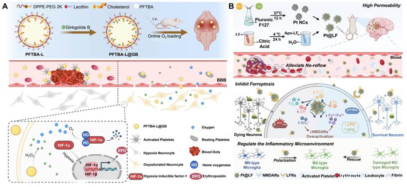
However, transcranial FUS application is limited due to distorting effects of skull bones on acoustic attenuation, scattering, and focal distortion. An elegant way out is to use laser-activated PFC NDs that are able to induce BBB permeabilization via local photothermal vaporization, thus achieving complete elimination of transcranial energy losses. Additionally, this system offers dual-modal photoacoustic and ultrasound imaging capabilities to track the biodistribution of the carriers and to confirm the localized barrier disruption [135] (Figure 7A). Oxygen and therapy can be precisely placed in the ischemic penumbra due to this multimodal platform [136]. This has led to the development of theranostics in ischemic stroke and subarachnoid hemorrhage by combining the great oxygen-carrying capacity of PFCs and targeted therapy. The nanoparticles restore oxygenation during ischemia-reperfusion and inhibit hypoxia-inducible factor-1α (HIF-1α) so as to stop the hypoxic-ischemic process [95,96,137].
Smart responsive nanomaterial strategies for precision diagnosis and treatment of HIBI. (A) Ginkgolide B-loaded perfluoro-tert-butyl lipid NPs (PFTBA-L@GB): Undergo online oxygen loading after intravenous injection, target ischemic brain tissue, alleviate hypoxia, regulate the HIF-1α pathway and erythropoietin expression, thereby reducing neuronal damage and protecting the BBB. Adapted with permission from [96], Copyright 2023 John Wiley and Sons. (B) Platinum-loaded lactoferrin NPs (Pt@LF): Cross the BBB with high permeability to alleviate the "no-reflow" phenomenon; their enzyme-mimetic activity scavenges ROS, inhibits ferroptosis, and regulates the inflammatory microenvironment to promote M2-type microglial polarization, ultimately achieving neuronal survival and functional recovery. Adapted with permission from [139], Copyright 2024 John Wiley and Sons.
By incorporating tunable responsiveness, high levels of oxygen supply, and stable drug encapsulation, multifunctional PFC platform enables an integrated therapeutic cascade of delivery agent accumulation, site-specific barrier permeability, and targeted co-delivery. This synergy resolves problems associated with monomodal deliverers and creates new opportunities for treatment of HIBI and other neurovascular diseases.
4.1.2. Hypoxia-responsive biomimetic nanobubbles
The key aspect of recovering the vulnerable brain tissue located in the ischemic core is rapid oxygen reperfusion [138]. For the implementation of targeted gas delivery, innovative PM-coated oxygen nanobubbles (PONBs) that provide a complete solution for effective oxygenation, combined therapy, and improved ultrasound imaging have been developed.
Ultrasonic cavitation and repeated compression-expansion processes around gas-liquid interface create encapsulated pure oxygen inside a dense PM shell, producing highly concentrated oxygen nanostructures with size 50-100 nm and very high dissolved oxygen (up to 29.3 ± 0.3 mg/L). Notably, the PM shell preserves the original targeting capabilities of the parent platelets and directs the PONBs to the damaged and inflamed neurovascular endothelium signature of acute IS. This advanced camouflage does not just specify the exact localization of oxygen but also boosts the use of nanobubbles as focused contrast agents for real-time monitoring through ultrasound. By proving its effectiveness in delivering enormous amounts of targeted oxygen to non-reperfused infarct cores, it can be said that PONBs create a reliable and novel way of starting the management of acute HIBI [139] (Figure 7B).
4.1.3. Oxygen-supporting hydrogels
Hydrogels refer to a type of 3D network created through the physical or chemical cross-linking of hydrophilic polymers. These materials are known for their high water content, biocompatibility, and great flexibility. In view of all these features, it is clear that they are perfect for brain extracellular matrix (ECM) modeling, drug delivery, and appropriate responding to pathological microenvironments [140]. Thus, hydrogel-based platforms can solve a number of crucial problems faced by HIBI therapy, including inefficient cellular delivery, persistent localized hypoxia, and multifaceted pathological cascades.
Early studies employed gelatinous hydrogels with improved porosity and swelling properties to imitate the ECM of the brain and provide preliminary neuroprotection [141]. However, these matrixes could not respond actively to the three core pathological aspects of IS: hypoxia, excess ROS, and neuroinflammation. To solve this issue, scientists have added active oxygen-delivery systems into the hydrogel structure. The perfluorotributylamine substance was loaded into a matrix based on fibrin, resulting in the generation of a stable oxygen-delivery system allowing for a constant oxygen release that can reverse the damage due to hypoxia in the surrounding glial cells such as Schwann cells [142].
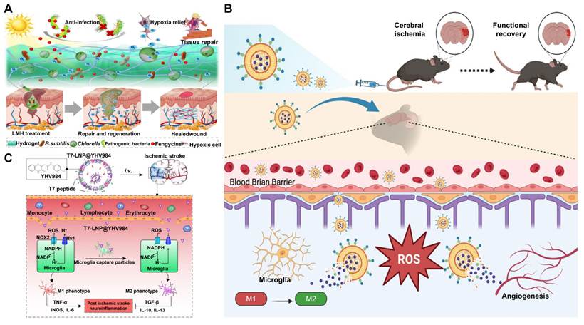
Innovations in micro-ecological hydrogels have gone further than chemical oxygenation. Temperature-sensitive materials such as pluronic F-127 and polydopamine were used to grow microalgae and Bacillus subtilis together in an ecosystem that produces oxygen while suppressing bacteria, which could be quite useful in infection HIBI treatment [143] (Figure 8A). Moreover, biomimetic soft scaffolds were developed through crosslinking hyaluronic acid and platelet-derived lysate, producing a matrix known as PLS Gel that contained MMP-sensitive triglycerides (TGMS@DHA). The addition of boronate ester bonds allows the scaffold to respond to both ROS and MMP signals. This type of architecture plays a role in altering the ischemic microenvironment and providing strong antioxidant and anti-inflammatory benefits, enabling a single therapeutic platform to act against multiple causes of HIBI [144].
Multimodal nanotherapeutic strategies targeting for HIBI. (A) Application of the living microecological hydrogel system in hypoxic tissue repair. This system achieves repair and healing of hypoxic wounds through anti-infection, hypoxia relief, and promotion of tissue regeneration. Adapted with permission from [143], Copyright 2023 The American Association for the Advancement of Science. (B) Mechanisms of nanomedicine-mediated neuroprotection and functional recovery after cerebral ischemia. This strategy achieves repair and functional reconstruction of ischemic brain tissue by regulating microglial polarization from M1 to M2, scavenging ROS, and promoting angiogenesis. Adapted with permission from [89], Copyright 2025 Springer Nature. (C) Schematic illustration of T7 peptide-modified lipid nanoparticles (T7-LNP@YHV984) for microglial-targeted inhibition of the Hv1 channel. This platform crosses the BBB to suppress microglial NLRP3 inflammasome activation and promote M1-to-M2 polarization, thereby alleviating neuroinflammation and enhancing neuronal survival. Adapted with permission from [90], Copyright 2025 Springer Nature.
4.2. BBB-crossing drug delivery materials
4.2.1. RVG29 peptide modification
The RVG29 peptide is a key targeting agent for HIBI theranostics. It binds to nicotinic acetylcholine receptors (nAChR), found on the surfaces of BMECs and neurons, which allows RVG29 to promote receptor mediated transcytosis and endocytosis [145]. Therefore, it is a vital agent to cross the BBB and connect microenvironment monitoring with targeted treatment.
Early implementations of RVG29 focused primarily on overcoming the fundamental barrier of cerebral delivery. For instance, modifying the surface of gelatin NPs with RVG29 enabled the efficient BBB penetration and enhanced neuronal uptake (specifically by SH-SY5Y cells) of natural therapeutics like curcumin (Cur), thereby establishing a baseline for targeted neuroprotection in clinical scenarios such as IS [84]. But most RVG29 modified nanoparticles undergo receptor-mediated endocytosis and enter the BMEC, with a large portion trapped in the endocytic compartment instead of completing a complete endocytosis reaction and being released into the brain parenchyma. This type of endosomal embedding represents a critical rate limiting step that greatly reduces the efficiency of subsequent neuronal targeting, as nanoparticles isolated within endothelial cells cannot enter the pathological damage in the brain interstitium. In response to this, researchers have developed a ROS responsive RVG29 targeted liposome puerarin delivery system, which connects PEG chains and RVG29 peptides through ROS sensitive TK bonds on the surface of liposomes. RVG29 specifically recognizes nAChR on the surface of BMEC, triggering receptor-mediated endocytosis and allowing liposomes to enter the intracellular structure of endothelial cells. In a high ROS environment, TK bonds undergo oxidative cleavage, and the PEG hydrophilic shell, along with the RVG29 ligand, falls off, exposing the hydrophobic properties of the liposome core and helping nanoparticles escape from intracellular retention, avoiding being trapped by endothelial cells and successfully released into the interstitial space of the brain parenchyma After entering the brain parenchyma, the high ROS environment in the ischemic penumbra further triggers the disintegration of the liposome structure, rapidly releasing encapsulated puerarin and achieving high concentration drug enrichment in the lesion area. This design breaks through the bottleneck of RVG29 mediated endocytosis that is easily intercepted by endosomes through cascade triggering of ROS response, while enhancing targeting specificity and reducing off target risk through microenvironment responsive drug release [89] (Figure 8B).
Researchers covalently modified RVG29 peptide on the surface of biodegradable mesoporous silica nanoparticles, and triggered receptor-mediated endocytosis by recognizing nAChR on the surface of BMEC, entering the endothelial cell endosome; The skeleton of mesoporous silica can degrade in an acidic (pH 5.0-6.0) environment of endosomes/lysosomes, assisting in the destruction of endosomal membrane structure and achieving endosomal escape, allowing the carrier to be released into the brain parenchyma. After entering the brain parenchyma, these multimodal carriers can actively disrupt the three pathological mechanisms of hypoxia, Aβ deposition, and excessive production of ROS, fully demonstrating the enormous potential of synchronously achieving multi lesion monitoring and dual targeted intervention [146].
The further improvement of the RVG29 functional system has pushed HIBI intervention to the subcellular level. The surface of nanostructured lipid carriers wrapped in RBCMs was modified with DSPE-PEG linked RVG29 peptides by post insertion method; RVG29 recognizes nAChR mediated endocytosis on the surface of BMEC, while CD47 protein on the RBCM mediates immune escape, prolongs circulation time, and enhances carrier enrichment at the BBB. After the carrier successfully crosses the BBB and enters the brain parenchyma, the triphenylphosphine (TPP) cationic group on the surface utilizes the high negative membrane potential of mitochondria to target the mitochondria inside neurons through electrostatic interactions, delivering the loaded resveratrol to the core site of ROS production, achieving mitochondrial targeting [147]. Taking this biomimetic integration to the extreme, a four-step cascade delivery system was designed recently. The current system leverages the natural inflammatory chemotaxis of MMs for early lesion targeting, employs RVG29 for BBB entry and neuronal uptake, and takes advantage of TPP for mitochondrial targeting. This advanced integration of biological membranes with synthetic targeting agents represents the pinnacle of biomimetic engineering in HIBI theranostics [148].
The RVG29 targeting system is used in combination with stimulus responsive release mechanisms, such as ROS cleavable bonds, for precise drug delivery. The integration of synthetic targeting agents with natural membranes enables one to achieve a multifunctional biomimetic interface involving active targeting, inflammatory chemotaxis, and immune evasion abilities.
However, multi-level and multifunctional architecture does create a major complexity in production. The significant quality parameters, such as RVG29 modification density, structural continuity of assembled lipid/polymer systems, and conservation of the cellular biomembrane components, have not been confirmed in terms of reproducibility in scale-up. The actual biological efficiency of endosomal escape and ligand release remains unmeasured in most of the studies. In addition, receptor mediated endocytosis triggered by RVG29 is severely constrained by subcellular pharmacokinetics within BMECs. When RVG29-modified nanocarriers undergo endocytosis into BMECs, only a small portion achieves effective endosomal escape, while the majority of the carriers are transported into lysosomes for enzymatic degradation. In most studies, the actual biological efficiency of endosome retention, endosome escape, and ligand release have not been quantitatively detected. This quantitative gap masks the actual proportion of the payload that can ultimately cross into the brain parenchyma. Although the integration of ROS-cleavable linkers and pH-responsive elements into the RVG29 system aims to alleviate this rate limiting bottleneck, the actual escape yield is highly dependent on nanoparticle size, surface charge, linker sensitivity, and local microenvironment pH/ROS levels of BMECs. Future preclinical evaluations should include quantitative analysis at the subcellular level in the BMEC model, distinguishing the proportion of carriers degraded by lysosomes from the proportion of successful endosomal escape, rather than relying solely on final brain tissue enrichment as an evaluation indicator. Simultaneously, whether the superimposition of various targeting ligands contributes to a truly synergistic clinical result, or only increases detrimental noise in the body and the risk of off-target actions, is a vital question that needs to be experimentally validated and thoroughly tested with pharmacodynamic trials.
4.2.2. T7 peptide modification
The T7 peptide binds directly to TfR, which is present at a high level in BMECs. This allows T7 to act as a key ligand in the transcytosis process across the BBB and helps to achieve the proper accumulation of nanotherapeutics in ischemic zones [149].
Researchers constructed a brain targeted static ligand modified targeted composite platform by encapsulating aspirin in hollow Prussian blue NPs and subsequently encapsulating them with T7 conjugated human serum albumin shell. This multifunctional structure can synergistically clear ROS and utilize T7 modification to promote drug penetration through the BBB. Meanwhile, the controlled release of aspirin can inhibit platelet aggregation and neutrophil infiltration, effectively preventing microthrombus formation. However, its BBB penetration only relies on endocytosis mediated by the binding of T7 and TfR. Due to the lack of an endosome escape mechanism, most carriers are easily "captured" in the endosomes of BMECs, and the proportion of drugs that are truly released into the brain parenchyma and act on neurons/glial cells is limited [150]. In addition, T7 modified ionizable lipid nanoparticles (T7-LNP@YHV984) LNP was constructed using ionizable lipids (Dlin-MC3-DMA), which possess pH responsive intracellular escape ability, and T7 modification significantly improved targeting accuracy and systemic pharmacokinetic properties. The delivered YHV984 can effectively promote the differentiation of microglia into anti-inflammatory M2 phenotype. However, the T7 peptide on the surface is covalently stable and lacks responsive dissociation design, making it impossible to achieve secondary targeting through ligand switching [90] (Figure 8C). Therefore, the traditional static T7 modification system can only achieve one of "BBB endothelial cell binding endocytosis" and "secondary targeting", which is prone to getting stuck in the bottleneck of endosome retention.
In response to this, researchers have shifted their design from static targeting to stimulus responsive active intervention, utilizing the acidic environment of endosomes and ischemic foci to simultaneously achieve endosome escape and secondary targeted activation. This cascade delivery system utilizes cationic liposomes to co load resveratrol and triggering receptor expressed on myeloid cells 2 (TREM2)-lowing antisense oligonucleotides (ASOs). A complex "acid reactive BBB penetration microglial targeting" sequence was formed by decorating the surface of liposomes with D-stereoisomers (D-T7) that can be cleaved by T7 acid and microglial cells targeting the MG1 peptide. This sequence mediates BBB endothelial cell recognition and endocytosis through T7. When the carrier enters the endosome of endothelial cells (pH 5.0-6.0) or reaches the acidic microenvironment of ischemic brain tissue, the acid sensitive hydrazone bond breaks, triggering endosome escape and breaking through the BBB parenchymal delivery barrier. Subsequently, ligand switching was performed to expose the secondary targeting element, the MG1 peptide, which specifically recognizes and binds to activated M1 microglia in ischemic lesions. The carrier is engulfed by microglia to help deliver the medicine needs of resveratrol and TREM2 ASOs [151].
Despite these advances, the current system for D-T7 peptide modification only uses a single pH responsive trigger and has not yet achieved coordinated regulation of endosomal escape and drug release from the lesion site. Integrating dual ROS/pH reaction bonds that match the ischemic microenvironment can further improve the synchronization of ligand separation and drug activation.
The major translational challenge, however, lies in the fact that TfR is not confined to brain microvessels, but it is also expressed in peripheral tissues, particularly in erythrocytes and hepatocytes, meaning that systemic administration of a TfR-targeting drug will inevitably lead to off-target accumulation organs. This non-specific distribution not only compromises the absolute brain-targeting efficiency but also introduces the potential for severe hematological and hepatotoxic risks. Surprisingly, the quantitative safety profile regarding this off-target accumulation is rarely evaluated in current literature. Therefore, future translational studies must rigorously assess the longitudinal systemic accumulation and chronic toxicity of T7-modified vectors following repeated administration, as resolving this safety bottleneck is an absolute prerequisite for clinical translation.
In addition, for T7 targeting TfR and acid-cleavable D-T7 modified nanocarriers, the competition between endosomal escape and lysosomal degradation is also the main limiting step for delivery to brain parenchyma. After TfR-mediated endocytosis in BMECs, the internalized nanocarriers are naturally be transported to lysosomes through the classical recycling pathway of transferrin receptors. At present, most functional evaluations in research are conducted at the whole-organ level, and it is impossible to distinguish how many drugs are degraded by lysosomes in brain endothelial cells before they are transported across cells. The uncertainty of subcellular pharmacokinetics constrains the rational optimization of T7 and D-T7 cascade delivery architectures, indicating the necessity of conducting quantitative subcellular tracing experiments using an in vitro BBB model based on BMEC. Thus, from a delivery design perspective, future systems should integrate hierarchical ligand shielding strategies and dual-stimuli responsive bonds to further improve the efficiency of endosomal escape and deep parenchymal infiltration while reducing non-specific peripheral binding-resolving this safety and efficacy bottleneck is an absolute prerequisite for clinical translation.
4.2.3. Nose-to-brain delivery platforms
The nose-to-Brain (N2B) delivery platform, as an invasive central drug delivery pathway, can bypass the BBB along the olfactory and trigeminal pathways and directly deliver drugs to the brain. The nasal cavity is rich in blood vessels, and drugs can be quickly absorbed through the olfactory mucosa area in the upper part of the nasal cavity, entering the cerebral cortex and striatum for targeted delivery [152].
MSC-derived exosomes represent a highly promising biological delivery modality for HIBI. Evidence from HIBI animal models and in vitro BV-2 microglial assays demonstrates that adipose-derived MSC exosomes (AMSCs) undergo rapid brain entry after intranasal administration: they penetrate the brain parenchyma within 1 h and reach peak cerebral accumulation at 24 h. By suppressing pro-inflammatory activation of microglia, AMSCs attenuate delayed neuronal death in the hippocampal CA1 subfield and consequently ameliorate HIBI-induced cognitive deficits [153]. Exosomes extracted from Wharton's jelly MSCs (WJ-MSCs) have two types of therapeutic effects performed in paracrine manner through N2B route as they both suppress neuroinflammation and foster endogenous neurogenesis reducing damage of the brain tissue and disabilities [154]. Different functionalized nanocarriers are very effective in providing neuroprotection when delivered by intranasal route in case of acute ischemic stroke. In terms of small-molecular drug delivery, both the black phosphorus-based biomimetic nanolayer system (MM@BPPF) and ROS-responsive mitochondria-targeting nanoparticles containing puerarin (SPNPs) have been proven effective in the MCAO/reperfusion (MCAO/R) models in rats. However, still, some important issues are not resolved yet: the final degradation products and long-term biosafety of black phosphorus nanosheets in vivo have not been studied yet; systematic studies are not available on the maximum drug loading capacity and possibilities of industrial production of puerarin nanoparticles [155,156]. Lipid nanoparticles containing circRNA circSCMH1 (circSCMH1@LNP1) have been shown to restore blood vessel integrity and promote nerve regeneration in a mouse model of photothrombotic stroke. This research adds to the existing data supporting N2B delivery of nucleic acid medications for cerebrovascular disease treatment [157] (Figure 9A). Stimulus-responsive smart delivery systems have confirmed the prospects for N2B route in the case of chronic neuroinflammatory diseases. The Ca2+-sensitive DNA nanocages used with hydrogel technology deliver drugs to neurons and glial cells via a single nasal application and maintain their efficacy and tolerance even over extended periods of treatment. It has been established that N2B-approaches are usable not only for the treatment of acute conditions but also for chronic conditions [158] (Figure 9B).
Nanotherapeutic strategies for neuroprotection and functional recovery in HIBI. (A) Schematic illustration of lipid nanoparticle-mediated delivery of circular RNA (circSCMH1@LNP1) via the intranasal route. Intranasal delivery facilitates efficient accumulation of circSCMH1 in the peri-infarct region, promoting synaptic plasticity, vascular repair, and myelin sheath formation to support comprehensive functional recovery. Adapted with permission from [157], Copyright 2025 John Wiley and Sons. (B) Schematic illustration of the Ca2+-responsive alginate hydrogel (SA@TSCC) system for N2B delivery. The hydrogel adheres to the nasal mucosa for sustained release of STING inhibitor-loaded DNA nanocages, which transport into the CNS, inhibit neuronal STING activation, and suppress inflammation-induced ferroptosis to protect the NVU without systemic immunosuppression. Adapted with permission from [158], Copyright 2026 John Wiley and Sons. (C) Mechanism of neuroprotection by E-A/P-CeO2 nanozymes. These nanozymes penetrate the BBB and exert antioxidant effects in the brain, scavenging ROS and reactive nitrogen species, while sustaining continuous antioxidant capacity via CeO2 redox regeneration. Adapted with permission from [164], Copyright 2018 American Chemical Society.
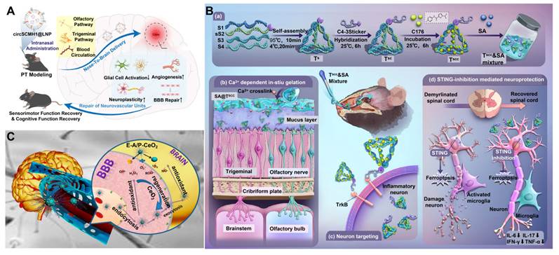
The growth of various delivery avenues in N2B, ranging from biological stem cell exosomes to synthetic nanoparticles, liposomes, and advanced hydrogels, has led to the growth of its therapeutic applications to include a variety of CNS disorders. Nevertheless, much of the current research is still largely confined to rodents. Some common challenges in clinically transporting N2B delivery technology involve the differences in the nasal morphology of different species (e.g., the olfactory epithelium is present in only about 3% of the overall surface area of the nasal cavity, which is comparatively lower than that observed in mice) and biosafety issues concerning N2B delivery methods. In future, more studies are needed to enhance the ability of delivering substances to the olfactory epithelium and the long-lasting ability of delivery systems, design a standardized procedure for safety assessment and encourage testing in higher-order animal models for effective use of N2B drug delivery technology.
4.2.4. Angiopep-2 modification
Angiopep-2 is a classic brain-targeting peptide targeting the LRP-1 receptor. It specifically binds to low-density lipoprotein receptors on the BBB, and endows various nanocarriers with efficient BBB-crossing capacity through surface modification. Multi-omics temporal analysis has revealed that Angiopep-2 enhances BBB penetration efficiency by mediating protein endocytosis-dependent transport [159].
Using baicalein-celastrol small-molecule complex (CB) as the core component, researchers performed surface modification via co-incubation of DSPE-PEG2k-Angiopep-2 and DSPE-mPEG2k, followed by self-assembly into carrier-free CB nanoparticles. After intravenous injection, Angiopep-2 modification mediates efficient brain accumulation of the nanoparticles. The nanoparticles simultaneously scavenge ROS and inhibit the HMGB1/NF-κB inflammatory pathway, ultimately reducing the MCAO model cerebral infarct area by 86.48%. The repair effect on neuromotor function is significantly superior to that of non-targeted preparations, further confirming the targeting property of Angiopep-2 [81]. In oral nanosystems, Angiopep-2 modification also exerts brain-targeting function. The drug concentration in brain tissue of Angiopep-2-modified metal-organic frameworks (MOFs) is significantly higher than that of non-targeted MOFs [160]. Building on the oral targeting strategy combining MOF with Angiopep-2, researchers further expanded the application scenarios of the peptide by incorporating spirulina. The outer spirulina layer prolongs intestinal retention and regulates gut-brain axis microbiota, while the inner Angiopep-2-modified MOF core achieves precise brain targeting. The two components form a two-stage delivery system of "intestinal absorption - BBB penetration", realizing therapeutic efficacy against combined brain injury induced by chemoradiotherapy [161].
Despite the progress outlined above, existing studies have only investigated the modification effect of Angiopep-2 at a single density, without systematic optimization of the peptide conjugation ratio. High modification density may induce endothelial cytotoxicity, whereas low density leads to insufficient targeting efficiency. The balance among modification dosage, safety and delivery efficiency still lacks systematic evaluation, which serves as a key entry point for subsequent nanocarrier optimization.
4.3. Antioxidant/anti-inflammatory nanozymes
4.3.1. CeO₂ and vanadium carbide nanozymes
Nanozymes, designed to mimic natural enzyme activity, are widely accepted in HIBI theranostics [162]. They act like conventional antioxidant enzymes and can eliminate excess ROS as well as be used as contrast agents. By using advanced delivery systems and responsive materials, nanozymes are becoming key components of new types of theranostic approaches [163].
Research has already shown that nanozymes, especially CeO2, can potentially assist in IS treatment. Initial research focused on improving their antioxidant effects. For example, monodispersed CeO2 cores modified with Angiopep-2 and PEG (E-A/P-CeO2) have been created to facilitate BBB penetration and further loaded with edaravone to increase the antioxidant action [164] (Figure 9C). While successful in broad BBB penetration and ROS reduction, these first-generation platforms lacked subcellular precision and robust anti-inflammatory modulation, thereby failing to fully address the complex inflammatory microenvironment inherent to IS. The later designs tried to act on the inflammation cascade on its origin. Encapsulating peptidylarginine deiminase 4 inhibitors in liposomal nanocarriers allowed researchers to utilize the synergistic mechanism where ROS scavenging of the nanozyme was combined with direct inhibition of downstream inflammatory pathways, creating the therapeutic closed-loop between antioxidant protection and inhibition of inflammation [165].
Despite the success of the functional aspects, clinical translation of naked CeO2 nanomaterials tends to be limited due to poor pharmacokinetic behavior such as short circulation half-life, systemic aggregation, and limited access to the catalytic active site [166]. In order to enhance these physiochemical properties, core-shell designs were developed. The in situ encapsulation of CeO2 with ZIF-8 modifies the particle size and surface charge, increasing the circulation time in the body and creating a protective microenvironment that stabilizes the reversible Ce3+/Ce4+ valence cycle and enhances intrinsic catalytic anti-inflammatory and antioxidant activities of the nanozyme [38]. The targeting hierarchy of CeO2 nanozymes has been further improved with a focus on targeting at the subcellular level. Researchers modified the CeO2 core through DSPE-PEG-TPP, allowing the nanozyme to stop ROS production and correct metabolic anomalies at the targeted site within the vulnerable organelles of HIBI [167] (Figure 10A). Meanwhile, a ceria nanosystem (MMTCe) integrating magnetic field-driven navigation, mitochondrial targeting and microglial cell membrane coating has been constructed. Researchers functionalize ultrasmall ceria nanoparticles by modifying their surface with TPP. Leveraging the negative mitochondrial membrane potential, the nanoparticles achieve precise organelle targeting and scavenge mitochondrial ROS directly at their generation source. Building on this functionalized core, a BV-2 microglial cell membrane is further coated onto the nanoparticle surface, followed by the conjugation of superparamagnetic iron oxide nanoparticles on the membrane. This hierarchical structure provides the nanosystem with the ability of immune evasion and extended circulation time. Meanwhile, taking the synergy provided by the intrinsic inflammation-homing ability of microglial membranes and magnetic fields directional guidance, the nanosystem effectively crosses the BBB, allowing precise delivery and accumulation at the ischemic brain injury sites [83]. As a result, CeO2 nanozymes have developed from being basic ROS scavengers to advanced theranostic tools that unite selective targeting, stimuli-triggered release, multi-mechanism action, and real-time imaging.
Nanotherapeutic strategies targeting vascular repair and neuroprotection in HIBI. (A) Construction of the TPP@(CeO2+ROF) nanoplatform and its neuroprotective effects in IS. The platform protects neurons and mitigates inflammation by targeting mitochondria to scavenge ROS, repairing the damaged BBB, and regulating microglial polarization. Adapted with permission from [167], Copyright 2024 American Chemical Society. (B) Schematic of a biochemical sequential-responsive system (DNase-RM@(PDA/IDB)) actively regulating the ischemic microenvironment. Following DNase I-mediated chemotaxis, intravascular NE triggers DNase I release to degrade NETs. The remaining carriers penetrate the BBB for ROS-responsive IDB release, achieving synergistic inside/outside NVU regulation rather than passive drug delivery. Adapted with permission from [88], Copyright 2025 John Wiley and Sons.
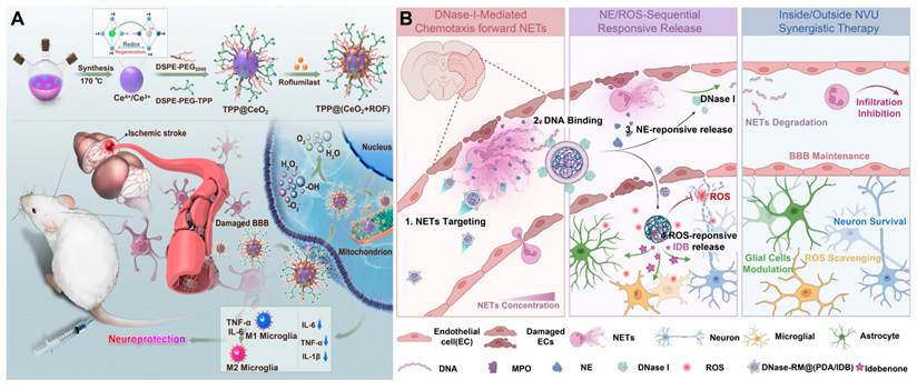
However, there is still a major barrier to overcome. While cerium oxide nanomaterials have good compatibility with living organisms, cerium itself has no biological or physiological significance for humans. CeO2 nanoparticles that enter the brain into a state that is poorly metabolized make them stay in the brain and accumulate there. Regarding their toxicity, the neurotoxicity of CeO2 nanoparticles depends on their size. Because of their smaller size, nanoscale CeO2 nanoparticles have a much larger specific surface area and a higher proportion of surface Ce3+, leading to augmented toxicity concerns. In addition to this, small-sized nanoscale CeO2 nanoparticles have a greater ability to penetrate the BBB. Animal studies also proved that nanosized CeO2 can cross the barrier and reach the brain parenchyma [168]. In addition, in vitro studies have shown that CeO2 nanoparticles can impact glial cells’ morphology, cell division, and viability, inhibit neural stem cells’ differentiation, and cause adverse effects in nervous system cells [169,170]. The concern of chronic toxicity and accumulation of CeO2 nanoparticles is a key obstacle for its clinical use [171]. This fact has led researchers to shift their focus to the nanomaterials that consist of the essential trace elements for humans like vanadium [172]. Importantly, two-dimensional V2C MXene-based nanozymes have been developed. These thin nanozymes exhibit significant enzyme-like activities (SOD, CAT, GPx) and great acute biocompatibility, thus preventing ROS-induced brain stroke [173]. In order to eliminate the long-term brain retention risk for these nanozymes, ROS-specific responsive V2C MXene nanozyme with controllable catalytic activities and degradation has been developed. Such design is expected to can suppress ROS-mediated inflammation and neurodegenerative pathological processes in the future, thus solving the problem of long-term brain bioaccumulation and chronic neurotoxicity [174].
Yet, viewed critically, the long-term in vivo biodegradation capabilities of MXene materials remain unexplored. It is vital to investigate nano-bio interactions of MXenes in brain tissues before these therapeutic systems move to clinical stages and analyze release kinetics of vanadium ions and their neurotoxic thresholds.
4.3.2. Other advanced antioxidant platforms
The accumulation of ROS is a major factor in the development of HIBI. In this way, regulating the local redox microenvironment is crucial for effective neuroprotection [175]. Therefore, modern approaches now emphasize the need not only for ROS scavenging but also for mimicking or upregulating endogenous antioxidant defense networks.
Researchers are exploring the multi-enzyme mimicking ability of Mn3O4 and improving the stability of the Mn3O4 NPs by using their functionalization with HSA. This allows researchers to overcome the problem of the inherent colloidal instability and short systemic circulation of the bare metal oxide. Most importantly, the HSA-functionalized nanoplatform retains SOD, CAT, and GPx-like activity and manages to perform efficient multi-enzyme synergistic removal of ROS [176].
Highly branched, ROS-responsive dendritic nanocarriers have been developed to further enhance their antioxidant capabilities. With the incorporation of the apoE-mimetic peptide COG1410, a targeting agent for the BBB, and loading with the natural antioxidant salvianolic acid A, these dendrimers can carry out three functions: effective transport across the BBB, releasing drug only where ROS is triggered, and restoring mitochondrial function on the ischemic site [177]. In addition, oxidative stress is directly associated with secondary neuroinflammation [178], and thus both processes require concurrent intervention. To accomplish this, innovative supramolecular structures that employ β-cyclodextrin have been created. By covalently attaching the multi-functional radical scavenger Tempol with an H2O2-dependent phenylboronic acid pinacol ester, scientists created a dual-purpose polymeric system. This design does not only reduce harmful ROS, but also reduces the inflammatory responses caused by ROS [179]. Therefore, these interesting forms of biomaterials increase the level of treatments from passive ROS scavenging to active and more efficient control of oxidative stress and neuroinflammation management caused by oxidative stress.
4.4. Neuroprotective and regenerative materials
4.4.1. Materials combined with extracellular vesicles from neural cells
Neural extracellular vesicles (EVs) are nanoscale, lipid-bilayer structures secreted by various cell types, including neural stem cells and MSCs [180]. Due to their inherent biocompatibility and critical role in intercellular communication, EVs hold immense promise for delivering regenerative signals and remodeling the pathological microenvironment in HIBI. However, the rapid in vivo clearance of free EVs necessitates protective delivery vehicles.
To achieve sustained and responsive EV delivery, researchers have developed dynamic hydrogel networks. For instance, by modifying HA with phenylboronic acid and cross-linking it with PEG, a dual-responsive hydrogel was engineered to encapsulate neural stem cell-derived EVs. This smart matrix degrades specifically in response to locally elevated glucose and ROS levels, seamlessly coupling EV release with the severity of the pathological microenvironment to improve cerebral perfusion and neurological function [181]. Likewise, 3D biological scaffolds responsive to ROS have been created to provide EVs from hypoxia-pretreated human umbilical MSCs. EV scaffolds can effectively alleviate neuroinflammation, which is a crucial pathophysiological contributing factor in both cerebral hemorrhage and HIBI, making their utilization significantly promising within neurovascular repair [182].
4.4.2. Biodegradable polymers promoting nerve repair
Biodegradable polymers are various functional substances that exhibit outstanding biocompatibility along with controllable degradation kinetics [183]. Through precise chemical modification and structural design, these polymers can be engineered to actively respond to specific pathological cues within the HIBI microenvironment.
Recent developments have targeted electrically conductive and structure-sustaining forms. Starting with metabolically degradable and π-conjugated conductive polymers with intrinsic ROS-responsiveness. These materials were originally created to induce overall nerve regeneration by removing localized oxidative damage but their responsive nature corresponds with important pathological processes occurring in HIBI [184].
Rebuilding the vascular system is essential to achieve functional recovery along with neural repair. To solve the problem, biodegradable matrices effective in use and non-swelling have been developed using clinical silk fibroin and HA in a combined method of covalent and non-covalent cross-linking. Such devices can make a contribution to restoration of local blood supply within the ischemic area for supplying oxygen and nutrients to the tissue. The usage of such techniques proceeds to the breakthrough outcomes in the IS models, allowing overcoming potential limitations of traditionally used HIBI repair technique [185].
4.5. Light- and sound-responsive materials
Operating within the NIR-II biological window, light-responsive materials offer exceptional deep-tissue penetration, minimized optical scattering, and exceedingly high spatiotemporal resolution. When coupled with the synergistic mechanisms of photothermal therapy and photodynamic therapy, these advanced optical platforms have emerged as core tools for the integrated theranostics of complex oncological and neurovascular pathologies.
NIR-II fluorescence imaging coupled with mild photothermal therapy has been used in previous applications of these dual-function systems for real-time imaging of vascular structure in the body and brain. In this context, IS lesions and brain hemodynamic features can be observed accurately and clearly for the first time. Thus, a basis for the new model of very accurate spatial imaging together with safe localized therapy for the sensitive brain activities was formed [186]. To customize these optical devices for oxygen-short microenvironments scientists developed advanced systems using small-molecule near-infrared photosensitizers (IT-IC). The products act much better under targeted NIR-II radiation providing proper improvement of local blood perfusion, proper ROS scavenging, and normalizing tissue edema. Importantly, this targeted photomodulation is performed properly making it harmless for surrounding healthy brain areas. Such design overcomes typical limitations of classical Type II phototherapy used for very hypoxic conditions [187].
HIBI is key pathogenic mechanisms-hypoxic microenvironments and excess ROS buildup-overlap with the pathologies that advanced optical systems target, leading to an enormous translational potential. Researchers can synthesize the two areas to create a full spectrum of theranostic protocols for HIBI. An ultimate goal of this line of treatment is NIR-II guided ischemic zones mapping combined with ROS neutralization and controlled neurovascular repair.
It is worth noting that the vast majority of the above-mentioned therapeutic materials are validated in adult acute focal ischemic stroke models (such as MCAO/R). However, HIBI also includes neonatal HIE and hypoxic brain injury at high altitudes, which exhibit fundamental physiological differences from adult focal stroke. For neonatal HIE in the developing immature brain, the BBB possesses unique structural-functional features, such as incompletely assembled tight-junction complexes and immature myeloid immune-cell phenotypes. Brain injury in neonatal HIE is mostly diffuse global hypoxic-ischemic damage, distinct from the well-demarcated focal lesions induced by arterial occlusion in adults. These physiological differences impose constraints on nanomaterials. BBB targeting peptides that work efficiently in adult models may significantly alter transcytosis efficiency within the neonatal brain. In addition, excessive inflammatory inhibition or potent ROS scavenging can interfere with normal developmental programs of the brain. Meanwhile, immature neural tissue imposes stricter requirements for material biocompatibility, degradation kinetics, and long-term neurotoxicity.
Chronic hypobaric hypoxia induced by the high-altitude environment does not stem from abrupt arterial occlusion. Its pathological manifestations mainly consist of diffuse cerebral edema, whole-brain persistent oxidative stress, and chronic inflammatory activation, without clearly demarcated focal lesions. Unlike acute reperfusion injury in MCAO models, sustained hypoxic stimulation alters the expression of hypoxia-responsive elements in endothelial and glial cells. Conventional nanocarriers struggle to achieve sufficient lesion enrichment under diffuse chronic hypoxic conditions. In summary, nanoplatforms optimized for adult focal stroke cannot be directly extrapolated to neonatal HIE or hypoxic brain injury at high altitudes. Subtype-specific physiological constraints reshape BBB permeability, lesion distribution, immune response, and material-tissue interactions.
5. Intelligent responsive systems: from passive delivery to active regulation
The HIBI microenvironment is complex and involves multiple pathological stimuli. The intelligent responsive nanosystem has achieved "pathological trigger activation" and abstracted the multi stimulus response design principle from the HIBI pathological cascade.
5.1. ROS-responsive nanocarriers
Nanocarriers sensitive to ROS belong to a very promising category of advanced drug delivery systems. They utilize the high oxidative stress level present in the ischemic microenvironment for achieving a very effective synchronicity between pathological signal initiation, delivery of therapeutic agents, and therapy modulation [22].
The early designs were focused on utilizing the own inherent catalytic properties for fighting oxidative bursts. For example, some biomimetic nanomotor platforms like Pt@LF were developed that were able to pass through the ischemic lesions and actively scavenge ROS thus significantly reducing the consequences of oxidative damage [139]. After that, more complex multi-component systems were developed that use ROS scavenging in combination with targeted drug release. A vivid example is a polydopamine-based nanocarrier, which is loaded with idebenone and functionalized with deoxyribonuclease I (DNase I). The polydopamine core serves two functions, being both a natural ROS scavenger and a responsive matrix for the controlled release of idebenone according to ROS concentration. At the same time, surface-anchored DNase I is responsible for the degradation of neutrophil extracellular traps (NETs) and destruction of the microthrombus framework. With the help of this system, it is possible to achieve a powerful repair of the NVU and brain tissue by saving neurons from apoptosis, keeping mitochondria intact, and getting rid of NETs [88] (Figure 10B).
To disrupt the self-amplifying "ROS-neuroinflammation" vicious cycle typical of severe HIBI, the ROS-specific approaches have been further improved. A new type of advanced nanocarriers based on a poly(propylene sulfide) (PPS)-responsive scaffold containing a brain-targeting peptide, cysteine-alanine-glutamine-lysine, has been invented for the synergistic application of Cur. When the PPS matrix meets highly oxidative conditions at the injury site, the PPS matrix experiences a phase transition, which allows curcumin to be released in a burst mode while simultaneously using inactivation of ROS. Implementation of this approach successfully interrupts the inflammation process that takes place in the body, which proves the great advancement in the complex treatment of HIBI [188].
Besides, ROS management is combined with state-of-the-art biomimicking targeting techniques. An important dual-drug platform (PP@PCL) comprising the antioxidant danazol and the healing drug metformin is developed using liposomes covered with a mixture of membranes of 4T1 tumors and blood platelets. This hybrid covering makes the vehicle both capable of penetrating BBB and able to target affected sites specifically. After localization, the platform implements several interventions that involve the scavenging of ROS as well as the polarization of microglia to the neuroprotective M2 phenotype simultaneously [189] (Figure 11A). Although the release is not entirely dependent on a chemically reactive bond, the biomimetic design considerably increases the number of advanced approaches that use ROS in HIBI theranostics.
Intelligent responsive systems for targeted nanotherapy: from passive delivery to active regulation. (A) Therapeutic mechanism of PP@PCL nanomedicine in rats subjected to MCAO/R. This agent targets damaged vasculature, promotes angiogenesis, penetrates the BBB, scavenges ROS and inhibits mitochondrial damage, while reprogramming microglial phenotypes to achieve neuroprotection. Adapted with permission from [189], Copyright 2024 John Wiley and Sons. (B) Scanning electron micrographs of microscale shear-activated nanotherapeutics (SA-NTs) and their constituent nanoparticles. (C) Quantitative validation demonstrating that SA-NTs act as an intelligent switch, dispersing into individual nanoparticles exclusively under pathological shear stress (≥100 dyne/cm2). (D) Fluid dynamics simulations mapping the elevated pathological shear stress in stenotic vessels that triggers this physical responsive release. Adapted with permission from [194], Copyright 2012 The American Association for the Advancement of Science. (E) Chemical structures of amphiphilic polypeptides used for vesicle construction. (F) Schematic of peptide vesicles designed for intelligent, shear stress-responsive drug release specifically at vascular stenosis sites. Adapted with permission from [195], Copyright 2025 John Wiley and Sons.
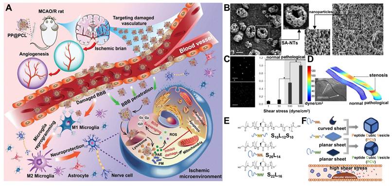
Currently, most nanocarriers still use M1 to M2 phenotype conversion as the core therapeutic target. However, single-cell transcriptome studies have shown that this binary division does not reflect the true functional diversity of macrophages in the complex microenvironment of lesions in vivo. Therefore, material design should break away from the single idea of "globally inducing a large number of M2 polarizations" and instead focus on targeting specialized subpopulations of myeloid cells, with typical representatives being lipid clearing repair related macrophages carrying GPNMB/CD63 molecular features [52]. On the one hand, nanocarriers can co deliver lipid metabolism regulators and antioxidant drugs, promoting the activation of repair related transcription programs while retaining the inherent phagocytic function of intermediate state myeloid cells, rather than forcing all myeloid cells to undergo M2 phenotype transition uniformly. On the other hand, a multi stimulus responsive drug release system can be constructed to simultaneously sense lipid fragments, redox levels, and hypoxic microenvironment signals in the lesion site, achieving spatiotemporal selective drug delivery to intermediate state macrophages/microglia subpopulations in situ, avoiding non-specific immune regulation of all myeloid cells.
5.2. Shear stress-responsive materials: exploiting hemodynamics for enhanced targeting
HIBI is fundamentally premised on neurovascular injury as well as significant hemodynamic anomalies [190]. As a result, various shear stress responsive nanomaterials have come along as a fundamental technology for targeted delivery, allowing to interpret abnormal hemodynamic signals in pathological blood vessels and convert the mechanical information into a system of control over drug release [191].
Subsequent to the HIBI event, the formation of microthrombi in the brain results in significant modifications in the parameters of shear stress in the blood vessels. Such variations affect margination of circulating nanoparticles which depends on the shape of NPs and consequently has influence over the formation and lysis of thrombus [192]. Initial studies comparing spherical, rod-like and disc-like PLGA nanoparticles using tissue-type plasminogen activator (tPA) have mentioned this effect of shapes on passive targeting. Discoidal NPs showed greater margination and attachment to thrombotic surfaces under excessive shear stress, leading to improved localized thrombolytic effectiveness [85]. Later, it was confirmed that margination applies to a variety of vascular diseases, showing that anisotropic structures improve localization in disturbed blood flow [193].
Researchers have created platforms in which shear stress acts as a direct trigger for active cargo release. For example, shear-activated nanotherapeutics (SA-NTs) have been developed as micro aggregates that can remain intact under physiological flow conditions. However, when they encounter pathologically raised shear stress brought about by vascular stenosis or microthrombi, the aggregates quickly break apart into nano-sized therapeutic fragments. Such mechanosensitive breakdown allows for the accurate and concentrated delivery of tPA at the blockage site [194] (Figure 11B-D).
Besides, this mechanosensitive concept has been developed even better molecularly. Researchers could build cubical vesicles with different angular designs by joining membranes with different curvatures. These complex structures stay stable at normal shear force but break quickly under pathological shear stress [195] (Figure 11E-F). By taking advantage of HIBI’s basic changes in hemodynamics, these mechanosensitive and shear-responsive systems create a very novel way to cross the systemic barriers of delivering drugs and conducting target-area-only treatments.
5.3. Ligand-receptor recognition-triggered in situ topological switching system
A general chemical strategy for ligand-triggered in situ topological switching is established, which converts transient, reversible ligand-receptor recognition into durable nanofibrous interfacial anchoring via a "touch-stay-act" mechanism, effectively overcoming the problem of drug washout caused by hemodynamic shear stress.
Researchers employ the amyloid KLVFF peptide motif as the structural basis for topological switching, which drives phase transition to form nanofibers through β-sheet stacking and π-π interactions. Using PEGylated lipid-NO donor (DSPE-PEG-L-Arg) as the kinetic stabilizer, the RGD peptide as the targeting anchoring moiety, and the SIPPK thrombolytic peptide as the functional component, a supramolecular assembly termed TRNO is rationally constructed. In normal blood circulation, TRNO exists as uniform spherical nanoparticles. The PEG corona prevents non-specific interactions and premature aggregation, endowing the system with excellent colloidal stability. Upon reaching the thrombotic lesion, the surface-exposed RGD specifically binds to integrin αIIbβ3. Multivalent ligand-receptor interactions alter the local free energy landscape and disrupt the kinetic stabilization conferred by PEG. Meanwhile, the KLVFF motifs become exposed and undergo intermolecular β-sheet stacking. The discrete nanospheres progressively assemble into high-aspect-ratio nanofibers, eventually forming an entangled dense nanofibrous network and triggering the in situ topological phase transition.
The nanofibrous network can adhere firmly to the thrombus site, resisting hemodynamic shear stress while activating localized NO release. In vivo evaluation in rats demonstrates no obvious toxic side effects of TRNO within a 14-day observation period [196].
This work breaks through the long-standing bottleneck of poor interfacial retention of supramolecular materials under non-equilibrium physiological conditions, and provides a novel paradigm for the design of adaptive biointerfacial materials. This strategy can be further extended to the treatment of HIBI.
5.4. Multimodal theranostics: imaging-guided therapy
Early strategies primarily focused on exploiting the inherent physicochemical properties of therapeutics for self-tracking. For instance, cytidine diphosphate choline (CDPC) liposomes were employed not only as a neuroprotective treatment for stroke but also as an intrinsic imaging tracer. By leveraging their natural chemical exchange saturation transfer (CEST) MRI signals, researchers successfully monitored real-time drug accumulation within areas of BBB disruption, establishing a foundational "treatment-as-diagnosis" model [197] (Figure 12A-D). However, this rudimentary design was largely restricted to passive pharmacokinetic tracking and lacked an active, functional synergy between the imaging modality and the therapeutic mechanism.
Multimodal theranostics based on nanocarriers in HIBI. (A) Schematic illustration of the fabrication of liposomes for CEST MRI-guided and vascular cell adhesion molecule 1 (VCAM-1)-targeted delivery of CDPC. (B) CEST signal of CDPC-loaded liposomes (~40 nm per liposome and 21.5 per CDPC), with 15 mm free CDPC solution (pH 7.4) as the reference. (C) Dynamic CEST signal changes at 1 ppm and 2 ppm during dialysis, respectively, associated with the release of CDPC from liposomes. (D) Drug release kinetics at different time points investigated by CEST (2 ppm) and ultraviolet absorption at 286 nm, respectively. Adapted with permission from [197], Copyright 2016 Ivyspring International Publisher. (E) Schematic diagram of Mn/quantum dot single-atom catalyst for imaging-guided traumatic brain injury therapy. The NIR-IIb emitting Mn single-atom catalyst enables the dynamic monitoring of the BBB status in a noninvasive way and repressing the ROS-mediated BBB breakdown. Adapted with permission from [198], Copyright 2023 Springer Nature.
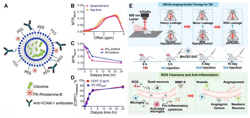
To solve this problem and attain real diagnostic-therapeutic synergy, very advanced optical systems have been created. One considerable progress has been the generation of silver (I) telluride (Ag2Te)-based quantum dots treated with single Mn catalysts. These quantum dots operate in the NIR-IIb range of wavelengths, allowing visualizing dynamically and accurately the BBB disruption. At the same time, the Mn atoms that stay on the surface are functioning as active catalysts for degradation of localized ROS. Thanks to this sophisticated architecture, the process of high-resolution pathological imaging and therapeutic intervention flow smoothly together with a real-time optical monitoring of the process of barrier restoration [198] (Figure 12E).
Apart from enhancing the diagnostic abilities in relation to optical windows, scientists have fabricated ultra-small iron gallate coordination polymer NPs (Fe-GA CPNs) aimed at dual-modality of PET/MRI. In material chemistry terms, these coordination polymers provide unparalleled anatomical and metabolic resolution. They validate recovery from cerebral metabolism in vivo via very sensitive multimodal mapping and show exceptional ROS-scavenging capabilities in alleviating ischemic injury at the cellular level [199].
Overall, the evolutionary progress of these advanced platforms, which involves moving from basic medication self-imaging to intelligent carrier-functionalized imaging, from single-target to multi-pathway approaches, and from monomodal monitoring to multimodal metabolic validation, proves the fast development of universal theranostic systems. These new multimodal technologies become an invaluable guide for the further development of precision management of HIBI.
6. Representative biomimetic implementation forms for HIBI theranostics
HIBI is an important type of CNS injury. Traditional treatment methods have many drawbacks, such as poor spatial targeting, difficulty in penetrating the blood-brain barrier, low biocompatibility, and causing serious adverse reactions.
To overcome these severe treatment challenges, biomimetic materials have emerged as a revolutionary treatment strategy. These innovative materials have excellent biocompatibility, strong immune tolerance to host systems, and autonomous targeting ability by accurately simulating the complex characteristics of natural cells and tissues; Most importantly, they are able to quickly adapt to the constantly changing microenvironment during the course of HIBI. Biomimetic nanotechnology can not only effectively improve the hemodynamic disorders caused by hypoxia, but also accurately target key parts of the injury pathway. Nanomaterials coated with cell membranes and carriers based on extracellular vesicles are important biomimetic implementations that achieve pathology driven material design principles, immune evasion, injury homing, and blood-brain barrier crossing performance. This article will explore in detail four types of biocompatibility technologies and their therapeutic effects and clinical application value.
6.1. Biomimetic RBCM nanomaterials
Constituting approximately 50% of total blood volume, RBCs exhibit a unique biconcave morphology and an exceptionally high specific surface area. The absence of a nucleus and most organelles in mature RBCs provides an expansive intracellular void, making them ideal candidates for drug loading and surface engineering [200]. As the primary vectors for systemic oxygen transport, RBCMs are inherently endowed with natural biocompatibility, prolonged circulation, and robust oxygen-binding capacities. Importantly, the RBCM lipid bilayer has a high level of immunomodulating proteins like CD47 [201]. The protein CD47 transmits a "don't eat me" signal when the RBCM interacts with signal regulatory protein α (SIRPα) on macrophages, isolating cells from taking up the NP. Therefore, RBCM-based camouflage has become an excellent biomimetic strategy for enabling NP circulation without prior clearance (Table 2).
Cell membrane-based natural membrane drug delivery systems for HIBI.
| Biomimetic membranes | Material names | Coating methods | Functions | Translational bottlenecks | Ref. |
|---|---|---|---|---|---|
| RBCM | RGD-RBCM@PPUNPs | Co-extrusion | Targeting, ultrasound response and immune escape | Orientation loss during extraction, impaired immune evasion, pathogen transmission risks and batch-to-batch heterogeneity | [219] |
| MOF-Hb/M-NPs | Sonication and extrusion | Efficient hemoglobin loading, oxygen binding and release with no blood toxcity and tissue residency | [220] | ||
| MOF-siRNA@Ang-RBCm | Sonication and co-extrusion | Efficient brain targeting, lysosome escape, BBB recovery and inflammation reduction | [202] | ||
| MNET | Extrusion | Neuroprotection and reduction of reperfusion injury before and after thrombolysis | [82] | ||
| SHp-RBC-NP | Co-sonication | Targeting ischemic area to reduce nerve injury and infarct volume with long circulation character | [203] | ||
| T-RBC-DTC NPs | Extrusion | Thrombus-targeted enrichment, ROS scavenging and thrombosis inhibition | [221] | ||
| NEMR | Extrusion | Microglial M2 polarization induction | [222] | ||
| PM | Cur@PLTM and Tid@PLTM | Sonication | Synergistically ROS scavenging and microglial M2 polarization induction | Pro-coagulant risks, limited harvesting yield, high mechanosensitivity and GMP manufacturing barriers | [207] |
| PP@PCL | Co-extrusion | Synergistically ROS scavenging, microglial M2 polarization induction and angiogenesis | [189] | ||
| PFNBs | Repeated compression-expansion | Microglial M2 polarization induction and protecting vascular endothelial integrity | [210] | ||
| PONBs | Repeated compression-expansion | Efficient oxygen loading and hypoxia-responsive oxygen release | [87] | ||
| tP-NP-rtPA/ZL006e | Sonication | Sequential targeted delivery and thrombin-responsive drug release to reduce ischemic area | [209] | ||
| RGD-PLT@PLGA-FE | Sonication | Cerebral blood flow and neurobehavioral recovery | [208] | ||
| PNP-PA | Sonication | Prolonged in vivo circulation time of rt-PA and retention time at thrombus site | [223] | ||
| miRNA/TPHL@PM | Sonication and extrusion | Reduced neuronal apoptosis and microglial M2 polarization induction | [48] | ||
| APLT-PA | Extrusion | Rapid release of tPA and potent thrombolysis | [224] | ||
| rPZDCu | Sonication | ROS response, potent thrombolysis and microglial M2 polarization induction | [225] | ||
| tPA/MNP@PM | Sonication | Potent thrombolysis, melanin particles release and nerves protection under near-infrared trigger | [226] | ||
| PAMNs | Extrusion | In situ NO production, vasodilation and anti-platelet effect | [227] | ||
| MM | M2M@BANPs | Extrusion | Synergistic anti-inflammatory and anti-apoptotic therapy with improved neurological function | Complex protein profiles, off-target immune activation and allogeneic immunogenicity | [211] |
| SiO2@MM@CORM-401 | Extrusion | Microglial M2 polarization induction and ferroptosis inhibition to reduce reperfusion injury | [228] | ||
| PPCs | Extrusion | Targeting ischemic area to reduce neuronal apoptosis and improve cognitive and memory functions | [215] | ||
| MM-Lip-Rg3/PNS | Co-extrusion | Non-invasive nasal administration to inhibit neuronal apoptosis and anti-inflammation | [216] | ||
| Ma@(MnO2+FTY) | Co-extrusion | ROS scavenging, O2 production and microglial M2 polarization induction | [213] | ||
| MM/ANPs | Co-sonication | Inhibition of pyroptosis and infarct volume to improve neurological function | [217] | ||
| MSAOR@Cur | Sonication and extrusion | ROS-responsive drug release and BBB penetration | [214] | ||
| MM-BA-LP | Extrusion | Enhanced brain targeting and prolonged circulation with improved neurological function | [229] |
To avoid the harmful cascade of immune activity, triggering MMP-9 production, and destruction of tight junctions, researchers developed a specially engineered combination of angiopep-2 (Ang), RBCM, and mesoporous organosilica nanoparticles carrying MMP-9 siRNA (MOF-siRNA@Ang-RBCm) [202]. This nanocarrier combines the natural immune evasion ability provided by RBCMs and the bioavailable substance targeting the BBB. The CD47 protein present allows the particle to evade destruction from the immune system in general, while Ang enables specific interaction with lipoprotein receptor-related protein 1 (LRP1), frequently present in blood-brain barrier endothelial cells, astrocytes, and microglia. This alteration made BBB penetration 1.5 times superior when compared to RBCMs without modifications. The experiments carried out in vivo on lipopolysaccharide (LPS)-induced neuroinflammation demonstrated a decrease in the amount of MMP-9 in the brain by 2.5 times. Besides, it restored the junctional proteins and reduced sodium fluorescein leakage.
A T7 peptide-modified, RBCM-coated Mn3O4 NP system (MNET) was created to tackle the problem of two-phase oxygen imbalance in IS, which comprises acute ischemic hypoxia and the hyperoxia that results from reperfusion [82] (Figure 13A-B). The RBCM coating layer is a crucial biomimetic shell for this nano system, retaining the CD47 "don't eat me" signal on the membrane surface, endowing the nano material with immune escape ability, prolonging systemic circulation, and providing a biocompatible storage space for hemoglobin and improving material stability. On the basis of RBCM biomimetic shell, the addition of T7 peptide and Mn3O4 enhances the ability of the nano system to cross BBB and scavenge reactive oxygen species.
Biomimetic erythrocyte membrane-based nanotherapeutic strategies for controlled oxygen release and drug delivery in HIBI. (A) Synthetic scheme of the T7 peptide-modified MNET. (B) Schematic diagram of MNET for acute IS therapy. The nanosponge achieves profound neuroprotection through continuous free radical scavenging and self-adapted oxygen regulation-supplying oxygen during pre-thrombolysis ischemia and storing excess oxygen during post-thrombolysis reperfusion. Adapted with permission from [82], Copyright 2020 American Chemical Society. (C) Schematic design of SHp-modified, RBCM-camouflaged nanoparticles (SHp-RBC-NP/NR2B9C). These biomimetic nanocarriers target ischemic brain tissues and undergo ROS-responsive intracellular release of the neuroprotectant NR2B9C to prevent excitotoxic damage. Adapted with permission from [203], Copyright 2018 American Chemical Society.
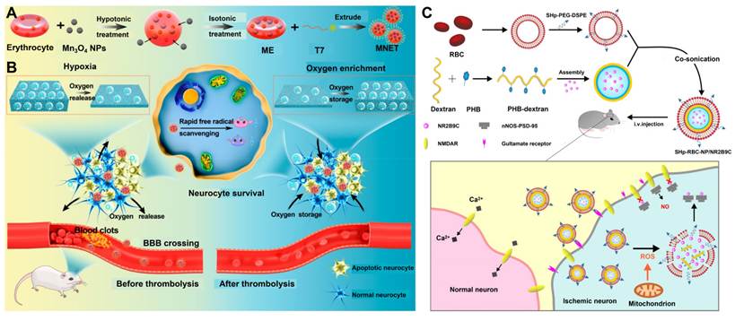
Researchers have created a new neuroprotectant delivery system designed to deal with the poor delivery efficiency and toxicity of standard neuroprotectants. They did this by creating ROS-responsive, stroke-targeting peptide (SHp)-modified RBCM NPs (SHp-RBC-NP), enabling them to deliver the neuroprotective agent NR2B subunit C-terminal 9-amino acid peptide (NR2B9c) [203] (Figure 13C). The RBCM is the core biomimetic skeleton of the nanoplatform, and the CD47 protein on the membrane surface endows the carrier with immune escape ability, significantly increasing the in vivo circulating half-life of the loaded NR2B9c. At the same time, the red blood cell membrane provides a modifiable substrate for anchoring stroke targeted SHp peptides, accessing boronic ester bonds that can be cleaved by reactive oxygen species, and encapsulating neuroprotective drug NR2B9c, resulting in a drug concentration in the injured hemisphere of MCAO model rats that is 1.5 times higher than that in the healthy hemisphere on the opposite side.
Collectively, the development of these advanced RBCM-camouflaged platforms illustrates a highly synergistic and multifaceted approach to HIBI intervention. MOF-siRNA@Ang-RBCm directly addresses the structural integrity of the NVU by halting BBB degradation [202]. MNET solves the problem of twofold oxygen and redox imbalance during ischemia-reperfusion [82]. And SHp-RBC-NP carries the precise stimulus-dependent rescue of damaged neurons [203]. The exploitation of the natural biomimetic features of RBCMs with special targeting and responsive release capabilities allows for an accurate and precise treatment of HIBI with these advanced nanotechnology platforms.
Despite these remarkable preclinical successes, the clinical translation of RBCM-camouflaged therapeutics faces significant hurdles. First, the cell membrane extraction process often leads to the loss of the right-side-out membrane configuration. During hypotonic lysis, repeated centrifugation and extrusion, mechanical shear and osmotic pressure changes can easily cause the flipping of the lipid bilayer. Inverted membranes expose intracellular phosphatidylserine and cytoplasmic protein domains, which not only significantly weakens CD47-mediated immune evasion but may also trigger severe immune responses. Currently, laboratory-scale detection methods such as acetylcholinesterase activity assay, protease protection immunoblotting and immunogold TEM have shortcomings including low throughput and poor quantitative accuracy. No pharmacopoeia-grade analytical method is available for GMP batch release. Second, maintaining the structural and functional integrity of fragile membrane components remains technically challenging. Native immunomodulatory proteins on the RBCM surface are prone to ectodomain shedding and conformational denaturation during purification, extrusion and long-term storage, resulting in inconsistent circulation half-life and off-target distribution. For engineered RBCM modified with targeting ligands, it is difficult to precisely control the coupling density on a large scale, and ligand detachment via hydrolytic cleavage during storage will further impair the targeting effect. Notably, residual free hemoglobin from incomplete membrane purification also poses potential safety risks of renal toxicity and oxidative stress in vivo. Third, the immunogenicity and biosafety risks of allogeneic RBCM require rigorous evaluation beyond current preclinical assessments. Numerous minor blood group antigens on RBCM can induce the production of alloantibodies after repeated administration, accelerate the systemic clearance of the nanoplatform, and may trigger subclinical hemolytic reactions. Meanwhile, the risk of pathogen transmission from human donor blood requires strict donor screening and validated viral inactivation steps, which may in turn compromise membrane protein activity.
Overall, addressing these CMC hurdles is critical for translating RBCM-based nanotherapeutics from the laboratory to clinical practice. Therefore, future innovative research and development must prioritize standardized large-scale membrane extrusion processes and coating technologies, or shift to fully synthetic artificial cell membranes to meet the stringent requirements of clinical pharmaceutical process standards.
6.2. PM-coated anti-inflammatory NPs
BBB disruption and endothelial injury leading to immediate platelet adhesion and aggregation can occur as a result of HIBI. The event initiates a chain reaction that forms microthrombi, worsening the collapse of cerebral microcirculation and further releasing pro-inflammatory elements, such as thromboxane A2, resulting in more severe neuroinflammatory injury [204]. Taking advantage of these processes, PMs are characterized by unique biomimetic properties: selective targeting of thrombus and inflamed endothelium, effective immune evasion, and vascular protection. PM-camouflaged NPs are able to steer the natural inflammation directed by the platelets, allowing them to precisely transport their cargo to the area of clot formation [205]. PMs, like RBCs, have CD47, which allows them to avoid immune system detection and prolong their stay in circulation. The unique feature of PMs is that they possess a natural endowment of growth factors such as vascular endothelial growth factor (VEGF) and platelet-derived growth factor (PDGF), which can stimulate growth of new blood vessels.
Acute thrombolysis is hampered by the narrow 4.5-hour time frame for treatment and severe bleeding risks with recombinant tissue plasminogen activator (rt-PA). Researchers developed a PM -coated PLGA cores loading lumbrokinase (PNPs/LBK) [206]. The PM shell provides efficient targeting of thrombus and an RBCM-like circulation half life. This design leads to high accumulation at the clot site, which helps eliminate the systemic distribution that usually causes bleeding problems. It is impressive that PNPs/LBK attained the same thrombolytic effect with only a third of the LBK dose without affecting normal blood clotting or increasing tail bleeding time in mice, thus creating a very safe biomimetic method for acute thrombolytic therapy.
Despite the use of thrombolysis, it remains ineffective in solving the problem of excessive ROS production as a consequence of reperfusion. To solve this issue, dual-NP technology was developed utilizing a dual-NP strategy utilizing PM-encapsulated Cur (Cur@PLTM) and alginate (Tid@PLTM) [207]. Cur@PLTM takes advantage of using P-selectin on PM to target ischemic blood vessels, thus getting rid of ROS. Tid@PLTM works in such a way that it inhibits glycogen synthase kinase 3β leading to the M1-to-M2 microglia transition. Neurovascular repair is essential after these acute interventions. Thus, arginine-glycine-aspartic acid (RGD)-modified PM-coated PLGA nanoparticles with fat extract (FE) (RGD-PLT@PLGA-FE) have been created [208]. This change of treatment approach goes beyond damage control as this peptide recognizes active angiogenesis areas by binding to integrins αvβ3 and the PM targets the injured endothelium. The two components work in unison to deliver neurogenic substances such as brain-derived neurotrophic factor and basic fibroblast growth factor found in fat extract to stimulate angiogenesis and neurogenesis.
To address the drawbacks of monotherapy and one-membrane operating technology, scientists created innovative multimodal systems. The dual-drug nanocarrier (PP@PCL) developed by scientists features a unique mix of 4T1 tumor cell membranes and PMs [189]. This unique coating provides a capacity for crossing the BBB using 4T1 cells and the ability to target ischemia with PMs by delivering the antioxidant danazol together with the healing metformin. This strategy works well to remedy the restricted BBB penetrability of unmodified PMs by producing a synergistic combination of antioxidant, anti-inflammatory, and pro-regenerative effects. To address the clinical need for sequential intervention, a thrombin-responsive platelet biomimetic NP (tP-NP-rtPA/ZL006e) has been developed [209] (Figure 14A-D). This compound delivers a spatiotemporally programmed release: rt-PA is released first at the thrombus site (82.1% release within a period of 2 hours under 1 U/mL of thrombin). As a consequence of enzymatic degradation, Tat peptide comes into the fore to cause BBB translocation of ZL006e (the neuroprotective agent) at three-fold the rate of the uncomplexed drug leading to the resolution of the problem of rapid recanalization against profound neuroprotection. At last, to broaden the alternatives of delivery methods, camouflaged PM nanobubble systems, which are namely fingolimod (FTY720)-loaded (also known as PFNBs) and oxygen-enriched (also known as PONBs) ones, are presented [87, 210]. PFNBs integrate PM targeting shell with FTY720 core to improve brain blood barrier-crossing abilities, on the other hand PONBs work against tissue hypoxia using local oxygen release and employing VEGF/PDGF for endothelial cell rescue. Thus, both technologies present a novel approach of "gas delivery combined with functional modulation" for HIBI treatment.
Schematic illustration of the multifunctional biomimetic nanoplatform for targeted therapy of IS. (A) Main components of tP-NP-rtPA/ZL006e. (B) After intravenous injection, tP-NP-rtPA/ZL006e targets the thrombus and releases rtPA triggered by thrombin. (C) The nanocarrier is transported into the brain via Tat-mediated transcytosis. Adapted with permission from [209], Copyright 2019 American Chemical Society. (D) Schematic of neuroprotective effects of Cur@PLTM and Tid@PLTM in transient MCAO (t-MCAO) rats. Adapted with permission from [207], Copyright 2025 Informa Healthcare.
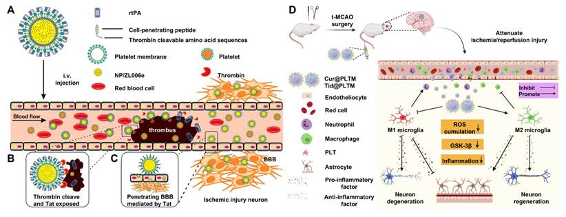
PM-camouflaged nanotherapeutics cannot be translated into clinical applications due to various challenges despite their impressive biomimetic targeting capabilities. The major obstacles within PM maneuvering are membrane consistency and the validation of the functional position of PM in delivery. PMs have a polarized distribution of proteins where adhesion and immunomodulatory proteins are located only on their outside surface for the purpose of thrombus home and immune evasion. But the extreme mechanosensitivity of platelets during membrane extraction and extrusion makes the lipid bilayer very likely to flip. Flipped membranes lose their ability to adhere correctly while exposing intracellular coagulant phospholipids and signaling molecules, thus potentially promoting systemic microthrombosis or worsening local coagulation cascades in vivo. Currently, high-throughput techniques allowing measuring of the level of correctly oriented membranes in the final product are not available. The activity of delicate surface proteins and engineered ligands is a huge challenge in manufacturing. Proteins found in native PM proteins that perform targeting, immune system evasion and vascular regeneration are prone to proteolytic degradation and conformational denaturation during purification and freeze-thawing processes. Even mild activation of platelets during the processing of these proteins can lead to excessive shedding and release of granules from the membrane. The use of synthetic ligands for the purpose of altering the PMs surface can hinder the proper functionality of the membrane and its proteins. Moreover, ligand density can cause quality control issues in production of membranes due to batch-to-batch variations. In addition to that, unlike erythrocytes, the variability in platelet donors relative to activation, reactivity under mechanical pressure and antigen markers is significantly high. Standardized blood collection protocols still lead to significant differences in the starting materials for membrane production due to the subjectivity of platelet physiology. In addition, both platelet isolation and membrane extraction procedures are very sensitive to operational parameters. Even minor differences in centrifugation speed, temperature, buffer composition or extruder pressure can lead to different membrane yields, protein retention and particle distribution. Thus, the extreme sensitivity of the process complicates scale-up for GMP production as the reproducibility of results is poor across batches and laboratories. Fourth, the safety risks of immunogenicity and thrombogenicity require rigorous clinically relevant evaluation. Allogeneic PMs carry HLA class I antigens and human platelet antigen systems, which can induce allogeneic immune responses after repeated administration. These responses not only accelerate the clearance of the nanoplatform from circulation but also raise the risk of adverse reactions such as febrile non-hemolytic reactions and even platelet transfusion refractoriness. In addition, residual pro-coagulant receptors on the membrane surface may interact with circulating platelets and endothelial cells in vivo even after extraction, which raises safety concerns for applications in IS where coagulation status is already dysregulated.
Therefore, future research and development should focus on synthesizing artificial PMs with fully synthetic lipid and peptide compositions, to decouple their targeted homing capacity from thrombogenic risks and production stability issues.
6.3. Macrophage membrane-wrapped "cytokine sponge"
The secondary brain injury in the later stage of HIBI is defined pathologically as inflammatory cytokine storm [211]. Traditional anti-inflammatory drugs are simply insufficient to precisely eliminate these excessive cytokines and often cause systemic immune suppression. The MM coated nano platform has a unique "cytokine sponge" effect, which is not easily achieved by RBCM or PM systems.
Macrophages have many immune receptors and operate as innate immune regulators. The macrophages’ various receptors bind to the inflammatory cytokines like Toll-like receptor 4 and tumor necrosis factor-α receptors. in the HIBI microenvironment. These receptors have created biomimetic nanoplatforms that bind to a variety of inflammatory cytokines since they work like receptors and they are used for the effective removal of inflammatory factors from the brain [212]. In addition, the CD47 proteins help these sponges evade the immune response and stay in the brain longer, and the MM's own chemotactic properties help them toward the ischemic core, making them perform optimally. Interestingly, unlike traditional drugs that require the drug to be taken up inside the cells for signaling, these sponges inhibit cytokines through the external route.
Different macrophage-camouflaged systems are being made to effectively address oxidative stress, microglial activation, and neuronal apoptosis. For example, Li and colleagues came up with manganese dioxide nanoparticles that had been camouflaged by macrophage and loaded with FTY720 [213]. The specific surface area of manganese dioxide allows scavenging of more than 80% of excess ROS and the catalysis of endogenous oxygen generation in an hour, providing effective relief from ischemic hypoxia in their target. At the same time, FTY720 affects the NF-κB pathway, inducing the M1 to M2 transition of microglia to avoid cytokine secretion. Liquid chromatography-mass spectrometry (LC-MS) results showed that this biomimetic coating increased the α-phase half-life (t1/2α) of FTY720. As a result, it accumulated in the t-MCAO/R rats’ lesions much more than the RBCM-coated ones and significantly reduced infarct volume and improved neurological scores.
Utilizing the ability of macrophages to penetrate the BBB, Su et al. have created a new nanoplatform called MSAOR@Cur [214]. This platform consists of a mixture of Angelica polysaccharides, resveratrol, and Cur inside of a nanomaterial shell. The successful penetration of this nanoplatform relies on CD44 proteins in the cell membrane, leading to its good performance in experiments, where the BBB penetration capability was shown to be 2.7 times more than that of unencapsulated Cur.
To further optimize translational viability, administration routes, and multipathway efficacy, researchers have continuously refined these designs. Maximizing localized antioxidant capacity, a MM-coated polydopamine NP loaded with puerarin (PPCs) was developed, with the polydopamine core achieving a 76.64% 2,2-diphenyl-1-picrylhydrazyl (DPPH) radical scavenging rate (at 100 μg/mL) [215]. An alternative strategy for complete BBB evasion involves direct N2B delivery, as demonstrated by the development of MM-camouflaged dual-drug liposomes (denoted as MM-Lip-Rg3/PNS) [216]. Furthermore, Ma et al. engineered RVG29-modified, MM-coated PLGA NPs loaded with Apelin-13 (denoted as MM/ANPs) to specifically target the severe pathological inflammation driven by reperfusion-induced necroptosis [217]. The RVG29 peptide actively binds to nAChR on brain endothelial cells to enhance BBB translocation, while the delivered Apelin-13 upregulates sirtuin 3 activity to block NLRP3 inflammasome assembly. This sophisticated design establishes a highly efficient, multi-mechanism paradigm for HIBI theranostics.
Ultimately, while erythrocyte and PMs excel in prolonging circulation and mediating thrombus-targeted delivery, they inherently lack the active immunomodulatory capacity required to quell the cytokine storm. In contrast, MM-coated systems uniquely bridge this gap through their intrinsic cytokine sponge effect. However, the complex membrane protein profile of macrophages also introduces a higher risk of off-target immune activation compared to the relatively 'inert' erythrocyte membranes. MMs carry abundant transmembrane receptors with well-defined extracellular domains. Correct right-side-out orientation is a prerequisite for the cytokine sponge effect, brain homing and immune evasion functions. If membrane flipping occurs during the extrusion process, it will not only abolish ligand binding capacity by orienting receptor domains inward, but also expose intracellular signaling molecules and pro-inflammatory phospholipids, thereby triggering aberrant immune activation and off-target toxicity. Second, the complex multi-domain structure of immune receptors renders them highly prone to denaturation, aggregation and ectodomain shedding during membrane extraction, sonication, extrusion and freeze-thaw cycles. Most of the existing MM encapsulated nanoplatforms rely on the overall homing characteristics of the MM, and efficacy evaluation is still based on macroscopic M1/M2 phenotype changes. The future biomimetic membrane strategy should target discrete transcriptome subgroups, rather than pursuing M2 polarization in a general way and artificially forcing phenotypic transformation of all myeloid cells. This strategy can better restore the in vivo complexity of HIBI neuroinflammation. For MMs engineered with brain-targeting ligands such as RVG29 peptide, achieving consistent control over coupling efficiency and ligand surface density at scale remains a major challenge. Overall, while MM nanotherapeutics have demonstrated excellent preclinical efficacy in neuroinflammation modulation, their clinical translation requires substantial process optimization and the development of robust quality control systems.
Establishing standardized analytical release assays is an important step in promoting GMP compliant clinical translation of nanoparticles encapsulated in cell membranes. At present, quantitative monitoring of membrane orientation, surface protein integrity, receptor binding activity, and coating integrity in laboratory scale preparation is still empirical. Conventional characterizations (TEM, DLS, SDS-PAGE, and Western blotting) can provide qualitative evidence of membrane coatings, but it is worth noting that these techniques are not sufficient to meet formal batch release specifications [218]. In addition, flow cytometry quantification of extracellular and intracellular epitopes is a powerful quality control tool for verifying the topology of the right outer membrane. This test can quantitatively calculate the proportion of correctly oriented membrane vesicles and detect the proportion of flipped or inverted membranes. This type of reading may serve as a batch release indicator to exclude batches with excessive double layer inversion. Meanwhile, surface plasmon resonance (SPR) can be implemented as another release related analytical strategy to quantitatively evaluate the binding affinity and functional retention of key membrane resident receptors. SPR directly measures real-time molecular interaction dynamics without the need for labeled reagents, enabling high-throughput screening of inter batch changes in receptor biological activity caused by extrusion, ultrasound treatment, or storage driven receptor shedding. It is worth noting that these technologies were originally developed for laboratory scale research and still need to be fully validated for performance stability and clear pass/fail criteria established in order to become mandatory GMP batch release requirements for cell membrane disguised nanomedicine.
6.4. EV-based nanocarriers
EVs (extracellular vesicles) are lipid bilayer structures that have bioactive materials from their parent cells, e.g. proteins, lipids, or nucleic acids [230]. EVs are excellent nanotherapeutic vehicles in HIBIs because they have the significant ability to cross the BBB, their biocompatibility, and their capability to reach the desired target. EVs can be divided into two main groups, depending on their source: those from plants and those from mammals [231]. Each of the groups shows unique biological properties so critical for its targeting efficiency, safety, and therapeutic effect.
To date, EVs produced from mammalian stem cells are leading in the research area due to their unique ability to precisely intervene in tissues, thanks to the homeotropic and functional properties of their progenitor cells. MSC-derived EVs (MSC-EVs) isolated from bone marrow, umbilical cord blood, or adipose tissue are central in this context because of their effective regenerative and immunomodulatory abilities [232]. MSC-EVs are characterized by the exosomal markers (CD81, CD63), as well as specific MSC receptors (e.g., CD29, CD44, CXCR4). The CXCR4/stromal cell-derived factor 1 axis is particularly important, as it ensures that these EVs are directed towards ischemic and hypoxic brain areas. Once they reach their destination, the EVs are able to activate certain signaling pathways by transferring the necessary miRNAs. In fact, the miR-760-3p transfers the function of the ChaC glutathione specific gamma-glutamylcyclotransferase 1 gene, which inhibits neuronal ferroptosis [233] (Figure 15A). At the same time, miR-22-3p helps to stop apoptosis and improve neuronal survival through the lysine-specific demethylase 6B/bone morphogenetic protein 2/Bcl-2 modifying factor (KDM6B/BMP2/BMF) axis [234]. Human umbilical cord blood MSC-EVs in MCAO murine models can help alleviate rt-PA-induced BBB disruption through astrocyte inhibition and the NF-κB inflammatory pathway. Thus, these EVs hold significant promise as adjuncts to rt-PA by minimizing thrombolysis-associated hemorrhagic risks [86]. They are also able to accumulate well in ischemic lesions after intranasal administration, thereby allowing for co-imaging that uses metal ions.
Active targeted drug delivery by engineered EVs. (A) Schematic illustration of M2exo@DNase 1 therapy. Engineered M2 macrophage-derived exosomes facilitate targeted delivery to the ischemic brain via transcytosis. Upon arrival, the encapsulated DNase 1 degrades NETs, thereby downregulating Toll-like and mTOR signaling while upregulating tight junction and TGF-β pathways. Concurrently, the M2 exosomes facilitate the M1-to-M2 microglial polarization, synergistically promoting BBB remodeling and neuroprotection. Adapted with permission from [237], Copyright 2024 John Wiley and Sons. (B) Schematic illustration of anti-CHAC1 ADSC-Exo-based therapy. Adipose-derived mesenchymal stem cell-derived exosomes (ADSC-Exo) are engineered to deliver miR-760-3p to the ischemic brain via intranasal administration. By targeting the ferroptosis-related gene CHAC1 in neurons, these engineered EVs effectively inhibit neuronal ferroptosis, improve neurological functional recovery, and mitigate ischemic brain injury. Adapted with permission from [233], Copyright 2023 Springer Nature.
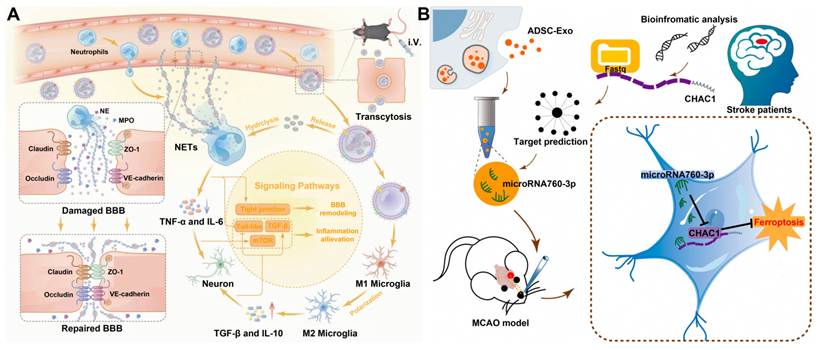
Beyond MSCs, other mammalian cell-derived EVs exhibit highly specialized therapeutic profiles. Neuron-derived EVs leverage their natural neuro-homing affinity to precisely target damaged brain parenchyma. Enriched with specific surface markers like neural cell adhesion molecule and p75 neurotrophin receptor, they deliver functional nucleic acids (e.g., miR-124a) to upregulate glutamate transporter 1 expression in astrocytes, thereby accelerating glutamate clearance and blunting excitotoxicity. Their carried synaptic-related proteins also hold potential as robust biomarkers for early HIBI diagnosis [235]. Similarly, astrocyte-derived EVs regulate synaptic glutamate concentrations by transporting excitatory amino acid transporter 1/2 and confer resistance to hypoxic injury by delivering HSP70 and miR-17-5p [236]. Conversely, M2 microglia-derived EVs function as potent anti-inflammatory vectors. Exploiting their inflammatory chemotaxis and BBB penetrability, they deliver anti-inflammatory nucleic acids (miR-124 and miR-7670-3p) to suppress M1 polarization and curb pro-inflammatory cytokine release. When loaded with DNase I, they can also cross the BBB to degrade NETs, effectively dismantling the vicious cycle of inflammation and vascular damage [237] (Figure 15B). Finally, endothelial cell/endothelial progenitor cell-derived EVs restore BBB integrity by upregulating ZO-1 and Claudin-5, while simultaneously delivering miR-126 to stimulate focal angiogenesis [238] (Table 3).
Extracellular vesicle-based drug delivery systems for HIBI.
| Derivations | Isolation methods | In vivo models | Results | Ref. |
|---|---|---|---|---|
| Immortalized mesenchymal stromal cells | PEG precipitation and ultracentrifugation | Neonatal mouse hypoxic-ischemic brain damage | Inhibit neuroinflammation, and promote neurogenesis and neurotrophic factors | [239] |
| Bone marrow MSCs/BMECs | Ultrafiltration | pMCAO | Inhibit Cav-1-dependent endocytosis, ZO-1 and Claudin-5 | [238] |
| DUC | ET-1 stroke | Track in combination with CT | [240] | |
| MSCs | DUC | MCAO/R | Deliver miR-22-3p | [234] |
| Ultracentrifugation | MCAO/R | Deliver melatonin and inhibit neuronal apoptosis | [86] | |
| Neural stem cells | Ultracentrifugation | MCAO/R | Inhibit neuronal apoptosis and alleviate oxidative stress | [241] |
| Ultracentrifugation | MCAO/R | Deliver VEGF and synergistically promote angiogenesis and neurogenesis | [242] | |
| Endothelial cells | DUC | MCAO/R | Enhance vascular recanalization and preserve BBB integrity | [243] |
| DUC | MCAO/R | Reduce inflammation and apoptosis, and restore astrocyte function | [244] | |
| Microvascular endothelial cells | Gradient centrifugation | MCAO/R | Inhibit apoptosis, and enhance BBB integrity and cerebral blood flow | [245] |
| Astrocytes | DUC | MCAO/R | Inhibit excessive neuronal autophagy and apoptosis | [246] |
| Human microglial clone 3 | Ultrafiltration | pMCAO | Promote endothelial cell function and vascular remodeling | [247] |
| M2 microglia | DUC | MCAO/R | Promote white matter structural remodeling and neurological function recovery | [248] |
| Ultracentrifugation | MCAO/R | Alleviate neuronal apoptosis | [249] | |
| EVs derived from M2 microglia | Ultracentrifugation | MCAO/R | Reduce astrocyte proliferation and migration, and glial scar formation | [250] |
| M2 macrophages | Gradient centrifugation | MCAO/R | Induce microglial M2 polarization | [237] |
| Adipose-derived stem cells | DUC | MCAO/R | Preserve BBB integrity | [251] |
| EVs derived from embryonic stem cells | Differential centrifugation and ultracentrifugation | MCAO/R | Reduce neuroinflammation and improve neurological function | [252] |
| SD rat plasma | Gradient centrifugation | MCAO/R | Scavenge ROS and attenuate neuronal injury | [253] |
Note: DUC, differential ultracentrifugation.
EVs derived from plant cells are a very safe type of natural nanomaterials. They have unique advantages over mammalian EVs, such as no human pathogen contamination, simplified extraction methods (for example, the differential centrifugation process gives high-purity product), and very high scalability. In HIBI, plant-derived EVs can have two different functions. The first function is that some plant-derived EVs, such as those from the plant Panax notoginseng, can work as therapeutic agents, where their unique lipid components help in the modulating process of microglial cells into M2 phenotype and effectively reduce the ischemia-reperfusion injury [254]. Indirectly, they have strong drug delivery capacities since ginseng-derived EVs have succeeded to deliver mtr-miR159a and some other select miRNAs which activate the phosphatidylinositol 3-kinase / Ras / extracellular signal-regulated kinase (PI3K/Ras/Erk) signalling pathway leading to the differentiation of neural stem cells and speeding up the regeneration of brain tissue [255]. Notably, despite the fact that investigations into the use of plant EVs in HIBI have only begun, their remarkable safety and delivery capacities allow them to be successful targets for further explorations.
Although EVs are generally useful for clinical therapy, there are some barriers in applying EV-based nano-systems into practice. If we talk about production and storage, the efficiency of obtaining natural EVs is very low and does not lead to profitable amounts of produced goods. Even though EVs derived from plants can be produced in large scales, their modifications are required for brain targeting. Furthermore, EVs have low physicochemical stability which results in their instability and loss of bioactive compounds during storage. Safety issues are present in case of using allogeneic EVs due to the influence of its immunogenic properties, while autologous EVs suffer from high preparation expenses and variability between donors. Moreover, standard methods for EV isolation, purification and assessment of EV quality have not been developed yet. Furthermore, there are no clinical studies demonstrating biodistribution data showing EVs’ pharmacokinetics, safety and immunogenicity in the long term perspective. Finally, biological differences between humans and rodents prevent one from applying the results of animal tests to humans.
Future efforts should be directed at moving beyond these translational barriers in three synchronized approaches. One is the design of multi-source chimeric EV platforms that combine the positive therapeutic capabilities of multiple parental cells. The other is designing combinatorial delivery cells loaded with thrombolytics, neuroprotective agents, and functional nucleic acids in order to achieve a complete inhibition of various overlapping pathological pathways. Number three is that methods must be developed systematically. Researchers have to develop inexpensive protocols for mass production. They must adhere to the standards of quality control regarding Good Manufacturing Practice. They also have to carry out comprehensive preclinical trials on suitable animal species.
7. Clinical translation and challenges
The pathogenesis of HIBI is exceptionally complex, driven by a multifaceted cascade encompassing hypoxic stress, inflammatory storms, BBB disruption, and neuroregenerative failure. Currently, advanced biomaterials - exemplified by oxygen-delivering hydrogels, EV-functionalized platforms, and stimuli-responsive smart carriers-have demonstrated robust neuroprotective and reparative potential across preclinical models. However, the translation of these benchtop innovations into clinical reality remains severely impeded by formidable interdisciplinary challenges. These include the poor reproducibility of preclinical efficacy, inadequate adaptation to dynamic pathological microenvironments, unresolved long-term biocompatibility concerns, complicated manufacturing-related CMC hurdles, and strict unmet regulatory requirements for CNS nanomedicines [256]. Therefore, systematically delineating the core translational bottlenecks of HIBI-targeted biomaterials and exploring evidence-based, viable strategies is imperative to bridge the substantial gap between basic research and clinical practice.
7.1. Dynamic progression of the BBB leads to uncontrollable targeting
Throughout the progression of HIBI, the BBB undergoes profound, spatiotemporally dynamic alterations in structural integrity, permeability, and surface receptor expression profiles [257]. Despite this biological reality, the vast majority of current nanotherapeutics are engineered based on static design principles and validated solely during the acute phase of BBB disruption in singular animal models. Consequently, many studies claim successful passive diffusion or receptor-mediated transcytosis without accounting for the subsequent temporal evolution of the barrier microenvironment.
This static validation strategy fundamentally ignores the clinical trajectory of HIBI. As the disease transitions from the acute to the subacute and chronic phases, the BBB undergoes dynamic remodeling, often resulting in the substantial downregulation or spatial redistribution of previously targeted surface receptors (e.g., LFA-1 or specific integrins). This temporal heterogeneity significantly diminishes the transcytotic efficiency of subsequent doses, precipitating a phenomenon akin to secondary drug resistance or therapeutic stagnation. Furthermore, while the field often reports short-term (e.g., 24 hours to 7 days) efficacy and biocompatibility [139], the BBB repair remains severely underexplored. This disconnect is further exacerbated by the widespread reliance on young, healthy animal models. In clinical reality, HIBI predominantly afflicts elderly patients or neonates, whose baseline BBB permeability, receptor expression, and endogenous repair capacities diverge drastically from those of standard preclinical models.
To bridge this critical translational gap, future research paradigms must pivot toward "dynamically adaptive targeting." By integrating in vivo imaging biomarkers (such as advanced MRI or positron emission tomography), clinicians could dynamically map the spatiotemporal permeability profile of a patient's BBB. Utilizing this real-time pathophysiological mapping to guide the administration of spatiotemporally responsive nanocarriers may help establish a prospective paradigm of "personalized, BBB-status-guided precise intervention". However, this conceptual strategy remains largely unvalidated in clinically-relevant animal models and human subjects, and cannot fully eliminate targeting heterogeneity risks within clinical cohorts.
7.2. Superficial safety evaluations and long-term biocompatibility blind spots
As a highly sensitive organ with severely restricted endogenous regenerative capacity, the CNS imposes exceptionally stringent demands on the long-term biocompatibility of targeted biomaterials [258]. The prolonged retention of synthetic nanocarriers, the focal accumulation of un-metabolizable degradation byproducts, or the elicitation of chronic foreign-body responses can precipitate irreversible neurotoxicity and exacerbate tissue devastation [259, 260].
Despite these high stakes, a pervasive limitation across current nanomedicine research is the brevity and superficiality of safety evaluations. Most toxicological assessments are constrained to standard acute panels - such as systemic blood biochemistry and gross pathological staining of peripheral organs - while conspicuously neglecting profound, brain-specific analyses. Consequently, critical parameters dictating long-term clinical viability remain largely uncharted. These encompass the in vivo degradation kinetics of materials within the brain parenchyma, the specific metabolic and excretory routes of their byproducts, the risk of lysosomal accumulation within neurons or glia, and the potential for chronic microglial activation or sustained neuroinflammation.
Alarmingly, numerous flagship studies report therapeutic efficacy and acute toxicity profiles based solely on truncated observation windows (e.g., 7 days to 4 weeks), fundamentally failing to capture the delayed neurological outcomes or late-onset toxicity associated with prolonged therapeutic exposure [84]. Furthermore, the specific safety profiles of these advanced nanoplatforms in high-risk clinical scenarios - such as the propensity for hemorrhagic transformation in IS models - are frequently overlooked [150].
To overcome this critical translational barrier, future research must mandate the establishment of rigorous, long-term safety evaluation frameworks that align with stringent regulatory guidelines, necessitating comprehensive in vivo follow-up periods extending well beyond six months. From a materials design perspective, priority must actively shift toward fully biodegradable architectures, endogenous trace elements (e.g., biologically metabolizable Fe or Mn), or highly homologous biomimetic membranes derived from autologous sources. Ultimately, engineering 'traceless' theranostic platforms that ensure complete systemic clearance without focal accumulation is paramount to averting chronic neurotoxic hazards and achieving authentic clinical translation.
7.3. Complex pathological cascades necessitate multi-modal and combinatorial therapeutics
The pathogenesis of HIBI is inherently multifaceted, driven by a catastrophic interplay of ischemic hypoxia, robust neuroinflammation, neuronal apoptosis, and neurovascular degradation [261]. While the profound sensitivity of the brain to oxygen deprivation dictates that restoring tissue oxygenation is paramount [262], simplistic oxygen-delivery strategies (e.g., bare PFC emulsions) are fundamentally ill - equipped to combat the severe oxidative stress and inflammatory cascades unleashed upon reperfusion. These reperfusion-induced cascades generate overwhelming surges of pro-inflammatory cytokines and ROS, which obliterate the BBB and inflict secondary damage upon surviving neurons and synapses [263]. Consequently, monomodal interventions - whether strictly oxygenating, solely anti-inflammatory, or uniquely neuroprotective - invariably fail to halt this progressive injury loop.
This pathophysiological complexity necessitates a paradigm shift toward intelligent, spatiotemporally synchronized theranostic platforms that integrate multiple therapeutic modalities. Pioneering studies have successfully engineered nanocarriers designed to simultaneously dismantle the vicious cycle of ROS overproduction and neuroinflammation, thereby significantly improving prognostic outcomes [84,188]. Building upon this, more advanced biomimetic architectures have been designed not only to exert dual antioxidant and anti-inflammatory effects but also to actively promote neurovascular remodeling and angiogenesis within the ischemic microenvironment [89,189]. These advancements indicate a definitive design transition from monomodal functionality toward multimodal synergy, establishing comprehensive biological platforms capable of intervening across multiple critical pathological nodes.
However, the relentless pursuit of "all-in-one" multifunctional integration introduces formidable translational hurdles, primarily concerning structural controllability and synthetic robustness. As the architectural complexity of these biomimetic nanoplatforms (e.g., multi-layered coatings, dual-drug loading) increases, the batch-to-batch coefficients of variation for critical parameters such as encapsulation efficiency, size distribution, membrane protein integrity, and responsive release kinetics are dramatically amplified. This structural heterogeneity poses severe challenges for scalable GMP production and severely complicates the regulatory evaluation of these highly complex formulations. Furthermore, for many purported multimodal platforms, the "synergistic effects" between the disparate components frequently lack rigorous, mechanism-driven pharmacological validation. Therefore, future translational designs must pivot from overly complex prototypes toward streamlined, scalable formulations, prioritizing rigorous experimental protocols that unequivocally validate true component synergy and long-term biosafety, while taking GMP-compliant manufacturing and regulatory evaluation criteria into account at the early material-design stage.
8. Conclusion
HIBI, a devastating pathology of the CNS, is driven by a highly intertwined injury network characterized by the cascading amplification of oxidative stress, neuroinflammation, BBB disruption, and excitotoxicity. Hampered by inherent bottlenecks such as monotargeted intervention, restricted BBB penetrability, and an inability to adapt to dynamic pathological evolution, conventional therapeutic strategies have consistently failed to achieve transformative clinical outcomes [98].
This review has systematically delineated the cutting-edge advancements in stimuli-responsive and biomimetic materials for HIBI theranostics. The deep integration of materials science and biomedical engineering offers an unprecedented solution to the historical diagnostic and therapeutic dilemmas of HIBI. Within the diagnostic realm, advanced platforms - ranging from NIR-II imaging agents to functional fluorescent probes - have catalyzed a paradigm shift. They transcend conventional structural imaging by enabling the quantitative visualization of the pathological microenvironment, facilitating the real-time, in vivo monitoring of cerebral oxygenation, oxidative stress, and BBB permeability. Therapeutically, engineered nanoplatforms achieve precise intervention at critical pathological nodes. By synergistically integrating biomimetic camouflage (e.g., native cell membranes and EVs) with stimuli-responsive architectures, these carriers orchestrate active homing, immune evasion, and spatiotemporally controlled drug release [115]. Ultimately, this synergy drives a fundamental transition in HIBI management from passive drug delivery to active and microenvironment-responsive regulation (Figure 16).
The three-layer conceptual framework of pathological nodes-material functions-therapeutic outcomes for HIBI proposed in this review. The upper panel outlines four core pathological cascades driving HIBI progression, namely oxidative stress, neuroinflammation, BBB disruption, and excitotoxicity. HIBI-targeted biomaterials are classified into three functional modules, including oxygen-carrying and antioxidant systems, BBB-crossing delivery and regenerative materials, and biomimetic intelligent therapeutic platforms. The lower panel summarizes corresponding therapeutic effects, covering antioxidant metabolic recovery, anti-inflammatory immunoregulation, BBB structural repair, and neuroprotection. Created with Figdraw.com.
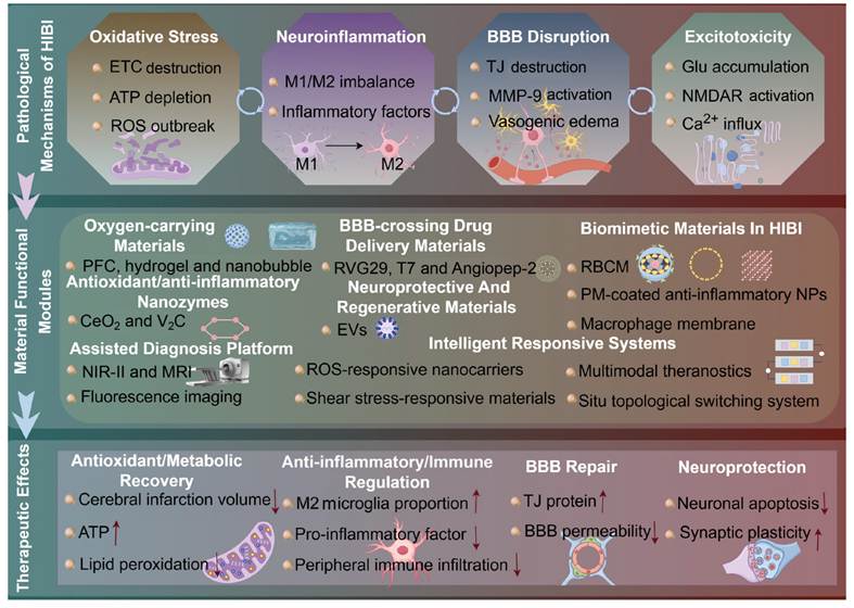
While accumulating preclinical evidence confirms that these multi-mechanism synergistic platforms can significantly ameliorate infarct volume and rescue long-term neurological deficits, the translational leap from bench to bedside remains fraught with formidable hurdles. Foremost, material designs must evolve from static models to dynamically adaptive systems capable of tracking the temporal evolution of BBB permeability. From the perspective of the whole HIBI spectrum, it should be emphasized that such promising material-based interventions are almost exclusively validated for adult ischemic stroke. There remains a notable lack of purpose-designed smart theranostic biomaterials targeting neonatal HIE and high-altitude hypoxic brain injury, even though these two conditions belong to important HIBI subtypes with huge clinical demands. Furthermore, the imperative for clinical safety demands rigorous in vivo investigations into the degradation kinetics and chronic immunotoxicity of these nanomaterials within the highly sensitive CNS. Finally, the inherent paradox between architectural complexity and manufacturing standardization must be resolved; streamlining multifunctional designs to ensure batch-to-batch consistency and scalable GMP production is the ultimate prerequisite for industrial translation [264].
In conclusion, stimuli-responsive and biomimetic materials are fundamentally redefining the landscape of HIBI theranostics through their unparalleled capacity for precision targeting, on-demand cargo release, and multidimensional synergistic repair. Rooted in a profound understanding of pathological cascades, this material-driven paradigm not only offers unprecedented strategies for combating HIBI but also establishes a robust technological foundation for the broader advancement of precision medicine in CNS disorders.
9. Future outlook
Research on stimuli-responsive and biomimetic materials for integrated HIBI theranostics has achieved monumental progress. Propelled by the profound convergence of materials science, neuroscience, and precision medicine, the field currently stands at a critical inflection point. However, surmounting the formidable translational chasm between laboratory breakthroughs and clinical reality remains a daunting endeavor. Under the guidance of the material design framework based on pathological nodes mentioned above, in order to break through these bottlenecks, promote clinical translation and practical applications, future material development should break the static research paradigm and focus on the key research directions detailed below.
Future material designs must transcend the conventional static paradigm and evolve into intelligent architectures capable of spatiotemporally synchronized interaction with disease progression. This necessitates a comprehensive elucidation of the dynamic BBB remodeling across the hyperacute, acute, and recovery phases. By coupling multistage responsive carriers with high-resolution in vivo imaging technologies, researchers can actively track material fate and disease resolution. Ultimately, this will catalyze the development of autonomous, closed-loop theranostic platforms-integrating microenvironment-sensing, computational decision-making, and on-demand actuation-to effectively combat dynamically evolving pathologies.
The multifactorial nature of HIBI renders monotherapies fundamentally inadequate. While constructing synergistic platforms that simultaneously execute oxygen delivery, ROS scavenging, and neuroregeneration is essential, researchers must actively confront the complexity paradox. The pivotal challenge lies in balancing the synergistic enhancement of these functional modules with structural controllability and reproducible synthesis. Future innovations must leverage modular assembly and orthogonal response designs to streamline fabrication, ensuring that these multi-target networks can satisfy the stringent CMC standards mandated for industrial-scale GMP.
Rigorously, longitudinal in vivo safety profiling constitutes the absolute bedrock of clinical translation. The field must move beyond acute toxicity panels and mandate systematic, multi-month investigations in clinically relevant large animal models. These evaluations must meticulously quantify parenchymal material retention, byproduct metabolic trajectories, and the risk of chronic microglial activation. Concurrently, material engineering should proactively mitigate long-term risks by utilizing fully bioresorbable architectures, endogenous trace elements (e.g., metabolizable Fe or Mn), and autologous biomimetic membranes, thereby establishing a truly traceless therapeutic clearance paradigm.
The clinical management of HIBI must not be confined to acute-phase recanalization and neuroprotection. Future strategies should emphatically pivot toward active functional remodeling during the extended rehabilitation phase. By deploying bioresorbable, implantable smart scaffolds coupled with precisely controlled electrophysiological stimulation and the sustained release of neurotrophic factors, researchers can actively guide neural circuit reconstruction and synaptic compensation. This will drive a definitive paradigm shift from acute neuronal salvage to comprehensive neurological functional reconstruction.
Conceptually, HIBI consists of adult ischemic stroke, neonatal HIE, and high-altitude hypoxic brain injury. Current progress of smart theranostic biomaterials is largely confined to adult IS research. Considering the distinct physiological features of the immature neonatal BBB, developmental immune microenvironment, and chronic cerebral adaptive changes induced by high-altitude exposure, developing subtype-adaptive biomaterials for neonatal HIE and high-altitude hypoxic brain injury represents an unmet and promising research niche for follow-up exploration.
Ultimately, stimuli-responsive and biomimetic materials present unprecedented opportunities for the advancement of HIBI theranostics. Through profound interdisciplinary collaboration and a relentless focus on translational viability, these innovations are poised to drive a revolutionary transformation in the diagnosis, treatment, and long-term management of a broad spectrum of CNS disorders.
Acknowledgements
This paper was funded by the National Natural Science Foundation of China (82574692), the 2025 Annual Open Project of Guangdong Provincial Laboratory of Traditional Chinese Medicine for Special Project of Ethnic Medicine Development Cooperation Alliance (HQL20250PA008), the Science and Technology Department of Xizang Autonomous Region (XZ202601ZY0173), the China Postdoctoral Science Foundation (2025MD784204 and GZB20250890), the Young Elite Scientists Sponsorship Program by CACM (CACM-2024-QNRC2-B07), the Science & Technology Department of Sichuan Province (2026NSFSC1839), and the Joint Innovation Fund of Health Commission of Chengdu and Chengdu University of Traditional Chinese Medicine (WXLH202403076 and WXLH202501037). The authors acknowledge the use of Figuredraw.com for the preparation of figures. Gemini 3.1 Pro (Google) was used solely as an AI-assisted language editing tool to improve English grammar and sentence clarity during manuscript preparation. Gemini 3.1 Pro was not used to generate scientific content, figures, images, data, analyses, interpretations, or conclusions.
Author contributions
X.L. Meng, Q.H. Ding and X.B. Wang conceived and supervised the project. J.L. Cui and X.P. Zhou wrote the manuscript and generated figures and table. S.W. Hua, J.R. Yan and Y. Hou polished the manuscript. All the authors read and approved the final manuscript.
Data availability
Data will be made available on request.
Competing interests
The authors have declared that no competing interest exists.
References
1. Meng XF, Zheng Z, Yang L, Yang C, Li XL, Hao YF. Dynamic mechanisms and targeted interventions in cerebral ischemia-reperfusion injury: pathological cascade from ischemia to reperfusion and promising therapeutic strategies. Front Neurosci. 2025;19:1649533
2. Sabir H. Neuroprotective therapies for neonatal hypoxic-ischemic brain injury-a contemporary update. Semin Perinatol. 2025;49:152128
3. Zhang ZY, Wu SJ, Jin TY, Duan ZX, Du XX. Comparative analysis of different stroke subtype burden and future trends over 15 years in China and globally from 1990 to 2021. Front Neurol. 2025;16:1631775
4. Luks AM, Swenson ER, Bärtsch P. Acute high-altitude sickness. Eur Respir Rev. 2017;26:160096
5. Peeples ES, Mietzsch U, Molloy EJ, Pilon B, Guez-Barber D, Soul J. et al. Expert consensus approach to developing inpatient common data elements for neonatal encephalopathy research. Neurology. 2026;106:e214658
6. Busl KM, Greer DM. Hypoxic-ischemic brain injury: pathophysiology, neuropathology and mechanisms. Neurorehabilitation. 2010;26:5-13
7. Panigrahi A, Das NR, Yadav N, Panda S, Sethy AS, Panda SK. et al. AI-driven quantitative metabolomics for early and precise HIE diagnosis: challenges and solutions. ACS Omega. 2025;10:57876-88
8. Achar A, Myers R, Ghosh C. Drug delivery challenges in brain disorders across the blood-brain barrier: novel methods and future considerations for improved therapy. Biomedicines. 2021;9:1834
9. Liu XN, Wu ZH, Lv HG, Li BY, Li CQ, Zhao JJ. et al. NIR-II-responsive biomimetic nanoplatform for precise inhibition of ischemic stroke via dual modulation of ion channels and microglia. ACS Appl Mater Interfaces. 2026;18:9497-511
10. Sun Y, Miao YY, Dai SY, Wang KL, Tan J, Jiao Z. et al. The use of xenon magnetic lipid bubbles to detect and treat intermittent hypoxia-induced cognitive impairmen. J Control Release. 2026;389:114478
11. Duan WJ, Yue Q, Liu Y, Zhang YF, Guo QH, Wang C. et al. A pH ratiometrically responsive surface enhanced resonance Raman scattering probe for tumor acidic margin delineation and image-guided surgery. Chem Sci. 2020;11:4397-402
12. Saha S, Yakati V, Shankar G, Jaggarapu M, Moku G, Madhusudana K. et al. Amphetamine decorated cationic lipid nanoparticles cross the blood-brain barrier: therapeutic promise for combating glioblastoma. J Mater Chem B. 2020;8:4318-30
13. Srivastava I, Xue R, Jones J, Rhee H, Flatt K, Gruev V. et al. Biomimetic surface-enhanced Raman scattering nanoparticles with improved dispersibility, signal brightness, and tumor targeting functions. ACS Nano. 2022;16:8051-63
14. Gong X, Li J, Xu XX, Wu Y, Lei Y, Liu HZ. et al. Microvesicle-inspired oxygen-delivering nanosystem potentiates radiotherapy-mediated modulation of tumor stroma and antitumor immunity. Biomaterials. 2022;290:121855
15. Stein JB, Zhang S, Roh EJ, Luo J, Chen M, Jang H. et al. Advanced biomaterial delivery of hypoxia-conditioned extracellular vesicles (EVs) as a therapeutic platform for traumatic brain injury. Adv Sci. 2025;12:e04147
16. Deng J, Lin D, Ding X, Wang Y, Hu YH, Shi H. et al. Multifunctional supramolecular filament hydrogel boosts anti-inflammatory efficacy in vitro and in vivo. Adv Funct Mater. 2022;32:2109173
17. Singh N, Sherin GR, Mugesh G. Antioxidant and prooxidant nanozymes: from cellular redox regulation to next-generation therapeutics. Angew Chem Int Ed Engl. 2023;62:e202301232
18. Cai X, Zhang H, Wei P, Liu Q, Sheng D, Li Z. et al. A wireless optoelectronic probe to monitor oxygenation in deep brain tissue. Nat Photonics. 2024;18:492-500
19. Zhang H, Wang J, Tian Y, Wang C, Hou L. Shear force-ROS sequential responsive drug delivery system for improving cerebral thrombosis microcirculation and neurological function. J Control Release. 2025;378:195-208
20. Ren JX, Ma HY, Yin WJ, Li YK, Lei SY, Liu JC. et al. Emerging targeted delivery strategies of nanosystems for ischemic stroke treatment. Int J Nanomedicine. 2025;20:8143-71
21. Kang C, Gwon S, Song CG, Kang PM, Park SC, Jeon J. et al. Fibrin-targeted and H2O2-responsive nanoparticles as a theranostics for thrombosed vessels. ACS Nano. 2017;11:6194-203
22. Wu F, Zhang Z, Ma S, He Y, He Y, Ma L. et al. Microenvironment-responsive nanosystems for ischemic stroke therapy. Theranostics. 2024;14:5571-95
23. Gollihue JL, Rabchevsky AG. Prospects for therapeutic mitochondrial transplantation. Mitochondrion. 2017;35:70-9
24. Tang YC, Tian HX, Yi T, Chen HB. The critical roles of mitophagy in cerebral ischemia. Protein Cell. 2016;7:699-713
25. Rekuviene E, Ivanoviene L, Borutaite V, Morkuniene R. Rotenone decreases ischemia-induced injury by inhibiting mitochondrial permeability transition in mature brains. Neurosci Lett. 2017;653:45-50
26. Reinhard L, Tidow H, Clausen MJ, Nissen P. Na+,K+-ATPase as a docking station: protein-protein complexes of the Na+,K+-ATPase. Cell Mol Life Sci. 2013;70:205-22
27. Paoletti P, Bellone C, Zhou Q. NMDA receptor subunit diversity: impact on receptor properties, synaptic plasticity and disease. Nat Rev Neurosci. 2013;14:383-400
28. Gaschler MM, Andia AA, Liu H, Csuka JM, Hurlocker B, Vaiana CA. et al. FINO2 initiates ferroptosis through GPX4 inactivation and iron oxidation. Nat Chem Biol. 2018;14:507-15
29. Ingold I, Berndt C, Schmitt S, Doll S, Poschmann G, Buday K. et al. Selenium utilization by GPX4 is required to prevent hydroperoxide-induced ferroptosis. Cell. 2018;172:409-22.e21
30. Brenner C, Moulin M. Physiological roles of the permeability transition pore. Circ Res. 2012;111:1237-47
31. Bauer TM, Murphy E. Role of mitochondrial calcium and the permeability transition pore in regulating cell death. Circ Res. 2020;126:280-93
32. Julien O, Wells JA. Caspases and their substrates. Cell Death Differ. 2017;24:1380-9
33. Yuan J, Najafov A, Py BF. Roles of caspases in necrotic cell death. Cell. 2016;167:1693-704
34. Dirks M, Niessen LW, Wijngaarden JDvan, Koudstaal PJ, Franke CL, Oostenbrugge RJvan. et al. Promoting thrombolysis in acute ischemic stroke. Stroke. 2011;42:1325-30
35. Kirby JP, Snyder J, Schuerer DJE, Peters JS, Bochicchio GV. Essentials of hyperbaric oxygen therapy: 2019 review. Mo Med. 2019;116:176-9
36. Brainina K, Stozhko N, Vidrevich M. Antioxidants: terminology, methods, and future considerations. Antioxidants. 2019;8:297
37. Li M, Wang W, Chen Y, Wang G, Yang H, Li T. et al. A single-atom FeCo-N6 nanozyme with dual enzyme-mimicking activity reverses redox imbalance and bioenergetic collapse in ischemic stroke. Adv Healthc Mater. 2025;15:e05735
38. He L, Huang G, Liu H, Sang C, Liu X, Chen T. Highly bioactive zeolitic imidazolate framework-8-capped nanotherapeutics for efficient reversal of reperfusion-induced injury in ischemic stroke. Sci Adv. 2020;6:eaay9751
39. Wu X, Lu S, Zhang Y, Wu K, Jiang J, Liu X. et al. Synergistic viscoelastic-oxygenation strategy in injectable granular hydrogels for neurovascular regeneration post-ischemic stroke. Adv Funct Mater. 2026;36:e11902
40. Bustamante A, Simats A, Vilar-Bergua A, García-Berrocoso T, Montaner J. Blood/brain biomarkers of inflammation after stroke and their association with outcome: from C-reactive protein to damage-associated molecular patterns. Neurotherapeutics. 2016;13:671-84
41. Wright-Jin EC, Gutmann DH. Microglia as dynamic cellular mediators of brain function. Trends Mol Med. 2019;25:967-79
42. Cherry JD, Olschowka JA, O'Banion MK. Neuroinflammation and M2 microglia: the good, the bad, and the inflamed. J Neuroinflammation. 2014;11:98
43. Lambertsen KL, Finsen B, Clausen BH. Post-stroke inflammation-target or tool for therapy? Acta Neuropathol. 2019;137:693-714
44. Kanazawa M, Ninomiya I, Hatakeyama M, Takahashi T, Shimohata T. Microglia and monocytes/macrophages polarization reveal novel therapeutic mechanism against stroke. Int J Mol Sci. 2017;18:2135
45. Ohtake Y, Li S. Molecular mechanisms of scar-sourced axon growth inhibitors. Brain Res. 2015;1619:22-35
46. Pereira M, Liang J, Edwards-Hicks J, Meadows AM, Hinz C, Liggi S. et al. Arachidonic acid inhibition of the NLRP3 inflammasome is a mechanism to explain the anti-inflammatory effects of fasting. Cell Rep. 2024;43:113700
47. Bijlsma JWJ, Buttgereit F. Adverse events of glucocorticoids during treatment of rheumatoid arthritis: lessons from cohort and registry studies. Rheumatology. 2016;55:ii3-5
48. Zhao C, Chen Q, Li W, Zhang J, Yang C, Chen D. Multi-functional platelet membrane-camouflaged nanoparticles reduce neuronal apoptosis and regulate microglial phenotype during ischemic injury. Appl Mater Today. 2022;27:101412
49. Shi J, Yang Y, Yin N, Liu C, Zhao Y, Cheng H. et al. Engineering CXCL12 biomimetic decoy-integrated versatile immunosuppressive nanoparticle for ischemic stroke therapy with management of overactivated brain immune microenvironment. Small Methods. 2022;6:e2101158
50. Duan R, Sun K, Fang F, Wang N, He R, Gao Y. et al. An ischemia-homing bioengineered nano-scavenger for specifically alleviating multiple pathogeneses in ischemic stroke. J Nanobiotechnology. 2022;20:397
51. Ma J, Zhan M, Sun H, He L, Zou Y, Huang T. et al. Phosphorus dendrimers co-deliver fibronectin and edaravone for combined ischemic stroke treatment via cooperative modulation of microglia/neurons and vascular regeneration. Adv Healthc Mater. 2024;13:e2401462
52. Wang W, Li T, Cheng Y, Li F, Qi S, Mao M. et al. Identification of hypoxic macrophages in glioblastoma with therapeutic potential for vasculature normalization. Cancer Cell. 2024;42:815-32.e12
53. Zhang X, Li H, Gu Y, Ping A, Chen J, Zhang Q. et al. Repair-associated macrophages increase after early-phase microglia attenuation to promote ischemic stroke recovery. Nat Commun. 2025;16:3089
54. Munoz-Rojas AR, Kelsey I, Pappalardo JL, Chen M, Miller-Jensen K. Co-stimulation with opposing macrophage polarization cues leads to orthogonal secretion programs in individual cells. Nat Commun. 2021;12:301
55. Zhang QY, Wang ZJ, Sun DM, Wang Y, Xu P, Wu WJ. et al. Novel therapeutic effects of leonurine on ischemic stroke: new mechanisms of BBB integrity. Oxid Med Cell Longev. 2017;2017:7150376
56. Warner DS, Sheng H, Batinić-Haberle I. Oxidants, antioxidants and the ischemic brain. J Exp Biol. 2004;207:3221-31
57. Pun PB, Lu J, Moochhala S. Involvement of ROS in BBB dysfunction. Free Radic Res. 2009;43:348-64
58. Brown RC, Davis TP. Calcium modulation of adherens and tight junction function: a potential mechanism for blood-brain barrier disruption after stroke. Stroke. 2002;33:1706
59. Chaturvedi M, Kaczmarek L. MMP-9 inhibition: a therapeutic strategy in ischemic stroke. Mol Neurobiol. 2014;49:563
60. Yang C, Hawkins KE, Doré S, Candelario-Jalil E. Neuroinflammatory mechanisms of blood-brain barrier damage in ischemic stroke. Am J Physiol Cell Physiol. 2019;316:C135-53
61. Sorby-Adams AJ, Marian OC, Bilecki IM, Elms LE, Yassi N, Hood RJ. et al. NK1 tachykinin receptor antagonist treatment reduces cerebral edema and intracranial pressure in an ovine model of ischemic stroke. J Cereb Blood Flow Metab. 2024;44:1759-73
62. Chen AQ, Fang Z, Chen XL, Yang S, Zhou YF, Mao L. et al. Microglia-derived TNF-α mediates endothelial necroptosis aggravating blood brain-barrier disruption after ischemic stroke. Cell Death Discov. 2019;10:487
63. Cheong EJY, Teo DWX, Chua DXY, Chan ECY. Systematic development and verification of a physiologically based pharmacokinetic model of rivaroxaban. Drug Metab Dispos. 2019;47:1291-306
64. Huang L, Chen Y, Liu R, Li B, Fei X, Li X. et al. P-glycoprotein aggravates blood brain barrier dysfunction in experimental ischemic stroke by inhibiting endothelial autophagy. Aging Dis. 2022;13:1546-61
65. Deng YY, Shen FC, Xie D, Han QP, Fang M, Chen CB. et al. Progress in drug treatment of cerebral edema. Med Chem. 2016;16:917-25
66. Wang A, Jia B, Zhang X, Huo X, Chen J, Gui L. et al. Efficacy and safety of butylphthalide in patients with acute ischemic stroke: a randomized clinical trial. JAMA Neurol. 2023;80:851-9
67. He WX, Zhang XY, Gong X, Luo KK, Ma XN, Lu EH. et al. Drug-free biomimetic oxygen supply nanovehicle promotes ischemia-reperfusion therapy in stroke. Adv Funct Mater. 2023;33:2212919
68. Puyal J, Ginet V, Clarke PG. Multiple interacting cell death mechanisms in the mediation of excitotoxicity and ischemic brain damage: a challenge for neuroprotection. Prog Neurobiol. 2013;105:24-48
69. Rossi DJ, Brady JD, Mohr C. Astrocyte metabolism and signaling during brain ischemia. Nat Neurosci. 2007;10:1377-86
70. Patabendige A, Singh A, Jenkins S, Sen J, Chen R. Astrocyte activation in neurovascular damage and repair following ischaemic stroke. Int J Mol Sci. 2021;22:4280
71. Zhang S, Shang D, Shi H, Teng W, Tian L. Function of astrocytes in neuroprotection and repair after ischemic stroke. Eur Neurol. 2021;84:426-34
72. Bettler B, Fakler B. Ionotropic AMPA-type glutamate and metabotropic GABAB receptors: determining cellular physiology by proteomes. Curr Opin Neurobiol. 2017;45:16-23
73. Brassai A, Suvanjeiev RG, Bán EG, Lakatos M. Role of synaptic and nonsynaptic glutamate receptors in ischaemia induced neurotoxicity. Brain Res Bull. 2015;112:1-6
74. Banerjee A, Larsen RS, Philpot BD, Paulsen O. Roles of presynaptic NMDA receptors in neurotransmission and plasticity. Trends Neurosci. 2016;39:26-39
75. Gazerani P. The neuroplastic brain: current breakthroughs and emerging frontiers. Brain Res. 2025;1858:149643
76. Steigerwald R, Epstein M, Chou TH, Simorowski N, Furukawa H. Molecular mechanism of calcium permeability and magnesium block in NMDA receptors. Nat Neurosci. 2026;29:1293-302
77. Dupuis JP, Nicole O, Groc L. NMDA receptor functions in health and disease: old actor, new dimensions. Neuron. 2023;111:2312-28
78. Pereira NAL, Sureda FX, Turch M, Amat M, Bosch J, Santos MMM. Synthesis of phenylalaninol-derived oxazolopyrrolidone lactams and evaluation as NMDA receptor antagonists. Monatsh Chem. 2013;144:473-7
79. Kopecky BJ, Liang R, Bao J. T-type calcium channel blockers as neuroprotective agents. Pflugers Arch. 2014;466:757-65
80. Cao XY, Shuai K, Wang P, Xu JC, Pan WQ, Y. Wang, et al. Biosponge-armored nanodots restore redox-calcium homeostasis to mitigate reperfusion-induced injury in ischemic stroke. Adv Funct Mater. 2025;50:e03183
81. Lu L, Bi X, Shan M, Liu D, Yang M, Li X. et al. Self-assembled carrier-free nanoplatforms for sequential modulation of the oxidative-inflammatory cascade in cerebral ischemia-reperfusion injury. Chem Eng J. 2025;526:171122
82. Shi J, Yu W, Xu L, Yin N, Liu W, Zhang K. et al. Bioinspired nanosponge for salvaging ischemic stroke via free radical scavenging and self-adapted oxygen regulating. Nano Lett. 2020;20:780-9
83. Liao J, He W, Zhang S, Wang J, Cheng H, Gong L. et al. Magnetic field driven ceria nanosystems for mitochondria targeted therapy of ischemic stroke. Adv Funct Mater. 2025;35:2423291
84. Yang Q, Li R, Hong Y, Liu H, Jian C, Zhao S. Curcumin-loaded gelatin nanoparticles cross the blood-brain barrier to treat ischemic stroke by attenuating oxidative stress and neuroinflammation. Int J Nanomed. 2024;19:11633-49
85. Liu C, Wang J, Wang Z, Fan X, Pan J, Guo X. et al. Discoidal nanoparticles exploit thrombus-induced shear gradients to enhance site-specific thrombolysis in stroke. Acta Biomater. 2025;201:471-84
86. Qiu L, Cai Y, Geng Y, Yao X, Wang L, Cao H. et al. Mesenchymal stem cell-derived extracellular vesicles attenuate tPA-induced blood-brain barrier disruption in murine ischemic stroke models. Acta Biomater. 2022;154:424-42
87. Li M, Miao L, Xu X, Liu Y, Wang Y, Yang F. Hypoxia-responsive biomimetic nanobubbles for oxygen delivery promote synergistic ischemic stroke protection. ACS Appl Mater Interfaces. 2024;16:52036-46
88. Zhang T, Liu P, Shen W, Li C, Zhao Z, Wu Y. et al. DNase I-mediated chemotactic nanoparticles for NETs targeting and microenvironment remodeling treatment of acute ischemic stroke. Adv Sci. 2025;12:e03689
89. Chen D, Jiang H, Sun L, Nurzat Y, Qin H, Zhao Z. et al. Neuron-targeted ROS-responsive liposomes for puerarin delivery remodel ischemic microenvironment via microglial modulation and neurovascular regeneration. J Nanobiotechnol. 2025;23:677
90. Yang Z, Jin L, Li L, Wu Y, Liu W, Feng X. et al. Brain targeted lipid nanoparticles with Hv1 inhibitors alleviate neuroinflammation post-ischemic stroke. J Nanobiotechnol. 2025;23:464
91. Dang Y, Shen F, Wang S, Zhang Y, Lu X, Qin D. et al. Dual-modified mannose/RVG29 peptide-functionalized lipid nanoparticles loaded with circHIPK2 siRNA ameliorate hypoxic-ischemic brain damage in neonatal mice by suppressing astrocyte activation. J Integr. Neurosci. 2025;24:45212
92. Liu Z, Xia Q, Wang C, Xu J, Tian K, Wang Z. et al. Biomimetic astrocyte cell membrane-fused nanovesicles for protecting neurovascular units in hypoxic ischemic encephalopathy. J Nanobiotechnology. 2024;22:766
93. Jiang Z, Wang W, Zhao Y, Li T, Xin D, Gai C. et al. Mitochondria-targeted cerium vanadate nanozyme suppressed hypoxia-ischemia injury in neonatal mice via intranasal administration. J Control Release. 2024;365:1074-88
94. Zhao J, Liang D, Xiong M, Deng J, Hu L, Jiang L. et al. Targeted delivery of IDR-1018 via biomimetic magnetic nanovesicles suppresses neurovascular cell death and promotes angiogenic repair after neonatal hypoxic-ischemic brain injury. Apoptosis. 2026;31:165
95. Peng Z, Ye QS, Li XJ, Zheng DY, Zhou Y, Hang CH. et al. Novel perfluorocarbon-based oxygenation therapy alleviates Post-SAH hypoxic brain injury by inhibiting HIF-1α. Free Radic Biol Med. 2024;214:173-83
96. Meng W, Ye H, Ma Z, Liu L, Zhang T, Han Q. et al. Perfluorocarbon nanoparticles incorporating ginkgolide B: artificial O2 carriers with antioxidant activity and antithrombotic effect. ChemMedChem. 2023;19:e202300312
97. Zuo JC, Liang J, Hu N, Yao B, Zhang QJ, Zeng XL. et al. Hypoxia preconditioned MSC exosomes attenuate high-altitude cerebral edema via the miR-125a-5p/RTEF-1 axis to protect vascular endothelial cells. Bioact Mater. 2025;52:541-63
98. Dong X, Gao J, Su Y, Wang Z. Nanomedicine for ischemic stroke. Int J Mol Sci. 2020;21:7600
99. Long Y, Zou Z, Wu Y, Feng H, Chen T, Yang Z. et al. Biomimetic membrane nanotechnology in cerebral ischemic stroke:a promising technology for therapeutic treatment and diagnosis. Front Bioeng Biotechnol. 2025;13:1605377
100. Lin D, Huang W, Yang H, Zhu J, Liu Y, Wang L. et al. Ultrabright NIR-II nanoparticles for high-resolution in vivo imaging: from systemic vasculature visualization to pathological microenvironment monitoring. Adv Mater. 2026;38:e10493
101. Sekhon MS, Ainslie PN, Griesdale DE. Clinical pathophysiology of hypoxic ischemic brain injury after cardiac arrest: a "two-hit" model. Crit Care. 2017;21:90
102. Qiu X, Yuan W, Jiang C, Feng W, Li F. NIR-II luminescent nanothermometer for precise assessment of ischemic stroke. ACS Appl Mater Interfaces. 2025;17:39967-77
103. Wang P, Zhang C, Meng L, Xu S, Zhang J, Wang H. et al. NIR-II emissive, ischemia targeted nanocomposites for localized therapeutic hypothermia of cerebral ischemia-reperfusion injury. Small. 2025;21:e05141
104. Bhaskar S, Bivard A, Parsons M, Nilsson M, Attia JR, Stanwell P. et al. Delay of late-venous phase cortical vein filling in acute ischemic stroke patients: associations with collateral status. J Cereb Blood Flow Metab. 2017;37:671-82
105. Sun YY, Zhu HJ, Du Y, Zhu S, Zhou SY, Pang SY. et al. A novel NIR-II albumin-escaping probe for cerebral arteries and perfusion imaging in stroke mice model. Biomaterials. 2024;311:122664
106. Chen S, Chen H, Li X, He S, Shou K, Qian K. et al. Dynamic pathophysiological insight into the brain by NIR-II imaging. Adv Sci. 2025;12:e2416390
107. Zhang X, Li S, Ma H, Wang H, Zhang R, Zhang XD. Activatable NIR-II organic fluorescent probes for bioimaging. Theranostics. 2022;12:3345-71
108. Zhang M, Wang Z, Wang C, Wu Y, Li Z, Liu Z. Visualizing oxidative stress level for timely assessment of ischemic stroke via a ratiometric near-infrared-II luminescent nanoprobe. ACS Nano. 2021;15:11940-52
109. Wang Q, Wang D, Liu P, Tang BZ, Li Y, Yin X. Inflammation-targeted nanoplatform: NIR-II imaging-guided encephalitis suppressing by dual antioxidant-ferroptosis action. Biomaterials. 2026;327:123741
110. Sun M, Wang Y, Xu H, Shen Y, Liu B, Ma Y. et al. Novel hypochlorous acid-activated near-infrared probe monitors the dynamic changes of myeloperoxidase activity in ischemic brain. J Med Chem. 2025;68:5382-99
111. Du E, Ma Y, Yang Y, Gao Y, Zhai J, Kan L. et al. Biomimetic non-conjugated segment-doped conjugated polymer nanoplatform for bright NIR-II imaging-guided phototherapy of choroidal melanoma. Adv Funct Mater. 2025;32:e20158
112. Xu J, Du Y, Zhu N, Li J, Zhang Y, Zhou D. et al. NIR-II fluorescent protein created by in situ albumin-tagging for sensitive and specific imaging of blood-brain barrier disruption. Adv Sci. 2025;12:e2500443
113. Li Z, Qiang Y, Chen D, Hu D, Gao D, Xu X. et al. Dual-modal super-resolution ultrasound and NIR-II fluorescence imaging of ischemic stroke with ICG-doped porous PLGA microspheres. Mater Today Bio. 2025;31:101513
114. Hao Y, Ji A, Xing R, Zhu W, Jiang B, Jian Y. et al. Capillaries segmentation of NIR-II images and its application in ischemic stroke. Comput Biol Med. 2022;147:105742
115. Yu K, Fu L, Chao Y, Zeng X, Zhang Y, Chen Y. et al. Deep learning enhanced near infrared-II imaging and image-guided small interfering ribonucleic acid therapy of ischemic stroke. ACS Nano. 2025;19:10323-36
116. Syzdoł B, Ćwik K, Woźniak M. Volumetric differences in the brain in children with hypoxic-ischemic encephalopathy after therapeutic hypothermia: a review of MRI findings. Pol J Radiol. 2026;91:e37-44
117. Khanduja S, Kim J, Kang JK, Feng CY, Vogelsong MA, Geocadin RG. et al. Hypoxic-ischemic brain injury in ECMO: pathophysiology, neuromonitoring, and therapeutic opportunities. Cells. 2023;12:1546
118. Zhao Y, Li Y, Jin W, Guo R, Ma C, Tang W. et al. Ultrafast J-resolved magnetic resonance spectroscopic imaging for high-resolution metabolic brain imaging. Nat Biomed Eng. 2025;9:1835-47
119. Li Y, Wang T, Zhang T, Lin Z, Li Y, Guo R. et al. Fast high-resolution metabolic imaging of acute stroke with 3D magnetic resonance spectroscopy. Brain. 2020;143:3225-33
120. Long C, Bao H. Study of high-altitude cerebral edema using multimodal imaging. Front Neurol. 2022;13:1041280
121. Zou P, Li T, Cao Z, Yang E, Bao M, Zhang H. et al. High-altitude hypoxia aggravated neurological deficits in mice induced by traumatic brain injury via BACH1 mediating astrocytic ferroptosis. Cell Death Discov. 2025;11:46
122. Bao H, He X, Wang F, Kang D. Study of brain structure and function in chronic mountain sickness based on fMRI. Front Neurol. 2021;12:763835
123. Ning Z, Zhang Y, Zhang S, Lin X, Kang L, Duan N. et al. Cellular fluorescence imaging technology for food risk and function evaluation. Trends Food Sci Technol. 2024;153:104742
124. Yang Y, Liu X, Xi D, Zhang Y, Gao X, Xu K. et al. Precision imaging of biothiols in live cells and treatment evaluation during the development of liver injury via a near-infrared fluorescent probe. Chem Biomed Imaging. 2024;3:169-79
125. Yang Y, Ma M, Shen L, An J, Kim E, Liu H. et al. A fluorescent probe for investigating the role of biothiols in signaling pathways associated with cerebral ischemia-reperfusion injury. Angew Chem Int Ed Engl. 2023;62:e202310408
126. Xing L, Wang B, Li J, Guo X, Lu X, Chen X. et al. A fluorogenic ONOO--triggered carbon monoxide donor for mitigating brain ischemic damage. J Am Chem Soc. 2022;144:2114-9
127. Ma X, Yao M, Shi J, Li X, Gao Y, Luo Q. et al. High intensity focused ultrasound-responsive and ultrastable cerasomal perfluorocarbon nanodroplets for alleviating tumor multidrug resistance and epithelial-mesenchymal transition. ACS Nano. 2020;14:15904-18
128. Arif WM, Elsinga PH, Gasca-Salas C, Versluis M, Martinez-Fernandez R, Dierckx R. et al. Focused ultrasound for opening blood-brain barrier and drug delivery monitored with positron emission tomography. J Control Release. 2020;324:303-16
129. Jung B, Huh H, Lee EH, Han M, Park J. An advanced focused ultrasound protocol improves the blood-brain barrier permeability and doxorubicin delivery into the rat brain. J Control Release. 2019;315:55-64
130. Purohit MP, Yu BJ, Roy KS, Xiang Y, Ewbank SN, Azadian MM. et al. Acoustically activatable liposomes as a translational nanotechnology for site-targeted drug delivery and noninvasive neuromodulation. Nat Nanotechnol. 2025;20:1688-99
131. Dauba A, Spitzlei C, Bautista KJB, Jourdain L, Selingue E, VanTreeck KE. et al. Low-boiling-point perfluorocarbon nanodroplets for adaptable ultrasound-induced blood-brain barrier opening. J Control Release. 2024;376:441-56
132. Vlatakis S, Zhang W, Thomas S, Cressey P, Moldovan AC, Metzger H. et al. Effect of phase-change nanodroplets and ultrasound on blood-brain barrier permeability in vitro. Pharmaceutics. 2023;16:51
133. Lehmler HJ. Perfluorocarbon compounds as vehicles for pulmonary drug delivery. Expert Opin Drug Deliv. 2007;4:247-62
134. Berard C, Desgranges S, Dumas N, Novell A, Larrat B, Hamimed M. et al. Perfluorocarbon nanodroplets as potential nanocarriers for brain delivery assisted by focused ultrasound-mediated blood-brain barrier disruption. Pharmaceutics. 2022;14:1498
135. Hallam KA, Nikolai RJ, Jhunjhunwala A, Emelianov SY. Laser-activated perfluorocarbon nanodroplets for intracerebral delivery and imaging via blood-brain barrier opening and contrast-enhanced imaging. J Nanobiotechnol. 2024;22:356
136. Xavierselvan M, Cook J, Duong J, Diaz N, Homan K, Mallidi S. Photoacoustic nanodroplets for oxygen enhanced photodynamic therapy of cancer. Photoacoustics. 2022;25:100306
137. Xu W, Xu R, Li X, Zhang H, Wang X, Zhu J. Downregulating hypoxia-inducible factor-1α expression with perfluorooctyl-bromide nanoparticles reduces early brain injury following experimental subarachnoid hemorrhage in rats. Am J Transl Res. 2016;8:2114-26
138. Siwicka-Gieroba D, Robba C, Gołacki J, Badenes R, Dabrowski W. Cerebral oxygen delivery and consumption in brain-injured patients. J Pers Med. 2022;12:1763
139. Wang R, Nie W, Yan X, Luo K, Zhang Q, Wang T. et al. Biomimetic nanomotors for deep ischemia penetration and ferroptosis inhibition in neuroprotective therapy of ischemic stroke. Adv Mater. 2024;37:e2409176
140. Jervis PJ. Hydrogels in regenerative medicine and other biomedical applications. Int J Mol Sci. 2022;23:3270
141. Wang Z, Wang J, Jin Y, Luo Z, Yang W, Xie H. et al. A neuroprotective sericin hydrogel as an effective neuronal cell carrier for the repair of ischemic stroke. ACS Appl Mater Interfaces. 2015;7:24629-40
142. Ma T, Wang Y, Qi F, Zhu S, Huang L, Liu Z. et al. The effect of synthetic oxygen carrier-enriched fibrin hydrogel on Schwann cells under hypoxia condition in vitro. Biomaterials. 2013;34:10016-27
143. Chen G, Wang F, Zhang X, Shang Y, Zhao Y. Living microecological hydrogels for wound healing. Sci Adv. 2023;9:eadg3478
144. Zhang W, Liu Y, Wu Y, Wang Z, Liu X, Hu Q. et al. ROS-triggered biomimetic hydrogel soft scaffold for ischemic stroke repair. Biomaterials. 2025;319:123217
145. Liu Y, Huang R, Han L, Ke W, Shao K, Ye L. et al. Brain-targeting gene delivery and cellular internalization mechanisms for modified rabies virus glycoprotein RVG29 nanoparticles. Biomaterials. 2009;30:4195-202
146. Zhang L, Cao K, Xie J, Liang X, Gong H, Luo Q. et al. Aβ42 and ROS dual-targeted multifunctional nanocomposite for combination therapy of Alzheimer's disease. J Nanobiotechnol. 2024;22:278
147. Han Y, Chu X, Cui L, Fu S, Gao C, Li Y. et al. Neuronal mitochondria-targeted therapy for Alzheimer's disease by systemic delivery of resveratrol using dual-modified novel biomimetic nanosystems. Drug Deliv. 2020;27:502-18
148. Han Y, Gao C, Wang H, Sun J, Liang M, Feng Y. et al. Macrophage membrane-coated nanocarriers co-modified by RVG29 and TPP improve brain neuronal mitochondria-targeting and therapeutic efficacy in Alzheimer's disease mice. Bioact Mater. 2021;6:529-42
149. Wang Z, Zhao Y, Jiang Y, Lv W, Wu L, Wang B. et al. Enhanced anti-ischemic stroke of ZL006 by T7-conjugated PEGylated liposomes drug delivery system. Sci Rep. 2015;5:12651
150. Song Y, Wang L, Peng T, Shan L, Wan B, Tang M. et al. Brain-targeting biomimetic nanozyme enhances neuroprotection in ischemic stroke by remodeling the neurovascular unit. J Control Release. 2025;382:113750
151. Wei Y, Xia X, Wang X, Yang W, He S, Wang L. et al. Enhanced BBB penetration and microglia-targeting nanomodulator for the two-pronged modulation of chronically activated microglia-mediated neuroinflammation in Alzheimer's disease. Acta Pharm Sin B. 2025;15:1098-111
152. Qiu Y, Huang S, Peng L, Yang L, Zhang G, Liu T. et al. The nasal-brain drug delivery route: mechanisms and applications to central nervous system diseases. MedComm. 2025;6:e70213
153. Ikeda T, Kawabori M, Zheng Y, Yamaguchi S, Gotoh S, Nakahara Y. et al. Intranasal administration of mesenchymal stem cell-derived exosome alleviates hypoxic-ischemic brain injury. Pharmaceutics. 2024;16:446
154. Theije CGMde, Palma STDe, Vaes JEG, Dallaglio K, Volpi G, Ardigo D. et al. Intranasal Wharton's jelly-derived mesenchymal stem cell therapy, alone or in conjunction with therapeutic hypothermia, alleviates neonatal hypoxic-ischemic brain injury in mice. Ann Neurol. 2025;98:1380-94
155. Yin Y, Li Z, Shu W, Liu H, Wang Z, Fu C. et al. Intranasal blood-brain barrier bypass enables sequential mitochondria-targeted bioengineered nanolamellar system for ischemic stroke therapy. Nat Commun. 2026;17:760
156. Zhang Y, Zhang H, Zhao F, Jiang Z, Cui Y, Ou M. et al. Mitochondrial-targeted and ROS-responsive nanocarrier via nose-to-brain pathway for ischemic stroke treatment. Acta Pharm Sin B. 2023;13:5107-20
157. Jia Y, Xu L, Leng S, Sun Y, Huang X, Wang Y. et al. Nose-to-brain delivery of circular RNA SCMH1-loaded lipid nanoparticles for ischemic stroke therapy. Adv Mater. 2025;37:e2500598
158. Shen X, Wang Y, Yang Y, Tang B, Bai L, Yao H. et al. Molecular gardening for neuroinflammation via nose-to-brain delivery: a Ca2+ responsive DNA nanocage-hydrogel system with neuron targeting and STING inhibiting. Adv Mater. 2026;38:e18814
159. Chang Y, Chen T, Wang M, Yuan H, Wang C, Liu Z. et al. Mass spectrometry-based multi-omics analysis reveals the mechanism of angiopep-2-functionalized nanoparticles crossing the blood-brain barrier. Chin Chem Lett. 2026 112770
160. Li X, Hua S, Zhong D, Zhou M, Ding Z. Metal-organic framework-edaravone nanoparticles for radiotherapy-induced brain injury treatment. Biomaterials. 2025;314:122868
161. Li X, Ye J, Zhong D, Xie J, Ding Z, Zhou M. Microalgae-MOF integrated drug delivery system for cranial radiotherapy and chemotherapy-induced brain injury. Biomaterials. 2026;327:123767
162. Wu J, Wang X, Wang Q, Lou Z, Li S, Zhu Y. et al. Nanomaterials with enzyme-like characteristics (nanozymes): next-generation artificial enzymes (II). Chem Soc Rev. 2019;48:1004-76
163. Zhang Y, Jin Y, Cui H, Yan X, Fan K. Nanozyme-based catalytic theranostics. RSC Adv. 2019;10:10-20
164. Bao Q, Hu P, Xu Y, Cheng T, Wei C, Pan L. et al. Simultaneous blood-brain barrier crossing and protection for stroke treatment based on edaravone-loaded ceria nanoparticles. ACS Nano. 2018;12:6794-805
165. Sun S, Lv W, Li S, Zhang Q, He W, Min Z. et al. Smart liposomal nanocarrier enhanced the treatment of ischemic stroke through neutrophil extracellular traps and cyclic guanosine monophosphate-adenosine monophosphate synthase-stimulator of interferon genes (cGAS-STING) pathway inhibition of ischemic penumbra. ACS Nano. 2023;17:17845-57
166. Rajeshkumar S, Naik P. Synthesis and biomedical applications of cerium oxide nanoparticles - A Review. Biotechnol Rep. 2017;17:1-5
167. Liao J, Li Y, Fan L, Sun Y, Gu Z, Xu QQ. et al. Bioactive ceria nanoenzymes target mitochondria in reperfusion injury to treat ischemic stroke. ACS Nano. 2024;18:5510-29
168. Hardas SS, Butterfield DA, Sultana R, Tseng MT, Dan M, Florence RL. et al. Brain distribution and toxicological evaluation of a systemically delivered engineered nanoscale ceria. Toxicol Sci. 2010;116:562-76
169. Gliga AR, Edoff K, Caputo F, Kallman T, Blom H, Karlsson HL. et al. Cerium oxide nanoparticles inhibit differentiation of neural stem cells. Sci Rep. 2017;7:9284
170. Villa S, Maggioni D, Hamza H, Nica VD, Magni S, Morosetti B. et al. Natural molecule coatings modify the fate of cerium dioxide nanoparticles in water and their ecotoxicity to Daphnia magna. Environ Pollut. 2020;257:113597
171. Thakur N, Manna P, Das J. Synthesis and biomedical applications of nanoceria, a redox active nanoparticle. J Nanobiotechnol. 2019;7:84
172. Anke M, Illing-Günther H, Gürtler H, Holzinger S, Jaritz M, Anke S. et al. In trace elements in man and animals 10. New York: Springer US. 2002
173. Hu H, Huang H, Xia L, Qian X, Feng W, Chen Y. et al. Engineering vanadium carbide MXene as multienzyme mimetics for efficient in vivo ischemic stroke treatment. Chem Eng J. 2022;440:135810
174. Feng W, Han X, Hu H, Chang M, Ding L, Xiang H. et al. 2D vanadium carbide MXenzyme to alleviate ROS-mediated inflammatory and neurodegenerative diseases. Nat Commun. 2021;12:2203
175. Dugan LL, Behrens MM, Ali SS. In contemporary clinical neuroscience. Totowa:Humana Press. 2009
176. Huang G, Zang J, He L, Zhu H, Huang J, Yuan Z. et al. Bioactive nanoenzyme reverses oxidative damage and endoplasmic reticulum stress in neurons under ischemic stroke. ACS Nano. 2022;16:431-52
177. Li Y, Zhang X, Qi Z, Guo X, Liu X, Shi W. et al. The enhanced protective effects of salvianic acid A: a functionalized nanoparticles against ischemic stroke through increasing the permeability of the blood-brain barrier. Nano Res. 2020;13:2791-802
178. Dash UC, Bhol NK, Swain SK, Samal RR, Nayak PK, Raina V. et al. Oxidative stress and inflammation in the pathogenesis of neurological disorders: mechanisms and implications. Acta Pharm Sin B. 2025;15:15-34
179. Yuan J, Li L, Yang Q, Ran H, Wang J, Hu K. et al. Targeted treatment of ischemic stroke by bioactive nanoparticle-derived reactive oxygen species responsive and inflammation-resolving nanotherapies. ACS Nano. 2021;15:16076-94
180. Jin S, Lv Z, Kang L, Wang J, Tan C, Shen L. et al. Next generation of neurological therapeutics: native and bioengineered extracellular vesicles derived from stem cells. Asian J Pharm Sci. 2022;17:779-97
181. Jiang Y, Wang R, Wang C, Guo Y, Xu T, Zhang Z. et al. Brain microenvironment responsive and pro-angiogenic extracellular vesicle-hydrogel for promoting neurobehavioral recovery in type 2 diabetic mice after stroke. Adv Healthc Mater. 2022;11:e2201150
182. Zhang A, Qiao D, Jia Z, Zhang Z, Yan D, Nan C. et al. ROS-responsive 3D biological scaffold delivers hypoxia-primed extracellular vesicles for targeted modulation of neuroinflammation in intracerebral hemorrhage. Stem Cell Res Ther. 2025;16:566
183. Kempe K, Nicolazzo JA. In nanomedicines for brain drug delivery. New York:Springer US. 2021
184. Qin Y, Qu X, Huang B, Sun D, Yin H, Ke M. et al. In vivo synthesis of metabolically degradable π-conjugated conductive polymers enabling seamless neural interface integration and tissue repair. Adv Funct Mater. 2025;37:2501813
185. Xiao D, Sun Y, Yang G, Yan W, Jiang M, Qin Z. et al. Nonexpansive biodegradable matrix promotes blood vessel organoid development for neurovascular repair and functional recovery in ischaemic stroke. Nat Biomed Eng. 2025
186. Yang Y, Fan X, Li L, Yang Y, Nuernisha A, Xue D. et al. Semiconducting polymer nanoparticles as theranostic system for near-infrared-II fluorescence imaging and photothermal therapy under safe laser fluence. ACS Nano. 2020;14:2509-21
187. Wang Q, Wang R, Wang X, Fu M, Gao Y, Feng J. et al. An NIR-II-emitting type-I photosensitizer for efficient hypoxic tumor phototheranostics. Chem Commun. 2024;60:5322-5
188. Fu X, Zhang Y, Chen G, Mao G, Tang J, Xu J. et al. Responsive nanoparticles synergize with curcumin to break the "reactive oxygen species-neuroinflammation" vicious cycle, enhancing traumatic brain injury outcomes. J Nanobiotechnol. 2025;23:172
189. Tang L, Yin Y, Liu H, Zhu M, Cao Y, Feng J. et al. Blood-brain barrier-penetrating and lesion-targeting nanoplatforms inspired by the pathophysiological features for synergistic ischemic stroke therapy. Adv Mater. 2024;36:e2312897
190. Moshayedi P, Liebeskind DS. In handbook of clinical neurology. Elsevier. 2021
191. Litman M, Azarpeykan S, Hood RJ, Martin K, Pepperall D, Omileke D. et al. Shear stress targeted delivery of nitroglycerin to brain collaterals improves ischaemic stroke outcome. Adv Sci. 2025;12:e06276
192. Müller K, Fedosov DA, Gompper G. Margination of micro- and nano-particles in blood flow and its effect on drug delivery. Sci Rep. 2014;4:4871
193. Zhao CR, Li J, Jiang ZT, Zhu JJ, Zhao JN, Yang QR. et al. Disturbed flow-facilitated margination and targeting of nanodisks protect against atherosclerosis. Small. 2023;19:e2204694
194. Korin N, Kanapathipillai M, Matthews BD, Crescente M, Brill A, Mammoto T. et al. Shear-activated nanotherapeutics for drug targeting to obstructed blood vessels. Science. 2012;337:738-42
195. Elafify MS, Elkasabgy NA, Sayed S, Ito Y, Ueda M. Shear stress-responsive peptide cubic vesicles assembled from membranes with different curvatures. Small. 2025;21:e2409582
196. Liao J, Liu H, Li N, Gong L, Wang J, Wu Y. et al. Ligand-triggered topology switching converts transient recognition into durable nanofibrillar anchoring. J Am Chem Soc. 2026;148:21123-35
197. Liu H, Jablonska A, Li Y, Cao S, Liu D, Chen H. et al. Label-free CEST MRI detection of citicoline-liposome drug delivery in ischemic stroke. Theranostics. 2016;6:1588-600
198. Huang B, Tang T, Chen SH, Li H, Sun ZJ, Zhang ZL. et al. Near-infrared-IIb emitting single-atom catalyst for imaging-guided therapy of blood-brain barrier breakdown after traumatic brain injury. Nat Commun. 2023;14:197
199. Du Y, Huo Y, Yang Q, Han Z, Hou L, Cui B. et al. Ultrasmall iron-gallic acid coordination polymer nanodots with antioxidative neuroprotection for PET/MR imaging-guided ischemia stroke therapy. Exploration (Beijing). 2023;3:20220041
200. Bhateria M, Rachumallu R, Singh R, Bhatta RS. Erythrocytes-based synthetic delivery systems: transition from conventional to novel engineering strategies. Expert Opin Drug Deliv. 2014;11:1219-36
201. Oldenborg PA, Zheleznyak A, Fang YF, Lagenaur CF, Gresham HD, Lindberg FP. Role of CD47 as a marker of self on red blood cells. Science. 2000;288:2051-4
202. Mao Z. Biomimetic nanoparticles loaded with siRNA for blood-brain barrier repair. Hubei University. 2024
203. Lv W, Xu J, Wang X, Li X, Xu Q, Xin H. Bioengineered boronic ester modified dextran polymer nanoparticles as reactive oxygen species responsive nanocarrier for ischemic stroke treatment. ACS Nano. 2018;12:5417-26
204. Smith SA, Travers RJ, Morrissey JH. How it all starts: initiation of the clotting cascade. Crit Rev Biochem Mol Biol. 2015;50:326-36
205. Olsson M, Bruhns P, Frazier WA, Ravetch JV, Oldenborg PA. Platelet homeostasis is regulated by platelet expression of CD47 under normal conditions and in passive immune thrombocytopenia. Blood. 2005;105:3577-82
206. Wang S, Wang R, Meng N, Guo H, Wu S, Wang X. et al. Platelet membrane-functionalized nanoparticles with improved targeting ability and lower hemorrhagic risk for thrombolysis therapy. J Control Release. 2020;328:78-86
207. Li J, Yang Y, Lin M, Du Y, Peng Y, Zhou Y. et al. Targeted strategy by curcumin and tideglusib biomimetic nano-systems alleviates oxidative stress and inflammation under ischemic stroke. Drug Deliv. 2025;32:2585599
208. Wang C, Yang X, Jiang Y, Qi L, Zhuge D, Xu T. et al. Targeted delivery of fat extract by platelet membrane-cloaked nanocarriers for the treatment of ischemic stroke. J Nanobiotechnol. 2022;20:249
209. Xu J, Wang X, Yin H, Cao X, Hu Q, Lv W. et al. Sequentially site-specific delivery of thrombolytics and neuroprotectant for enhanced treatment of ischemic stroke. ACS Nano. 2019;113:8577-88
210. Li M, Xu X, Tang G, Wu H, Wang Y, Cheng C. et al. Modular assembled biomimetic nanobubbles for synergistic therapy of ischemic stroke via cascade modulation thrombo-inflammatory network. Bioact Mater. 2025;52:753-72
211. Zhang S, Li R, Song M, Han J, Fan X. Exploration of M2 macrophage membrane as a biotherapeutic agent and strong synergistic therapeutic effects in ischemic stroke. J Control Release. 2025;378:476-89
212. Kovuru N, Raghuwanshi S, Sangeeth A, Malleswarapu M, Sharma DS, Dahariya S. et al. Co-stimulatory effect of TLR2 and TLR4 stimulation on megakaryocytic development is mediated through PI3K/NF-ĸB and XBP-1 loop. Cell Signal. 2021;80:109924
213. Li C, Zhao Z, Luo Y, Ning T, Liu P, Chen Q. et al. Macrophage-disguised manganese dioxide nanoparticles for neuroprotection by reducing oxidative stress and modulating inflammatory microenvironment in acute ischemic stroke. Adv Sci. 2021;8:e2101526
214. Su Y, Guo C, Chen Q, Guo H, Wang J, Kaihang M. et al. Novel multifunctional bionanoparticles modified with sialic acid for stroke treatment. Int J Biol Macromol. 2022;214:278-89
215. Li T, Wang W, Liu W, Sun M, Wang Q, Li Z. et al. Macrophage membrane coated functionalized nanoparticles for targeted drug delivery and neural function repair in cerebral ischemia-reperfusion injury. Int J Pharmacol. 2025;672:125329
216. Liu T, Wang Y, Zhang M, Zhang J, Kang N, Zheng L. et al. The optimization design of macrophage membrane camouflaging liposomes for alleviating ischemic stroke injury through intranasal delivery. Int J Mol Sci. 2024;25:2927
217. Ma CS, Ma YP, Han B, Duan WL, Meng SC, Bai M. et al. Apelin-13-loaded macrophage membrane-encapsulated nanoparticles for targeted ischemic stroke therapy via inhibiting NLRP3 inflammasome-mediated pyroptosis. Int J Nanomedicine. 2024;19:9175-93
218. Guo JT, Yuan Z, Chen S, Liu LZ. Evolution of cell membrane coating technology: from qualitative studies to quantitative engineering. Acc Mater Res. 2025 7
219. Xu Z, Huang J, Zhang T, Xu W, Liao X, Wang Y. et al. RGD peptide modified RBC membrane functionalized biomimetic nanoparticles for thrombolytic therapy. J Mater Sci Mater Med. 2023;34:18
220. Liu X, Jansman MMT, Hosta-Rigau L. Haemoglobin-loaded metal organic framework-based nanoparticles camouflaged with a red blood cell membrane as potential oxygen delivery systems. Biomater Sci. 2020;8:5859-73
221. Zhao Y, Xie R, Yodsanit N, Ye M, Wang Y, Gong S. Biomimetic fibrin-targeted and H2O2-responsive nanocarriers for thrombus therapy. Nano Today. 2020;35:100986
222. Yin N, Zhao Y, Liu C, Yang Y, Wang ZH, Yu W. et al. Engineered nanoerythrocytes alleviate central nervous system inflammation by regulating the polarization of inflammatory microglia. Adv Mater. 2022;34:e2201322
223. Xu J, Zhang Y, Xu J, Liu G, Di C, Zhao X. et al. Engineered nanoplatelets for targeted delivery of plasminogen activators to reverse thrombus in multiple mouse thrombosis models. Adv Mater. 2020;32:e1905145
224. Quan X, Han Y, Lu P, Ding Y, Wang Q, Li Y. et al. Annexin V-modified platelet-biomimetic nanomedicine for targeted therapy of acute ischemic stroke. Adv Healthc Mater. 2022;11:e2200416
225. Kong J, Zou R, Chu R, Hu N, Liu J, Sun Y. et al. An ultrasmall Cu/Cu2O nanoparticle-based diselenide-bridged nanoplatform mediating reactive oxygen species scavenging and neuronal membrane enhancement for targeted therapy of ischemic stroke. ACS Nano. 2024;18:4140-58
226. Yu W, Yin N, Yang Y, Xuan C, Liu X, Liu W. et al. Rescuing ischemic stroke by biomimetic nanovesicles through accelerated thrombolysis and sequential ischemia-reperfusion protection. Acta Biomater. 2022;140:625-40
227. Li M, Li J, Chen J, Liu Y, Cheng X, Yang F. et al. Platelet membrane biomimetic magnetic nanocarriers for targeted delivery and in situ generation of nitric oxide in early ischemic stroke. ACS Nano. 2020;14:2024-35
228. Ma J, Tian Y, Chen Y, Zhang X, Ding C, Lin Z. Loaded endogenous CO mimetic nanomedicine mitigates ischemic stroke ischemia-reperfusion injury. Colloid Interface Sci Commun. 2025;65:100820
229. Long Y, Xiang Y, Liu S, Zhang Y, Wan J, Ci Z. et al. Macrophage membrane modified baicalin liposomes improve brain targeting for alleviating cerebral ischemia reperfusion injury. Nanomedicine. 2022;43:102547
230. Zhu A, Jiang Y, Pan L, Li J, Huang Y, Shi M. et al. Cell inspired delivery system equipped with natural membrane structures in applications for rescuing ischemic stroke. J Control Release. 2025;377:54-80
231. Ueno Y, Hira K, Miyamoto N, Kijima C, Inaba T, Hattori N. Pleiotropic effects of exosomes as a therapy for stroke recovery. Int J Mol Sci. 2020;21:6894
232. Pathipati P, Lecuyer M, Faustino J, Strivelli J, Phinney DG, Vexler ZS. Mesenchymal stem cell (MSC)-derived extracellular vesicles protect from neonatal stroke by interacting with microglial cells. Neurotherapeutics. 2021;18:1939-52
233. Wang Y, Niu H, Li L, Han J, Liu Z, Chu M. et al. Anti-CHAC1 exosomes for nose-to-brain delivery of miR-760-3p in cerebral ischemia/reperfusion injury mice inhibiting neuron ferroptosis. J Nanobiotechnol. 2023;21:109
234. Zhang Y, Liu J, Su M, Wang X, Xie C. Exosomal microRNA-22-3p alleviates cerebral ischemic injury by modulating KDM6B/BMP2/BMF axis. Stem Cell Res Ther. 2021;12:111
235. Morel L, Regan M, Higashimori H, Ng SK, Esau C, Vidensky S. et al. Neuronal exosomal miRNA-dependent translational regulation of astroglial glutamate transporter GLT1. J Biol Chem. 2013;288:7105-16
236. Du L, Jiang Y, Sun Y. Astrocyte-derived exosomes carry microRNA-17-5p to protect neonatal rats from hypoxic-ischemic brain damage via inhibiting BNIP-2 expression. Neurotoxicology. 2021;83:28-39
237. Wang ZH, Qing HR, Li R, Li X, Guo X, Zhou SB. M2 macrophage-derived exosomes inhibiting neutrophil extracellular traps for ischemic stroke therapy. Adv Funct Mater. 2024;34:2402724
238. Li Y, Liu B, Zhao T, Quan X, Han Y, Cheng Y. et al. Comparative study of extracellular vesicles derived from mesenchymal stem cells and brain endothelial cells attenuating blood-brain barrier permeability via regulating Caveolin-1-dependent ZO-1 and Claudin-5 endocytosis in acute ischemic stroke. J Nanobiotechnol. 2023;21:70
239. Labusek N, Mouloud Y, Köster C, Diesterbeck E, Tertel T, Wiek C. et al. Extracellular vesicles from immortalized mesenchymal stromal cells protect against neonatal hypoxic-ischemic brain injury. Inflamm Regen. 2023;43:24
240. Perets N, Betzer O, Shapira R, Brenstein S, Angel A, Sadan T. et al. Golden exosomes selectively target brain pathologies in neurodegenerative and neurodevelopmental disorders. Nano Lett. 2019;19:3422-31
241. Wang J, Zhao M, Fu D, Wang M, Han C, Lv Z. et al. Human neural stem cell-derived extracellular vesicles protect against ischemic stroke by activating the PI3K/AKT/mTOR pathway. Neural Regen Res. 2025;20:3245-58
242. Shi V, Wu S, Lian Q, Shi R, Liu Z, Xu T. et al. Dual-functionalized extracellular vesicles promote brain repair and remodeling following ischemic stroke in mice. CNS Neurosci Ther. 2025;31:e70597
243. Li C, Wang C, Zhang Y, Alsrouji OK, Chebl AB, Ding G. et al. Cerebral endothelial cell-derived small extracellular vesicles enhance neurovascular function and neurological recovery in rat acute ischemic stroke models of mechanical thrombectomy and embolic stroke treatment with tPA. J Cereb Blood Flow Metab. 2021;41:2090-104
244. Gao X, Gao H, Yue K, Cao X, Yang E, Zhang Z. et al. Observing extracellular vesicles originating from endothelial cells in vivo demonstrates improved astrocyte function following ischemic stroke via aggregation-induced emission luminogens. ACS Nano. 2023;17:16174-91
245. Pan Q, He C, Liu H, Liao X, Dai B, Chen Y. et al. Microvascular endothelial cells-derived microvesicles imply in ischemic stroke by modulating astrocyte and blood brain barrier function and cerebral blood flow. Mol Brain. 2016;9:63
246. Chen W, Wang H, Zhu Z, Feng J, Chen L. Exosome-shuttled circSHOC2 from IPASs regulates neuronal autophagy and ameliorates ischemic brain injury via the miR-7670-3p/SIRT1 axis. Mol Ther Nucleic Acids. 2020;22:657-72
247. Testa AM, Vignozzi L, Corallo D, Aveic S, Viola A, Allegra M. et al. Hypoxic human microglia promote angiogenesis through extracellular vesicle release. Int J Mol Sci. 2024;25:12508
248. Li Y, Liu Z, Song Y, Pan JJ, Jiang Y, Shi X. et al. M2 microglia-derived extracellular vesicles promote white matter repair and functional recovery via miR-23a-5p after cerebral ischemia in mice. Theranostics. 2022;12:3553-73
249. Zhang D, Cai G, Liu K, Zhuang Z, Jia K, Pei S. et al. Microglia exosomal miRNA-137 attenuates ischemic brain injury through targeting Notch1. Aging (Albany NY). 2021;13:4079-95
250. Li Z, Song Y, He T, Wen R, Li Y, Chen T. et al. M2 microglial small extracellular vesicles reduce glial scar formation via the miR-124/STAT3 pathway after ischemic stroke in mice. Theranostics. 2021;11:1232-48
251. Liang HB, Chen X, Zhao R, Li SJ, Huang PS, Tang YH. et al. Simultaneous ischemic regions targeting and BBB crossing strategy to harness extracellular vesicles for therapeutic delivery in ischemic stroke. J Control Release. 2024;365:1037-57
252. Xia YG, Hu GW, Chen Y, Yuan J, Zhang JT, Wang SF. et al. Embryonic stem cell derived small extracellular vesicles modulate regulatory T cells to protect against ischemic stroke. ACS Nano. 2021;15:7370-85
253. Guo L, Huang Z, Huang L, Liang J, Wang P, Zhao L. et al. Surface-modified engineered exosomes attenuated cerebral ischemia/reperfusion injury by targeting the delivery of quercetin towards impaired neurons. J Nanobiotechnol. 2021;19:141
254. Yan YY, Na T, Feng XZ, Jian TZ, Tao W, Min LH. et al. Cross-kingdom miRNA delivery by Panax notoginseng-derived extracellular-like nanoparticles vesicles restores neuronal function after ischemic injury. J Nanobiotechnol. 2026;24:242
255. Xu XH, Yuan TJ, Dad HA, Shi MY, Huang YY, Jiang ZH. et al. Plant exosomes as novel nanoplatforms for microRNA transfer stimulate neural differentiation of stem cells in vitro and in vivo. Nano Lett. 2021;21:8151-9
256. Joyce P, Allen CJ, Alonso MJ, Ashford M, Bradbury MS, Germain M. et al. A translational framework to deliver nanomedicines to the clinic. Nat Nanotechnol. 2024;19:1597-611
257. Meng J, Zhang J, Fang J, Li M, Ding H, Zhang W. et al. Dynamic inflammatory changes of the neurovascular units after ischemic stroke. Brain Res Bull. 2022;190:140-51
258. Zheng W, Jiang M, Dong N, Fang H, Tian H, Chen X. Opportunities and challenges of biomaterials in brain disease. Adv Healthc Mater. 2025;14:e2500746
259. Hussain Z, Thu HE, Elsayed I, Abourehab MAS, Khan S, Sohail M. et al. Nano-scaled materials may induce severe neurotoxicity upon chronic exposure to brain tissues: a critical appraisal and recent updates on predisposing factors, underlying mechanism, and future prospects. J Control Release. 2020;328:873-94
260. Liao J, Huang Q, Li R, Gong L, Li T, Li Z. Long-acting nanomedicine for brain diseases. J Control Release. 2026;392:114665
261. Greco P, Nencini G, Piva I, Scioscia M, Volta CA, Spadaro S. et al. Pathophysiology of hypoxic-ischemic encephalopathy: a review of the past and a view on the future. Acta Neurol Belg. 2020;120:277-88
262. Kristián T. Metabolic stages, mitochondria and calcium in hypoxic/ischemic brain damage. Cell Calcium. 2004;36:221-33
263. Liu F, McCullough LD. Inflammatory responses in hypoxic ischemic encephalopathy. Acta Pharmacol Sin. 2013;34:1121-30
264. Younis MA, Tawfeek HM, Abdellatif AAH, Abdel-Aleem JA, Harashima H. Clinical translation of nanomedicines: challenges, opportunities, and keys. Adv Drug Deliv Rev. 2022;181:114083
Author contact
Corresponding authors: E-mail addresses: VitaDrwangedu.cn (X. Wang); dingqihangedu.cn (Q. Ding); xlm999edu.cn (X. Meng)