Impact Factor




Theranostics 2027; 17(1):16-36. doi:10.7150/thno.138291 This issue Cite
Research Paper
An organoid tissue patch preserves regenerative permissiveness at the remnant-liver boundary after hepatectomy
Jiong Yu1,2,3†, Jing He4,5†, Qigu Yao1†, Xiaowei Shi1, Jing Ma1, Jiahao Weng5,6, Guoqiang Cao1, Haoyu Fang1, Qiaoling Pan1, Jinfeng Yang1, Lanjuan Li1,3, Yong He5,6,7  , Hongcui Cao1,2,3
1. State Key Laboratory for the Diagnosis and Treatment of Infectious Diseases, National Clinical Research Center for Infectious Diseases, National Medical Center for Infectious Diseases, Collaborative Innovation Center for Diagnosis and Treatment of Infectious Diseases, The First Affiliated Hospital, Zhejiang University School of Medicine, Hangzhou City 311121, China.
2. Zhejiang Key Laboratory for Diagnosis and Treatment of Physic-Chemical and Aging-Related Injuries, Hangzhou City 311121, China.
3. Yuhang Institute of Medical Science Innovation and Technology Transformation, Hangzhou City 311112, China.
4. Center for Reproductive Medicine, Department of Gynecology, Zhejiang Provincial People’s Hospital, Affiliated People’s Hospital, Hangzhou Medical College, Hangzhou City 310014, China.
5. State Key Laboratory of Fluid Power and Mechatronic Systems & Liangzhu Laboratory, School of Mechanical Engineering, Zhejiang University, Hangzhou City 310027, China.
6. Zhejiang Key Laboratory of Additive Manufacturing Technology and Equipment, College of Mechanical Engineering, Zhejiang University, Hangzhou City 310027, China.
7. Dr. Li Dak Sum & Yip Yio Chin Center for Stem Cells and Regenerative Medicine, Zhejiang University, Hangzhou City 310027, China.
†Equal contribution.
Received 2026-5-25; Accepted 2026-9-18; Published 2027-1-1
Abstract
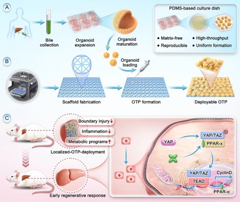
Background: Early recovery after major hepatectomy may depend not only on remnant liver volume, but also on whether the injury-adjacent remnant-liver boundary can withstand the narrow postoperative convergence of mechanical disruption, inflammatory amplification, and transient metabolic insufficiency that may otherwise divert the remnant liver away from productive regeneration.
Methods: We engineered an organoid tissue patch (OTP) by integrating matrix-free expanded human bile-derived liver organoids with a reticulated microfibrous scaffold for localized deployment on the remnant liver surface. OTP was evaluated in a murine 70% partial hepatectomy model using survival, serum biochemical, histological, and molecular analyses, together with pharmacologic PPARα inhibition.
Results: OTP treatment was associated with improved survival and biochemical recovery, together with attenuated tissue injury in the predefined patch-placement region. At Day 3, OTP-treated tissue showed reduced NF-κB-linked inflammatory activity, lower inflammatory mediator expression, and an increased CD206-associated macrophage signal. Metabolism- and proliferation-related programs were concurrently enriched, accompanied by increased PPARα immunoreactivity, YAP-positive nuclei, and hepatocyte proliferative entry. Systemic PPARα inhibition partially attenuated these effects, supporting a functional contribution of PPARα-responsive signaling.
Conclusions: These findings support localized OTP deployment as a strategy to provide temporary bioactive support to the vulnerable remnant-liver boundary during endogenous recovery, without evidence of bulk donor tissue replacement.
Keywords: organoid tissue patch, injury-adjacent remnant-liver boundary, hepatectomy, liver regeneration, PPARα, localized tissue support
Introduction
Liver regeneration after major hepatectomy is commonly interpreted through remnant liver volume and the intrinsic proliferative capacity of the residual parenchyma [1, 2]. Yet early recovery may also depend on the condition of the newly exposed injury-adjacent remnant-liver boundary, where local tissue injury may evolve before regenerative responses are fully engaged [3, 4]. At this boundary, mechanical disruption, inflammatory amplification, and transient metabolic insufficiency converge within a narrow early window, potentially shifting the remnant liver away from productive regeneration before organ-level recovery is established [5-8]. Failure to stabilize this vulnerable region may exacerbate local tissue injury, propagate inflammatory responses, and delay functional recovery [9-11]. This perspective reframes early postoperative recovery as a problem not only of whether the remnant liver can regenerate, but also of whether the local boundary environment can remain regenerative-permissive during its most vulnerable interval [12, 13].
Current regenerative strategies are not specifically designed to address this spatially restricted challenge. Many regenerative approaches still prioritize cell potency, delivery dose, or post-transplant survival as the main determinants of repair [1, 14]. However, in the setting of hepatectomy, such approaches do not directly address the spatially restricted, mechanically stressed, and metabolically burdened surface region of the remnant liver [15, 16]. Injection-based delivery suffers from poor spatial control and extensive post-transplant cell loss [17, 18], while bulk hydrogels and larger tissue constructs are constrained by diffusion limitations, limited conformability, and deployment complexity in acute surgical settings [19-25]. Collectively, these limitations reveal a persistent mismatch between current regenerative strategies and the early postoperative boundary environment [26, 27]. What appears to be missing is not simply greater cellular abundance [28, 29], but a surgically deployable format that can remain locally positioned and provide temporary biological support until endogenous repair is effectively engaged [16, 30, 31]. This need is particularly relevant to organoid-based therapeutics, whose functional potential may be undermined by poor perioperative retention and the lack of suitable delivery formats.
We therefore developed a deployable organoid tissue patch (OTP) by integrating human bile-derived organoids expanded under matrix-free microwell conditions with a reticulated microfibrous scaffold for application to the remnant liver surface adjacent to the resection plane. OTP was designed not simply as a carrier for transplanted cells or as a passive physical barrier, but as a temporary source of localized bioactive support during the early postoperative phase. The scaffold provides conformability, local retention, and surgical handling, whereas the organoid component is intended to provide localized biological cues during the narrow interval in which the remnant liver is exposed to pronounced mechanical, inflammatory, and metabolic stress [32-35]. The purpose is not to replace resected liver mass, but to support the injury-adjacent remnant-liver boundary when mechanical stress, inflammatory spillover, and transient functional overload are most likely to precipitate early collapse [36-38]. To suit this application, the organoids were expanded under matrix-free microwell conditions, generating a more uniform dBDO population with enhanced hepatocyte-like and metabolism-associated features before scaffold integration.
We next tested whether localized OTP deployment could improve early postoperative recovery in a murine 70% hepatectomy model, which imposes abrupt loss of functional liver mass together with a narrow postoperative window of inflammatory, mechanical, and metabolic stress [39-42]. We assessed local OTP retention, biochemical and histological recovery, and inflammatory, metabolic, and regenerative responses within the predefined patch-placement region of the remnant liver. We further used pharmacological PPARα inhibition to examine the functional contribution of PPARα-responsive signaling to the OTP-associated recovery phenotype. The resulting findings support a boundary-directed model in which OTP treatment is associated with a less inflammatory and more regeneration-compatible local host-tissue response, rather than bulk donor tissue replacement.
Materials and Methods
Fabrication and mechanical characterization of bioengineered microfibrous scaffolds
Bioengineered microfibrous scaffolds featuring a reticulated node microarchitecture were fabricated using melt electrowriting (MEW), an additive manufacturing technique that enables predefined control over fiber diameter and in-plane fiber spacing. Patches were printed using a MEW system (BP6601, Yongqinquan Intelligent Equipment, China) with medical-grade poly (ε-caprolactone) (PCL; EFL-PCL-80K). To mitigate inter-fiber electrostatic interactions during fabrication, a stepwise in-plane translation strategy was applied. For the 0° orientation, fibers were initially deposited at a spacing of 150 μm, followed by two successive in-plane passes with lateral offsets of 50 μm each. This staged deposition strategy reduces electrostatic interference between adjacent fibers and promotes uniform node formation. The resulting microarchitecture consisted of microfibers (diameter 5.32 ± 0.70 μm) interconnected by eight-rayed asterisk-shaped reticulated nodes, yielding characteristic pore dimensions of 50.01 ± 0.53 μm (0° orientation) and 34.08 ± 2.50 μm (45° orientation). Pore areas were quantified using Fiji and analyzed with Origin 2025b. Structural stability was assessed by incubating scaffolds in phosphate-buffered saline (PBS) at 37 °C under agitation (200 rpm), followed by metallurgical microscopy evaluation on days 0, 3, and 7. Tensile properties were measured using a universal testing machine (Zwick GmbH & Co. KG, Germany).
Bile-derived organoid (BDO) culture and hepatic differentiation
Human bile-derived organoids (BDOs) were established from bile aspirates obtained during clinically indicated endoscopic retrograde cholangiopancreatography (ERCP). The protocol was adapted from previously reported bile-derived and cholangiocyte organoid culture methods [43]. Bile samples were collected from adult patients with benign, non-malignant biliary diseases, mainly choledocholithiasis or benign biliary obstruction. Patients with known or suspected hepatobiliary or pancreatic malignancy were excluded. The available donor information, including underlying disease, inflammatory or obstructive status, and exclusion criteria, is summarized in Supplementary Table S1. All protocols for human bile aspirates were approved by the Clinical Research Ethics Committee of The First Affiliated Hospital, Zhejiang University School of Medicine (reference No. 2021–158).
Briefly, human bile samples (3–10 mL) were collected under sterile conditions and kept on ice until processing within 1 h. Samples were centrifuged at 400 × g for 5 min at 4 °C to recover bile-associated cellular material, followed by one wash with cold PBS containing 1% bovine serum albumin (BSA). The cell pellets were filtered through a 70 µm strainer, counted, and assessed for viability using Trypan Blue exclusion.
The cell pellet was resuspended in Matrigel (Corning), and 30–50 μL droplets were plated into 24-well plates and allowed to polymerize at 37 °C for 30 min. After gelation, 500 μL of human liver initiation medium (h-IM) was added to each well. The culture medium was subsequently replaced with human liver expansion medium (EM) after 5–7 days. For hepatic differentiation, proliferative BDOs were switched to human liver differentiation medium (DM) for 5 days, leading to the formation of hepatocyte-like, functionally differentiated organoids (dBDOs). For matrix-free aggregation and subsequent scaffold loading, organoids were transferred to microwell culture as detailed below.
Matrix-free microwell culture of bile-derived liver organoids
To enable scalable and translationally oriented organoid expansion, Matrigel-embedded BDOs were dissociated into single cells using TrypLE Express (Gibco, 37 °C, 5–7 min), neutralized with EM, and resuspended at 2.5 × 10⁵ cells per 35-mm plate. Cells were seeded onto PDMS-based microwell plates (EFL-SP102, 595 microwells per plate; well diameter 520 µm, depth 500 µm; EFL & Liangzhu Laboratory) pre-treated to remove trapped air (< -0.08 MPa, 60 s) and UV-sterilized. A 200 µL cell suspension was evenly spread over the microwell array region and allowed to settle by gravity for 10 min before adding 2 mL EM gently along the dish wall. Plates were maintained undisturbed for the first 24 h to promote uniform spheroid formation. Medium was changed every 2–3 days. After 3–5 days, compact Matrigel-free spheroids (eBDOs or dBDOs) were harvested by gentle pipetting and brief centrifugation (800 rpm, 3–5 min) for downstream expansion, differentiation, or scaffold loading.
Preparation of organoid tissue patches (OTPs) and seeding of dBDOs
OTP scaffolds were sterilized and surface-modified prior to organoid seeding. After fabrication, scaffolds were immersed in 0.1 mol/L sodium hydroxide solution for 5 min to increase fiber surface roughness, followed by three washes with sterile phosphate-buffered saline (PBS). To enhance cell attachment, scaffolds were incubated with a cell-adherent reagent (Cat# C1010; diluted 1:250 in PBS; Cellcook, China) for 1 h at room temperature. Scaffolds were then immersed in 75% ethanol for at least 4 h for sterilization and subsequently rinsed thoroughly with sterile PBS. Organoids were collected, counted, and resuspended at a density of approximately 1 × 10⁷ cells/mL. Approximately 100 μL of the organoid suspension was evenly seeded onto each scaffold and incubated at 37 °C for 45–60 min to allow initial attachment. During this period, organoids settled into the inter-fiber gaps under surface tension. After initial adherence, excess liquid was gently removed, and scaffolds were transferred into 2 mL of culture medium for continued incubation.
Viability assessment of long-term cryopreserved OTPs
To evaluate long-term viability following cryopreservation, assembled OTPs were stored in a commercial serum-free cryopreservation medium (CS10) at –196 °C in liquid nitrogen. Viability was assessed at three predefined time points: 1 week, 3 months, and 6 months post-freezing. At each time point, OTPs were rapidly thawed in a 37 °C water bath and transferred to a cell culture incubator for 2 h to allow stabilization prior to analysis. Control groups included: (i) freshly cultured dBDOs without scaffolds (Control), and (ii) freshly prepared OTPs analyzed 1 h after seeding. For viability assessment, OTPs were gently washed with PBS and incubated with Live/Dead staining solution containing Calcein-AM and propidium iodide (PI) according to the manufacturer’s instructions (Beyotime Biotechnology, China). Viability was assessed based on the relative abundance of Calcein-AM-positive viable signals and PI-positive dead-cell signals within organoid structures loaded on the scaffold.
Periodic acid–Schiff staining of liver organoids
Periodic acid–Schiff (PAS) staining was performed to assess glycogen accumulation in liver organoids. Organoids were released from Matrigel by washing three times with ice-cold basal medium and directly fixed in 4% paraformaldehyde (PFA) for 30 min at room temperature. Following fixation, samples were washed with PBS and stained using a commercial PAS staining kit (Solarbio, Beijing, China) according to the manufacturer’s instructions. Briefly, organoids were incubated with periodic acid solution for 10 min, rinsed with distilled water, then treated with Schiff reagent for 15 min in the dark. After washing, samples were directly imaged under a bright-field microscope (Leica Microsystems) without hematoxylin counterstaining.
Indocyanine green uptake and release assay
Indocyanine green (ICG) uptake and release assays were performed to assess hepatic functional capacity. Organoids were isolated from Matrigel and resuspended in basal medium containing 1 mg/mL ICG (MCE, Shanghai, China). After incubation at 37 °C for 1 h, organoids were washed three times with cold PBS and imaged under a bright-field microscope to assess ICG uptake. For release assessment, organoids were transferred to ICG-free medium and incubated for an additional 6 h. Residual ICG signal was visualized and quantified by imaging at the indicated time points. Representative images were captured using identical exposure settings across all groups.
Quantification of secreted albumin by ELISA
The level of albumin secreted by organoids under different culture conditions was measured using a commercial ELISA kit (Cloud-Clone Corp., China), according to the manufacturer’s instructions. Organoids were cultured under five distinct conditions: embedded in Matrigel and maintained in expansion medium (EM); embedded in Matrigel and cultured in differentiation medium (DM); seeded on PDMS-based microwells in EM or DM, respectively; and control conditions. Conditioned medium was collected and centrifuged at 300 × g for 5 min at 4 °C to remove debris. Samples and standards were added to 96-well plates pre-coated with capture antibodies and incubated for 1–2 h at 37 °C. After washing, detection antibody solution was added, followed by HRP-conjugated secondary antibody. The TMB substrate was added and allowed to react for 10–20 min in the dark, and the enzymatic reaction was stopped using stop solution. Absorbance was read at 450 nm using a microplate reader (BioTek Synergy). Albumin concentrations were calculated based on a standard curve generated from known concentrations.
Immunofluorescence and immunohistochemical staining of human organoids
Human organoids were harvested and washed three times with ice-cold basal medium (BM) to remove residual Matrigel. Organoids were then fixed in 4% paraformaldehyde for 1 h at room temperature. After fixation, samples were permeabilized with 0.1% Triton X-100 in PBS and blocked for 1 h in the same buffer supplemented with 2% fetal calf serum (FCS). Primary antibodies were added and incubated overnight at 4 °C on a shaker in 24-well plates. The following day, samples were washed three times in PBS (40 min per wash), incubated with secondary antibodies for 2 h at room temperature, and subsequently washed again. Organoids were resuspended in PBS, counted, and mounted for imaging. Fluorescent images were acquired using a Zeiss LSM 710 confocal microscope under consistent exposure settings. Antibodies used in this study are listed in Supplementary Table S2.
RNA isolation and quantitative real-time PCR (qRT-PCR)
Total RNA was extracted from mouse liver tissues using TRIzol reagent (Life Technologies, Carlsbad, CA, USA) according to the manufacturer’s protocol. Complementary DNA (cDNA) was synthesized using the HiScript III RT SuperMix with gDNA wiper (Vazyme, Nanjing, China). Quantitative real-time PCR was carried out using the ChamQ Universal SYBR qPCR Master Mix (Vazyme) on an Applied Biosystems QuantStudio 5 instrument (Thermo Fisher Scientific, Waltham, MA, USA). Gene expression levels were calculated using the 2^–ΔΔCT method. Primer sequences are provided in Supplementary Table S3.
RNA sequencing and bioinformatics analysis
Total RNA was extracted from liver organoids cultured under different conditions and from mouse liver tissues using TRIzol reagent (Thermo Fisher Scientific, 15596018) according to the manufacturer’s instructions. RNA quantity and purity were assessed using a NanoDrop ND-1000 spectrophotometer, and RNA integrity was evaluated using an Agilent Bioanalyzer 2100. Samples with RNA concentration > 50 ng/μL, total RNA amount > 1 μg, and RNA integrity number (RIN) > 7.0 were used for library construction. Four biological replicates were included for each organoid RNA-seq group.
Poly(A)+ mRNA was enriched from total RNA using oligo(dT) magnetic beads and fragmented at 94 °C for 5–7 min. First-strand cDNA was synthesized using random primers and reverse transcriptase, followed by second-strand cDNA synthesis with E. coli DNA polymerase I, RNase H, and dUTP. After end repair, A-tailing, adapter ligation, size selection, and PCR amplification, strand-specific cDNA libraries with an average insert size of approximately 300 ± 50 bp were generated. Libraries were sequenced using a 2 × 150 bp paired-end strategy on an Illumina NovaSeq 6000 platform by LC-Bio Technology Co., Ltd. (Hangzhou, China). Sequencing generated 32.4–41.9 million valid reads per sample, with 97.67–98.27% of reads mapped to the reference genome and 89.88–94.94% uniquely mapped.
Raw sequencing reads were processed for quality control before downstream analysis. Adapter sequences, polyA/polyG-containing reads, reads with more than 5% unknown nucleotides, and low-quality reads containing more than 20% bases with Q ≤ 20 were removed using Cutadapt v1.9. Sequencing quality metrics, including Q20, Q30, and GC content, were evaluated using FastQC v0.11.9. Clean reads from human organoid samples were aligned to the human reference genome, and mouse liver tissue reads were aligned to the mouse reference genome using HISAT2 v2.2.1.
Mapped reads were assembled and quantified using StringTie v2.1.6, and transcriptome comparison was performed using gffcompare v0.9.8. Gene expression abundance was calculated as FPKM values for visualization and exploratory analyses. Differential expression analysis was performed using the DESeq2 package. Differentially expressed genes (DEGs) were defined as genes with an absolute fold change ≥ 2 and a false discovery rate (FDR) < 0.05. Data visualization included volcano plots, hierarchical clustering heatmaps, and principal component analysis (PCA) to evaluate inter-sample variation and group clustering.
Functional annotation and pathway enrichment analyses were performed using the Gene Ontology (GO) and Kyoto Encyclopedia of Genes and Genomes (KEGG) databases. Significantly enriched GO terms and KEGG pathways among DEGs were identified using a hypergeometric test. Gene Set Enrichment Analysis (GSEA) was performed using GSEA software v4.1.0 and MSigDB gene sets. Genes were ranked using the Signal2Noise method, and enriched pathways were evaluated using the normalized enrichment score (NES), nominal P value, and FDR q value. Gene sets with |NES| > 1, nominal P < 0.05, and FDR q < 0.25 were considered enriched.
The RNA-seq data generated in this study have been deposited in the National Genomics Data Center (NGDC), China National Center for Bioinformation, under accession number HRA019744 and CRA046568, respectively.
Partial hepatectomy mouse model
FRG mice (Fah⁻/⁻Rag2⁻/⁻Il2rg⁻/⁻) used in this study were purchased from BioRigino (Zhejiang, China). Throughout the experimental period, FRG mice were continuously maintained on nitisinone (NTBC; PHR1731, Supelco, Merck, Darmstadt, Germany) administered in the drinking water at a working concentration of 4 μg/mL. NTBC was not withdrawn before or after partial hepatectomy. All animal procedures were performed in accordance with the Guide for the Care and Use of Laboratory Animals (NIH publication No. 80-23) and were approved by the Animal Care Ethics Committee of The First Affiliated Hospital, Zhejiang University School of Medicine (no. 2020–1088). Mice were housed in a controlled environment (20–26 °C, 40–70% humidity, 12-h light/dark cycle starting at 6:00 a.m.) with ad libitum access to autoclaved chow and purified water. After overnight fasting, mice underwent 70% partial hepatectomy (ePH) under anesthesia, as previously described by Mitchell and Willenbring, with removal of the median and left lateral liver lobes. Animals were randomly assigned to four groups: CTRL (sham), Model (ePH), Scaffold (ePH + scaffold), OTP (ePH + OTP). Immediately after surgery, scaffold-only controls or assembled OTPs were gently applied to the exposed surface of the remnant liver. The abdominal cavity was then closed, and mice were recovered under temperature-controlled conditions. Subsets of mice were sacrificed on Day 3 for early inflammatory, regenerative, and transcriptomic analyses, on Day 14 for short-term histological and tissue-repair assessment, and at 2 months after surgery for long-term safety evaluation.
For tissue sampling, the predefined patch-placement region of the remnant liver was defined as the subcapsular remnant liver tissue corresponding to the standardized scaffold/OTP placement site. Because the external scaffold frame was removed before implantation and the implanted material was biodegradable, sampling was not guided by residual scaffold edges at harvest. Instead, liver tissue was collected according to the standardized intraoperative placement site and anatomical landmarks, while avoiding large vessels, hilar structures, and ligation stumps. For histological and immunostaining analyses, tissue blocks were oriented to include the liver capsule and underlying subcapsular parenchyma from this predefined region. For qPCR, western blotting, and bulk RNA-seq, tissue was collected from the same standardized anatomical region. In ePH-only mice, the anatomically corresponding remnant liver surface region was sampled using the same criteria.
Pharmacologic inhibition of PPARα using GW6471
To examine the functional contribution of PPARα-responsive signaling to OTP-associated recovery, mice subjected to 70% partial hepatectomy were randomly assigned to the OTP, OTP + GW6471, and GW6471-only groups. For OTP treatment, dBDO-loaded organoid tissue patches were placed onto the capsular surface of the remnant liver adjacent to the resection plane immediately after hepatectomy, as described above. GW6471, a selective pharmacological antagonist of PPARα, was administered once by intraperitoneal injection at 20 mg/kg immediately after surgery. In the OTP + GW6471 group, GW6471 was administered immediately after OTP placement. Mice in the OTP group received an equal volume of vehicle. Serum and tissue from the predefined patch-placement region of the remnant liver were collected at Day 3 after surgery for serum biochemical analysis, qPCR, immunofluorescence staining, and histological evaluation. In the GW6471-only group, the anatomically corresponding remnant-liver surface region was collected using the same standardized anatomical landmarks.
In vivo tracking of OTPs in mice
Human bile-derived organoids (1 × 10⁶ cells in 100 μL PBS) were labeled with 5 μM DiR dye (Thermo Fisher Scientific, D12731) according to the manufacturer’s instructions prior to assembly. DiR-labeled organoids were then incorporated into organoid tissue patches (OTPs) as described above. Following a 70% partial hepatectomy performed under isoflurane anesthesia, DiR-labeled OTPs were placed onto the capsular surface of the remnant liver adjacent to the resection plane in FRG mice. Age- and sex-matched untreated FRG mice served as controls. In vivo fluorescence signals were acquired using an IVIS Spectrum imaging system (PerkinElmer) at Day 3 post-transplantation. For ex vivo analysis, major organs—including the heart, liver, spleen, lungs, and kidneys—were harvested and imaged immediately after sacrifice. Fluorescence signals were detected using excitation and emission filters set at 750 nm and 800 nm, respectively.
Histological, immunofluorescence, immunohistochemical, and serum biochemical analyses
Mouse liver tissues were harvested at the indicated time points and fixed in 4% (w/v) paraformaldehyde (PFA) for 24 h. Fixed tissues were dehydrated, embedded in paraffin, and sectioned at 5 μm thickness. For histological evaluation, sections were stained with hematoxylin and eosin (H&E) according to standard protocols. For immunofluorescence (IF) and immunohistochemical (IHC) analyses, paraffin sections were deparaffinized, rehydrated, and subjected to heat-mediated antigen retrieval. IF and IHC staining were performed following standard protocols, and primary antibodies are listed in Supplementary Table S2.
For serum biochemical analyses, whole blood was collected from mice and centrifuged at 3,500 rpm for 10 min at 20 °C to obtain serum. Serum levels of aspartate aminotransferase (AST), alanine aminotransferase (ALT), blood urea nitrogen (BUN), creatinine (CRE), and total bilirubin (TBIL) were measured using a dry chemistry analyzer (DRI-CHEM 4000i; Fujifilm Corp., Tokyo, Japan) according to the manufacturer’s instructions.
Western blot analysis
Total protein was extracted from tissue collected on Day 3 from the predefined patch-placement region of the remnant liver using RIPA lysis buffer supplemented with protease and phosphatase inhibitors. Protein concentrations were determined using a BCA protein assay kit. Equal amounts of protein were separated by SDS-PAGE and transferred onto PVDF membranes. After blocking with 5% bovine serum albumin in TBST, membranes were incubated overnight at 4 °C with primary antibodies against p-p65, total p65, and β-actin, followed by HRP-conjugated secondary antibodies. Detailed information on the antibodies used is provided in Supplementary Table S2. Protein bands were visualized using enhanced chemiluminescence and quantified with ImageJ. NF-κB-linked signaling activity was assessed by the p-p65/p65 ratio, with β-actin used as a loading control.
Statistical analysis
All quantitative data are expressed as mean ± standard error of the mean (SEM) from at least three independent experiments, unless otherwise specified. Statistical comparisons between two groups were performed using two-tailed unpaired Student’s t-test, while multiple-group comparisons were assessed using one-way analysis of variance (ANOVA) followed by appropriate post hoc tests. Survival curves were analyzed using the log-rank (Mantel–Cox) test. Statistical analyses were conducted using GraphPad Prism version 10 (GraphPad Software, San Diego, CA, USA). A P value of less than 0.05 was considered statistically significant.
Results
Microwell confinement establishes a uniform dBDO baseline with enhanced hepatocyte-like and metabolism-associated features
To develop a reproducible and translationally oriented strategy for organoid expansion, we established bile-derived organoids (BDOs) from patient bile samples. When maintained in expansion medium (EM), these organoids were defined as expansion-stage BDOs (eBDOs), whereas transfer to differentiation medium (DM) promoted the acquisition of hepatocyte-like features, generating differentiated BDOs (dBDOs). Consistent with an expansion-stage biliary epithelial state, eBDOs displayed the typical cystic morphology of proliferative bile-derived organoids and expressed the progenitor markers SOX9 and CK19 together with the proliferation markers Ki67 and PCNA (Figure S1). A schematic of the matrix-free microwell workflow and the design of the PDMS-based hexagonal microwell plate are shown in Figure 1A–B.
Matrix-free microwell culture supports more uniform organoid growth and enhanced hepatocyte-like features of bile-derived organoids. (A) Schematic of the matrix-free microwell workflow for establishing bile-derived organoids (BDOs) from patient bile samples, followed by organoid expansion and hepatic differentiation. (B) Representative images of the PDMS-based microwell culture plate and magnified view of individual hexagonal microwells. (C) Representative bright-field images of organoids cultured in the microwell system and in Matrigel, showing improved morphological uniformity under microwell confinement. Scale bars, 100 μm. (D) Relative cell number of organoids cultured in Matrigel and in the microwell system over 9 days. (E) Diameter measurements of organoids cultured in the microwell system and in Matrigel at Day 4. (F) Albumin levels in culture supernatants from Matrigel- and microwell-cultured organoids at the indicated time points. Cell-free differentiation medium (DM) served as the baseline control. (G) Representative images of indocyanine green (ICG) uptake and release, and periodic acid–Schiff (PAS) staining in Matrigel- and microwell-cultured organoids. Scale bars, 50 μm. (H) Quantification of PAS signal intensity and ICG uptake in Matrigel- and microwell-cultured organoids. (I) Immunofluorescence staining of microwell-cultured differentiated BDOs (dBDOs) for SOX9 and ALB, and for HNF4A and CYP3A4. Scale bars, 50 μm. (J) Volcano plot of differentially expressed genes between microwell-cultured and Matrigel-cultured dBDOs. (K) Selected gene set enrichment analyses showing enrichment of metabolic programs in microwell-cultured dBDOs, including PPAR signaling, bile acid biosynthetic process, xenobiotic metabolic process, and the citrate cycle (TCA cycle). Data are presented as mean ± s.e.m. *P < 0.05, **P < 0.01, ****P < 0.0001; ns, not significant.
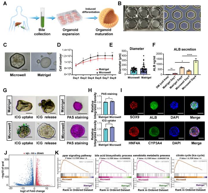
Time-course imaging further documented organoid behavior within this confined system: during the expansion phase, eBDOs underwent progressive epithelial compaction and growth from Day 1 to Day 4 (Figure S2A), whereas after induction with DM, dBDOs continued to enlarge while maintaining a stable spheroid morphology through Day 9 (Figure S2B). Compared with Matrigel-grown organoids, microwell-cultured organoids displayed smoother contours and markedly reduced size heterogeneity (Figure 1C). Cell growth assays confirmed a progressive increase in cell numbers in both systems, with microwell-cultured organoids exhibiting greater cell yields and lower inter-sample variability over the nine-day culture period. During early expansion, microwell culture generated spheroids with a smaller mean diameter and a narrower size distribution than Matrigel culture (Figure 1D-E).
Microwell confinement was associated with enhanced hepatocyte-like functional features. Microwell-cultured dBDOs showed higher concentrations of secreted albumin in the culture supernatant than Matrigel-cultured controls (Figure 1F). They also demonstrated ICG uptake followed by release and showed stronger PAS staining (Figure 1G–H), consistent with enhanced hepatocyte-like transport activity and glycogen-associated features. Immunofluorescence staining further demonstrated that these organoids retained expression of the epithelial/progenitor-associated marker SOX9 while concomitantly expressing hepatocyte-associated markers, including ALB, HNF4A, and CYP3A4 (Figure 1I). These findings indicate acquisition of hepatocyte-like features while retaining SOX9 expression.
To further characterize the transcriptional programs associated with these hepatocyte-like features, bulk RNA sequencing was performed on microwell-cultured and Matrigel-cultured dBDOs collected from both groups after 5 days of hepatic differentiation followed by 3 additional days under the indicated culture conditions. Differential expression analysis revealed broad transcriptomic remodeling in microwell-cultured organoids (Figure 1J), and heatmap visualization further showed coherent segregation of the two transcriptional states (Figure S3A). Gene Ontology enrichment analysis highlighted terms related to lipid metabolic processes, oxidoreductase activity, and cytosolic/extracellular remodeling (Figure S3B), whereas KEGG analysis indicated pathway-level shifts involving metabolism- and organization-related programs (Figure S3C). Selected gene set enrichment analyses further showed enrichment of liver-associated metabolic programs, including PPAR signaling, bile acid biosynthetic processes, xenobiotic metabolism, and the citrate cycle (TCA cycle) (Figure 1K), and additional supplementary GSEA plots supported enrichment of hepatocyte-associated metabolic pathways in microwell-cultured dBDOs (Figure S3D). Collectively, these findings indicate that microwell confinement generates a more uniform dBDO population with enhanced hepatocyte-like functional readouts and transcriptional enrichment of multiple metabolism-associated programs before scaffold integration.
A reticulated microfibrous scaffold enables deployable OTP assembly while preserving selected hepatocyte-like features
Having established a matrix-free platform for organoid expansion and hepatocyte-like differentiation, we next sought to define structural and translational design rules for an assembled organoid tissue patch (OTP) suitable for transplantation. Using melt electrowriting (MEW), we engineered a reticulated-node poly (ε-caprolactone) (PCL) microfibrous scaffold, enabling precise control over fiber orientation, spacing, and pore geometry (Figure 2A). For scaffold-based organoid delivery, two structural constraints are critical: an open architecture compatible with organoid loading and medium access, and mechanical robustness to preserve architectural integrity under physiological deformation. To compare these properties, three geometric configurations—square, triangular, and reticulated-node configurations—were fabricated with identical fiber spacing and diameter (Figure 2B) and systematically compared. Quantitative analysis revealed pronounced differences in pore architecture (Figure 2C), with the reticulated-node configuration exhibiting the smallest mean pore area (1211.28 ± 425.96 μm²), substantially lower than both square (4011.14 ± 166.83 μm²) and triangular (2033.84 ± 515.58 μm²) configurations. Mechanical testing further demonstrated that tensile resistance scaled with fiber support density and orientation (Figure 2D). The square configuration showed the lowest Young’s modulus at 45° (1.83 ± 0.20 kPa), reflecting its anisotropic alignment, whereas the reticulated-node configuration displayed superior mechanical performance in both tested orientations (20.28 ± 0.70 kPa at 0° and 24.32 ± 1.43 kPa at 45°) (Figure 2E). Together, these properties supported the selection of the reticulated-node configuration, which combined relatively small pore dimensions with orientation-resistant mechanical performance, for subsequent optimization. Based on the selected reticulated-node configuration, path optimization was subsequently applied to generate the final scaffold architecture. The optimized reticulated-node scaffold exhibited a fiber diameter of 5.32 ± 0.70 μm, a spacing of 50.01 ± 0.53 μm at 0°, and 34.08 ± 2.50 μm at 45°. This refinement reduced the mean pore area to 426.06 ± 286.40 μm² and shifted the pore size distribution toward smaller pores (Figure 2F). Mechanical testing revealed a ductile tensile response with stress increasing nonlinearly with engineering strain, and a maximum tensile load of 0.16 ± 0.01 N corresponding to a nominal tensile stress of 0.47 ± 0.14 MPa (Figure 2G). Continuous agitation testing at 200 rpm and 37 °C showed no obvious loss of scaffold architecture over time, supporting the structural stability of the optimized reticulated-node scaffold under handling-related conditions (Figure 2H).
A reticulated microfibrous scaffold enables construction of a deployable organoid tissue patch (OTP). (A) Schematic of the melt electrowriting (MEW)-based workflow used to fabricate poly (ε-caprolactone) (PCL) microfibrous scaffolds for OTP construction. (B) Representative micrographs of three scaffold unit-cell architectures printed with matched fiber spacing and diameter: square, triangular, and reticulated-node configurations. (C) Quantification of pore area across the square, triangular, and reticulated-node configurations shown in B. (D) Representative stress–strain curves showing orientation-dependent mechanical behavior of the three scaffold configurations. (E) Young’s modulus values of the indicated scaffold designs, supporting selection of the reticulated-node configuration for subsequent optimization. (F) Representative overview image of the final optimized reticulated-node PCL scaffold, together with the corresponding pore-area distribution. (G) Representative stress–strain curve of the optimized reticulated-node scaffold. (H) Structural stability of the optimized reticulated-node scaffold under continuous agitation at 200 rpm and 37 °C over time. (I) Bright-field images showing progressive attachment and coverage of scaffold-loaded dBDOs over 3 days. Scale bars, 100 μm. (J) Immunofluorescence staining of scaffold-associated dBDOs for HNF4A and CYP3A4. Scale bars, 100 μm. (K) Quantification of HNF4A⁺ and CYP3A4⁺ cells in scaffold-associated dBDOs. (L) Confluence rates of OTPs seeded at different loading densities. (M) Live/dead quantification of fresh OTPs and post-thaw OTPs after 1 week, 3 and 6 months of cryopreservation in CS10 medium. n = 3 per group. Data are presented as mean ± s.e.m. for quantified panels. **P < 0.01, ***P < 0.001.
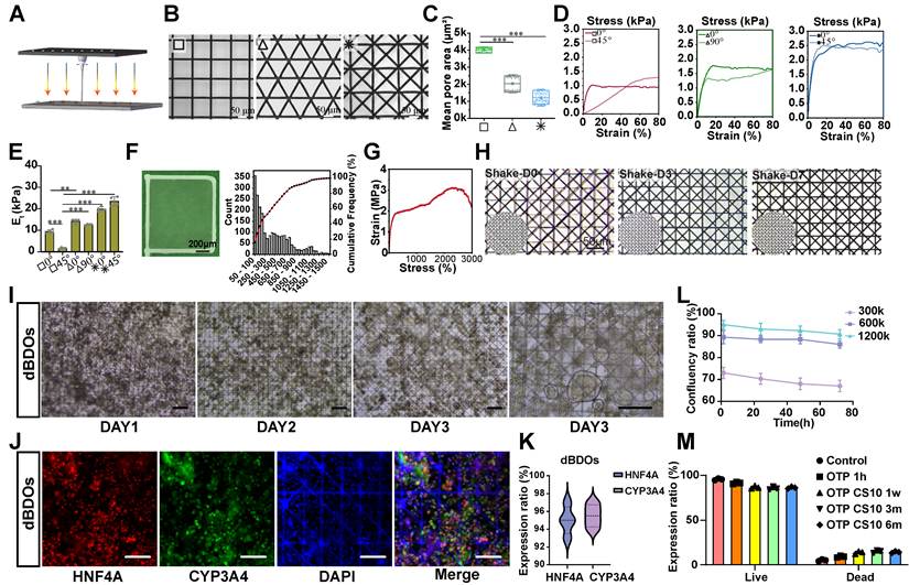
We next examined whether the optimized reticulated-node scaffold could support high-density dBDO loading while preserving organoid morphology and hepatic marker expression. After seeding onto the optimized scaffold, dBDOs attached to the fiber network and maintained spheroid-like morphology over several days (Figure 2I). Immunofluorescence staining demonstrated continued expression of the hepatic markers HNF4A and CYP3A4 in scaffold-associated dBDOs (Figure 2J–K). Confluence analysis further showed that the highest seeding density (1.2 × 10⁶ cells) achieved the most complete and stable surface coverage over 72 h, indicating efficient scaffold colonization under high loading conditions (Figure 2L). Organoid-loaded patches also retained high live-cell percentages after cryopreservation for up to 6 months, supporting the feasibility of post-thaw OTP recovery for on-demand preparation (Figure 2M).
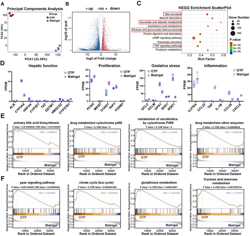
To characterize the transcriptional state of OTP-associated dBDOs relative to conventionally Matrigel-cultured dBDOs, bulk RNA sequencing was performed on OTP-associated dBDOs and compared with Matrigel-cultured dBDOs. Principal component analysis revealed clear separation between the two conditions, indicating a distinct transcriptional state in OTP-associated dBDOs (Figure 3A). Consistently, volcano plot analysis and heatmap visualization showed broad transcriptomic remodeling between OTP-associated and Matrigel-cultured dBDOs (Figure 3B and Figure S4A). KEGG pathway analysis highlighted pathway-level remodeling involving hepatic metabolic and adaptive signaling programs, including bile secretion, porphyrin metabolism, arachidonic acid metabolism, ascorbate and aldarate metabolism, pentose and glucuronate interconversions, TNF signaling, and cAMP signaling (Figure 3C). Complementary GO enrichment analysis showed remodeling of cellular programs linked to membrane-, cytoskeleton-, extracellular-, endoplasmic-reticulum-, and metal ion-binding–related terms in OTP-associated dBDOs (Figure S4B). Analysis of selected genes showed that representative hepatocyte-associated transcripts remained expressed in OTP-associated dBDOs, together with differences in genes related to oxidative stress responses and inflammatory signaling (Figure 3D). Gene set enrichment analyses supported enrichment of hepatocyte-associated metabolic programs, including primary bile acid biosynthesis, cytochrome P450–mediated drug metabolism, xenobiotic metabolism, and drug metabolism by other enzymes (Figure 3E). Additional enrichment analyses indicated metabolic and stress-adaptive features, including PPAR signaling, the citrate cycle, glutathione metabolism, and fructose/mannose metabolism (Figure 3F). RNA-seq–derived SNV annotation did not reveal obvious shifts in expressed variant category distributions between OTP-associated and Matrigel-cultured dBDOs (Figure S4C). Supplementary GSEA further suggested modulation of Hippo-associated and inflammatory mediator/TRP channel–related signaling programs (Figure S4D–E).
Scaffold-associated dBDOs retain selected hepatocyte-like features and exhibit metabolic and stress-adaptive transcriptional differences. (A) Principal component analysis showing separation between OTP-associated dBDOs and Matrigel-cultured dBDOs. (B) Volcano plot of differentially expressed genes between OTP-associated and Matrigel-cultured dBDOs. (C) KEGG enrichment analysis showing pathway-level remodeling in OTP-associated dBDOs, including bile secretion, pentose and glucuronate interconversions, porphyrin metabolism, arachidonic acid metabolism, ascorbate and aldarate metabolism, TNF signaling, and cAMP signaling. (D) Expression levels of representative genes associated with hepatic function, proliferation, oxidative stress defense, and inflammatory signaling in OTP-associated and Matrigel-cultured dBDOs. (E) Gene set enrichment analyses showing enrichment of hepatocyte-associated metabolic programs in OTP-associated dBDOs, including primary bile acid biosynthesis, cytochrome P450–mediated drug metabolism, xenobiotic metabolism, and drug metabolism by other enzymes. (F) Gene set enrichment analyses showing enrichment of metabolic and stress-adaptive programs in OTP-associated dBDOs, including PPAR signaling, the citrate cycle (TCA cycle), glutathione metabolism, and fructose and mannose metabolism.
Because microwell culture itself induced hepatocyte-like transcriptional changes, we further compared OTP-associated dBDOs with PDMS microwell-cultured dBDOs at the same differentiation stage to distinguish scaffold-associated effects from matrix-free aggregation alone. PCA showed clear separation between the two groups, and differential expression analysis identified 1,873 upregulated and 2,151 downregulated genes in OTP-associated dBDOs (Figure S5A–C). This comparison indicated that scaffold loading did not simply amplify the microwell-induced hepatic-like program. Instead, OTP-associated dBDOs showed additional changes related to matrix/interface adaptation, epithelial organization, and stress-adaptive remodeling, while hepatocyte-associated genes displayed a mixed pattern (Figure S6A). GSEA further showed enrichment of lysosome- and autophagy-related programs, whereas ribosome/translation and selected metabolic pathways were reduced (Figure S6B–C). These results indicate that scaffold association was accompanied by an additional interface- and stress-adaptive transcriptional state, rather than broad further enhancement of hepatic differentiation.
Together, these findings demonstrate that the reticulated microfibrous scaffold supports mechanically stable and high-density OTP assembly, while scaffold-associated dBDOs retain selected hepatocyte-like features and exhibit additional transcriptional adaptations associated with scaffold loading before application to the remnant liver surface.
Localized OTP deployment improves early postoperative recovery and attenuates injury in the patch-placement region after hepatectomy
To evaluate the in vivo performance of the engineered construct, dBDO-loaded PCL scaffolds were assembled as organoid tissue patches (OTPs) and applied onto the capsular surface of the remnant liver in FRG mice subjected to 70% partial hepatectomy (Figure 4A–B). This placement strategy was designed to provide local support to the injury-adjacent remnant-liver surface after hepatectomy, without direct application to the ligation stump or transection site. Survival analysis showed higher 14-day survival in OTP-treated mice than in the untreated ePH model and scaffold-only groups (Figure 4C). Detailed survival information, including the number of animals, number and timing of deaths, and available cause-of-death assessment, is provided in Table S4. In vivo fluorescence imaging showed a robust localized DiR signal from OTP-associated material at Day 3 after implantation (Figure 4D). Ex vivo imaging further confirmed predominant liver localization of the fluorescent signal, without obvious extrahepatic enrichment in the heart, spleen, lung, or kidney (Figure 4E–F). Macroscopic examination was consistent with these findings: compared with the pale and irregular appearance of model livers at Day 3, OTP-treated livers displayed improved gross morphology at the same time point and maintained a more normalized appearance at Day 14 (Figure 4G).
Localized OTP deployment improves early postoperative recovery and attenuates injury in the patch-placement region. (A) Schematic of the experimental design showing OTP placement on the remnant liver surface and longitudinal analysis following 70% partial hepatectomy in FRG mice. (B) Representative intraoperative images showing the hepatectomy field, scaffold placement, and OTP implantation on the capsular surface of the remnant liver adjacent to the resection plane. (C) Kaplan–Meier survival curves for CTRL, model, scaffold-only, and OTP-treated groups over the 14-day observation period. (D) In vivo fluorescence imaging showing localized DiR signal from OTP-associated material at Day 3 after implantation. (E, F) Ex vivo fluorescence imaging and quantitative analysis of DiR signal in major organs, showing predominant liver localization without obvious extrahepatic enrichment at Day 3. (G) Representative gross images of remnant livers from ePH model, ePH + scaffold, and ePH + OTP mice at the indicated time points. Scale bar, 1 cm. (H) Serum ALT, AST, and TBIL levels in the indicated groups at Day 3 after hepatectomy. (I) H&E staining of Day 3 patch-placement region or the anatomically corresponding region in non-patch groups. Scale bar, 200 μm. (J, K) Immunohistochemical staining of HMGB1 and CK19 in the same tissue regions at Day 3. Scale bar, 200 μm. (L, M) Quantification of HMGB1 integrated optical density (IOD) and CK19-positive area in Day 3 liver tissue. For biochemical analyses, n = 5 per group; for histological quantification, n = 6 per group. Data are presented as mean ± s.e.m. One-way ANOVA. *P < 0.05, **P < 0.01, ***P < 0.001, ****P < 0.0001; ns, not significant.
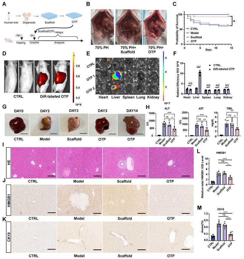
We next examined whether localized OTP deployment was associated with early biochemical and structural recovery. Serum biochemical analyses at Day 3 showed that OTP treatment significantly reduced ALT, AST, and total bilirubin levels relative to both model and scaffold-only groups (Figure 4H), consistent with reduced hepatocellular injury and improved bilirubin handling during early recovery. Histological examination of Day 3 tissue from the predefined patch-placement region of the remnant liver showed reduced necrotic and congestive changes, with better preservation of local parenchymal architecture in OTP-treated livers (Figure 4I). Immunohistochemical analyses at Day 3 further demonstrated reduced HMGB1 signal intensity and diminished CK19-positive ductular reaction in OTP-treated livers compared with control groups (Figure 4J–M), supporting attenuation of local injury and the associated ductular response within the injury-adjacent remnant-liver region.
To determine whether donor-derived human signals remained detectable after the early postoperative phase, we performed a series of donor-specific analyses at Day 14. Scaffold-only controls showed no evident hALB-positive signal, whereas OTP-treated livers displayed weak hALB immunoreactivity in the OTP placement region (Figure S7A–B). To further assess donor-derived persistence at Day 14, we performed serum hALB measurement, human-specific Alu DNA qPCR, and human-specific qPCR for lineage-associated transcripts. Serum hALB was detectable in OTP-treated mice but absent or nearly undetectable in scaffold-only and model groups (Figure S7C). Human-specific Alu DNA was also detectable in liver tissue from the OTP placement region, whereas scaffold-only and model tissues showed no detectable Alu signal (Figure S7D). In parallel, human-specific transcripts associated with cholangiocyte identity, hepatocyte-like features, epithelial state, albumin production, and proliferation, including SOX9, KRT19, HNF4A, CYP3A4, ALB, CDH1, and KI67, were detectable in OTP-treated liver tissue but not in scaffold-only or model controls (Figure S7E–K). These assays supported the local presence of donor-derived human signals at Day 14, without evidence of bulk donor tissue replacement.
Collectively, localized OTP deployment improved early postoperative survival and biochemical recovery and attenuated structural injury in the predefined patch-placement region after major hepatectomy.
OTP attenuates NF-κB-linked inflammatory programs and increases a CD206-associated macrophage signal at Day 3
To characterize the early host-tissue response associated with localized OTP deployment, transcriptomic profiling was performed on tissue from the predefined patch-placement region of the remnant liver collected at Day 3 after surgery. Principal component analysis showed clear separation between OTP-treated and model samples, indicating distinct tissue transcriptional profiles between the two groups (Figure 5A). Consistent with this shift, volcano plot analysis identified broad differential gene expression, with a predominance of downregulated immune- and inflammation-associated transcripts in OTP-treated livers (Figure 5B). Heatmap visualization further showed a coordinated downregulation pattern of inflammatory gene modules across OTP-treated samples (Figure S8A). KEGG enrichment analysis showed that the downregulated genes were enriched in immune- and inflammation-related pathway annotations, including cytokine–cytokine receptor interaction, neutrophil extracellular trap formation, and B cell receptor and T cell receptor signaling (Figure 5C). In the immunodeficient FRG background, the latter annotations likely reflect shared signaling-gene enrichment within bulk liver tissue rather than adaptive lymphocyte activity. Complementary Gene Ontology analysis further supported broad attenuation of immune-associated biological processes, including inflammatory response, leukocyte migration, immune effector activation, and related extracellular/immune-associated compartments (Figure S8B). Gene set enrichment analysis additionally demonstrated significant negative enrichment of the NF-κB signaling pathway, consistent with reduced NF-κB-linked inflammatory activity in OTP-treated tissue (Figure 5D). Protein–protein interaction analysis further identified interconnected clusters containing TNF-family members, CCL/CXCL chemokines, and other cytokine-associated mediators among the downregulated genes (Figure S8C).
OTP attenuates NF-κB-linked inflammatory programs and increases a CD206-associated macrophage signal on Day 3. (A) Principal component analysis of bulk RNA-seq data from tissue collected from the Day 3 patch-placement region, showing separation between model (M) and OTP-treated groups. (B) Volcano plot of differentially expressed genes between OTP-treated and model groups. (C) KEGG enrichment analysis of genes downregulated in OTP-treated tissue, showing enrichment of immune- and inflammation-related pathway annotations among genes downregulated in OTP-treated tissue. (D) Gene set enrichment analysis showing negative enrichment of the NF-κB signaling pathway in OTP-treated tissue from the patch-placement region. (E) Relative mRNA expression levels of TNF-α, IL-6, IL-1β, CCL2, CCL5, CXCL1, and CXCL2 in Day 3 tissue from the patch-placement region. (F) Immunoblot analysis of p-p65, total p65, and β-actin, with corresponding quantification of the p-p65/p65 ratio. (G) Immunofluorescence staining of F4/80 and CD206 in the patch-placement remnant liver region on Day 3. Scale bar, 100 μm. (H) Quantification of the CD206⁺/F4/80⁺ fraction in the patch-placement remnant liver region. For qPCR and histological quantification, n = 6 per group; for immunoblotting, n = 3 per group. Data are presented as mean ± s.e.m. One-way ANOVA. *P < 0.05, **P < 0.01, ***P < 0.001, ****P < 0.0001; ns, not significant.
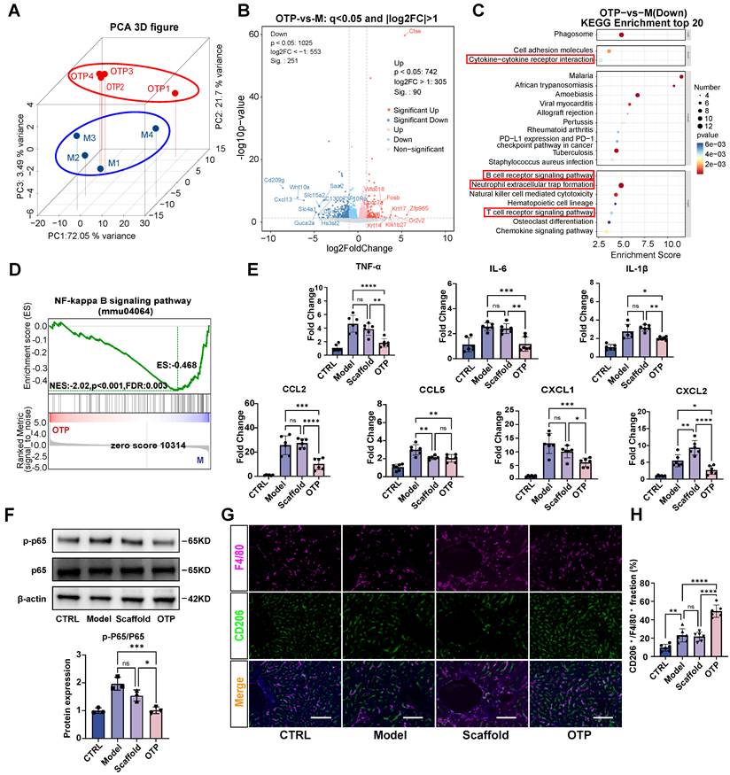
These transcriptomic findings were further examined by qPCR and protein analysis. At Day 3, OTP-treated liver tissue showed reduced expression of TNF-α, IL-6, IL-1β, CCL2, CCL5, CXCL1, and CXCL2 compared with the model and scaffold-only groups (Figure 5E). Immunoblotting also showed a reduced p-p65/p65 ratio in the OTP group (Figure 5F), consistent with decreased NF-κB pathway activity. Immunofluorescence staining for F4/80 and CD206 further demonstrated an increased CD206-associated signal within the F4/80-positive macrophage compartment in OTP-treated livers relative to model and scaffold-only controls (Figure 5G–H). Collectively, OTP treatment was associated with reduced inflammatory mediator expression, lower NF-κB-linked activity, and an increased CD206-associated macrophage signal in the patch-placement region at Day 3.
OTP treatment is associated with metabolic pathway enrichment and increased hepatocyte proliferative entry at Day 3
Having observed reduced inflammatory activity following OTP treatment, we next examined metabolism- and regeneration-associated responses in the Day 3 patch-placement region of the remnant liver. Bulk transcriptomic analysis of the same tissue samples revealed enrichment of lipid metabolic and regenerative signaling programs in OTP-treated livers compared with model controls, with prominent representation of the PPAR signaling pathway together with Wnt-related networks and cytochrome P450–associated oxidative metabolism (Figure 6A). GSEA further identified positive enrichment of DNA replication, cell-cycle progression, homologous recombination, and PPAR signaling gene sets (Figure 6B). Consistently, selected cell-cycle-associated transcripts were increased in OTP-treated samples (Figure S9).
OTP treatment is associated with metabolic pathway enrichment and increased hepatocyte proliferative entry at Day 3. (A) WikiPathways enrichment analysis of genes upregulated in OTP-treated tissue from the Day 3 patch-placement region compared with model tissue at Day 3, highlighting lipid metabolic, nuclear receptor, Wnt-related, and PPAR signaling programs. (B) Gene set enrichment analyses showing positive enrichment of DNA replication, cell cycle, homologous recombination, and PPAR signaling pathways in OTP-treated tissue. (C) Relative mRNA expression levels of PCNA and Ki67 in tissue from the Day 3 patch-placement region. (D) Immunofluorescence staining for PCNA and HNF4A in the same tissue regions at Day 3. Scale bar, 100 μm. (E) Quantification of PCNA and HNF4A fluorescence intensity. (F) Immunohistochemical staining of Ki67 in the same tissue regions at Day 3. Scale bar, 200 μm. (G) Quantification of Ki67-positive cells in liver sections. (H) Immunohistochemical staining of PPARα in the same tissue regions at Day 3. Scale bar, 200 μm. (I) Quantification of relative PPARα integrated optical density (IOD). (J) Immunohistochemical staining of YAP in the same tissue regions at Day 3. Scale bar, 200 μm. (K) Quantification of YAP nuclear-positive cells. For qPCR and histological quantification, n = 6 per group. Data are presented as mean ± s.e.m. One-way ANOVA. *P < 0.05, **P < 0.01, ***P < 0.001, ****P < 0.0001; ns, not significant.
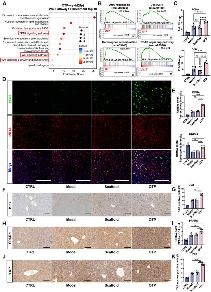
These transcriptional differences were accompanied by increased proliferation-associated signals in host liver tissue. qPCR showed higher expression of Pcna and Ki67 in OTP-treated livers than in the model and scaffold-only groups (Figure 6C). HNF4A/PCNA co-immunofluorescence further showed increased PCNA fluorescence in HNF4A-positive hepatocyte regions following OTP treatment (Figure 6D–E). Consistently, Ki67 immunohistochemistry demonstrated a higher fraction of proliferating cells in OTP-treated livers than in model or scaffold-only controls (Figure 6F–G). Immunohistochemical analysis additionally showed increased PPARα immunoreactivity and a higher proportion of cells with nuclear YAP staining in OTP-treated tissue (Figure 6H–K). These parallel changes were consistent with enhanced PPARα-associated metabolic signaling and YAP-associated regenerative activity, although the present data do not establish a direct regulatory relationship between the two pathways. Collectively, OTP treatment was associated with metabolism-related transcriptional enrichment and increased hepatocyte proliferative entry in the patch-placement region at Day 3.
Pharmacologic PPARα inhibition partially attenuates OTP-associated early recovery
To test whether PPARα activity functionally contributes to OTP-associated metabolic–regenerative recovery, we examined the effects of pharmacologic PPARα inhibition using GW6471 in the hepatectomy model at Day 3 after surgery (Figure 7A). qPCR analysis showed that expression of the PPARα-responsive genes Acox1 and Cpt1a was lower in the OTP + GW6471 group than in the OTP group and remained low in mice receiving GW6471 alone (Figure 7B), consistent with pharmacologic suppression of the PPARα-responsive transcriptional program. Compared with OTP treatment alone, OTP + GW6471 treatment was associated with higher serum ALT, AST, and total bilirubin levels (Figure 7C), indicating partial attenuation of the biochemical recovery associated with OTP treatment.
PPARα inhibition partially attenuates OTP-associated Day 3 regenerative recovery. (A) Schematic of the experimental design for pharmacologic PPARα inhibition using GW6471 in the 70% partial hepatectomy model. (B) qPCR analysis of the PPARα-responsive genes Acox1 and Cpt1a in the indicated groups. (C) Serum ALT, AST, and TBIL levels at Day 3 after hepatectomy, showing attenuation of OTP-associated biochemical recovery after PPARα inhibition. (D) Immunofluorescence staining of Ki67 and PCNA in tissue from the Day 3 patch-placement region from OTP, OTP + GW6471, and GW6471 groups. Scale bar, 100 μm. (E) Quantification of Ki67 and PCNA fluorescence intensity. (F) Immunofluorescence staining of F4/80 and CD206 in the same tissue regions at Day 3. Scale bar, 100 μm. (G) Quantification of the CD206⁺/F4/80⁺ fraction. (H) Working model summarizing PPARα-responsive signaling as one functional contributor to OTP-associated recovery, together with additional inflammatory, metabolic, and regenerative processes. n = 6 per group unless otherwise indicated. Data are presented as mean ± s.e.m. One-way ANOVA. *P < 0.05, **P < 0.01, ***P < 0.001, ****P < 0.0001; ns, not significant.
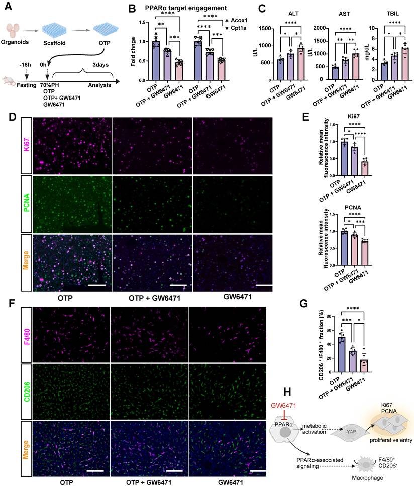
At the tissue level, Ki67- and PCNA-positive signals were increased in OTP-treated livers but were reduced following GW6471 co-administration (Figure 7D–E), indicating attenuation of proliferation-associated responses. The CD206-associated signal within the F4/80-positive macrophage compartment was also lower in the OTP + GW6471 group than in the OTP group (Figure 7F–G). Collectively, these findings indicate that systemic PPARα inhibition partially blunted the biochemical, proliferative, and macrophage-associated responses observed following OTP treatment. Thus, PPARα-responsive signaling represents a functional contributor to OTP-associated early recovery, although the systemic pharmacologic design does not establish an OTP-specific or exclusive PPARα mechanism.
OTP deployment is not associated with overt biochemical or extrahepatic histological abnormalities during short- and intermediate-term follow-up
To assess the short- and intermediate-term tolerability and biodistribution of OTP deployment, serum biochemical indices and major-organ histology were evaluated at Day 14 and 2 months after surgery (Figure S10A). At Day 14, serum BUN and creatinine levels were comparable among the control, model, scaffold-only, and OTP-treated groups (Figure S10B), indicating no overt renal biochemical abnormality associated with OTP treatment. Histological examination of the heart, spleen, lung, and kidney showed no obvious group-specific histopathological abnormalities in the examined sections (Figure S10C). These findings indicated that OTP deployment was not associated with overt renal dysfunction or extrahepatic histological injury during the 14-day observation period.
At 2 months, serum ALT and AST levels were not significantly different among the control, model, scaffold-only, and OTP-treated groups, and BUN and creatinine levels were also comparable among groups (Figure S11A–B). Human-specific Alu DNA was detectable in liver tissue from OTP-treated mice but was not detected in the heart, spleen, lung, or kidney; control liver tissue was also negative for human Alu DNA (Figure S11C). These findings indicated liver-restricted detection of donor-derived human DNA among the examined organs. The Alu signal was interpreted as a biodistribution marker rather than evidence of viable long-term donor-cell engraftment. H&E staining of the liver, heart, spleen, lung, and kidney showed no obvious histopathological abnormalities or ectopic tissue-like lesions in OTP-treated mice compared with the other groups (Figure S11D).
Collectively, these findings provide preliminary short- and intermediate-term tolerability and biodistribution evidence, with no overt biochemical or extrahepatic histological abnormalities identified at the assessed endpoints.
Discussion
In this study, our findings support the concept that early recovery after major hepatectomy may depend not only on remnant liver mass, but also on the local condition of the newly exposed injury-adjacent remnant-liver boundary during the immediate postoperative window. By integrating matrix-free expanded human bile-derived organoids into a reticulated microfibrous scaffold, we established an OTP that could be deployed onto the remnant liver surface and retained locally within the predefined patch-placement region. In this setting, OTP treatment was associated with improved survival and organ-level biochemical recovery, together with reduced structural injury and coordinated inflammatory, metabolic, and proliferative responses within the predefined patch-placement region. Importantly, the overall pattern of evidence argues against extensive donor-driven parenchymal replacement as the dominant explanation for these effects. Instead, sparse hALB-positive signals detected at later time points are more consistent with limited local donor-derived signals, whereas the major biological response is more consistent with an OTP-associated change in the adjacent host-tissue response [44, 45]. Conceptually, these findings support a boundary-centered model of early liver recovery, in which the local condition of the injury-adjacent remnant-liver region may influence whether endogenous repair is effectively engaged after major hepatectomy.
A central finding of this study is that OTP treatment was associated with a shift in the Day 3 injury-adjacent remnant-liver environment toward reduced inflammatory activity and greater regenerative permissiveness. Transcriptomic profiling showed coordinated suppression of inflammatory pathways in OTP-treated tissue, including negative enrichment of NF-κB-associated programs, and this was supported by reduced expression of inflammatory mediators together with a reduced p-p65/p65 ratio. Histologically, this inflammatory restraint was accompanied by attenuation of HMGB1 signal and reduced local injury in the patch-placement remnant liver region. In parallel, OTP-treated tissue exhibited an increased CD206⁺/F4/80⁺ fraction, consistent with an increased CD206-associated macrophage signal in the injury-adjacent remnant-liver region [45-47]. CD206/F4/80 staining alone does not establish macrophage polarization or reparative function, particularly in the immunodeficient FRG background. The weaker responses observed with the scaffold-only control further suggest that the effects of OTP cannot be explained by mechanical coverage alone, although the present data do not identify the specific organoid-derived mediators or cellular interactions involved. Taken together, these findings suggest that the scaffold-only condition did not reproduce the full OTP-associated phenotype and that OTP treatment was associated with a less inflammatory and more regeneration-compatible local host-tissue response during the early postoperative period. Rather than serving as a replacement graft, the patch may therefore provide temporary localized support at the remnant-liver boundary while endogenous repair becomes established [48, 49].
Beyond inflammatory restraint, our data support the emergence of a coordinated metabolism- and regeneration-associated response in adjacent host tissue. At Day 3, OTP-treated livers showed enrichment of lipid metabolic and proliferative programs, together with increased PPARα expression, increased YAP-positive nuclei, and enhanced proliferative entry, including increased PCNA-positive hepatocytes and Ki67-positive cells. These findings are consistent with the view that early regenerative activation is not driven by proliferative signaling alone, but is gated by metabolic competence at the tissue level [32, 50, 51]. Notably, the in vivo host response showed partial convergence with the transcriptional state observed in scaffold-associated dBDOs in vitro, which retained hepatic identity while acquiring metabolism-associated and stress-adaptive features. This correspondence raises the possibility that scaffold-associated conditioning may help maintain an organoid state compatible with the stressed remnant-liver surface environment after deployment. At the same time, our pharmacologic perturbation experiments further examined the functional relevance of PPARα-responsive signaling within this response. Pharmacologic inhibition with GW6471 reduced expression of PPARα-responsive genes, attenuated OTP-associated biochemical improvement, diminished Ki67 and PCNA signals, and weakened the CD206-associated macrophage signal. These results support a functional contribution of PPARα-responsive signaling to OTP-associated recovery. Nevertheless, given the systemic administration of GW6471, this experiment should be viewed as a pharmacologic sensitivity test of the PPARα-linked response rather than as proof of an OTP-specific PPARα mechanism. Thus, the present data support PPARα-responsive signaling as one functional contributor to OTP-associated recovery rather than an exclusive mediator, while locally restricted or cell-type-specific PPARα perturbation will be required to determine whether OTP engages an organoid-to-host PPARα axis.
From a design perspective, the present work places the OTP in a distinct space relative to existing regenerative delivery formats. The reticulated scaffold was not selected simply as a passive carrier, but because it reconciled several competing requirements relevant to acute surgical use: local conformability, mechanical robustness, open porosity, high-density organoid loading, and preliminary feasibility of cryopreservation and post-thaw recovery [34, 35]. This is important because many cell-based regenerative approaches remain limited by poor spatial control, low post-delivery retention, diffusion constraints, or impracticality in the operative setting [19]. In contrast, the OTP format separates the problem of local placement and structural handling from the problem of biological payload activity, allowing the patch to function as a surgically manageable, remnant-liver surface adjunct rather than an attempt at bulk tissue replacement. Direct application of unscaffolded organoids to the exposed remnant-liver surface would be more susceptible to dispersion and loss of local positioning; however, this was not directly compared in the present study. Accordingly, the specific contribution of scaffold-mediated retention relative to free organoid delivery remains to be quantified. In this respect, our findings suggest that the condition of the injury-adjacent remnant-liver boundary may represent a complementary determinant of early recovery, and that engineering constructs for local compatibility, retention, and bioactive support under postoperative stress may provide a practical translational strategy alongside approaches aimed at increasing donor-cell survival or transplanted tissue mass [52, 53].
Several limitations of the present study should be acknowledged. Although Day 14 serum hALB, human-specific Alu DNA, and human-specific lineage-associated transcripts demonstrated continued local detectability of donor-derived human material and signals, these findings do not establish viable long-term engraftment, substantial donor-cell expansion, anatomical integration, or complete in vivo differentiation into functional hepatocytes or cholangiocytes [54, 55]. Similarly, bulk RNA-seq of the patch-placement remnant liver region identifies an OTP-associated host response, but cannot resolve whether this response is initiated by paracrine signaling, donor–host contact, scaffold-mediated local retention, or secondary host-intrinsic adaptation after reduced injury. The FRG model was necessary for xenogeneic human organoid delivery, but its immunodeficient background also limits the interpretation of immune-related findings, particularly adaptive immune pathway annotations and CD206-associated macrophage signals. In addition, although GW6471 partially attenuated OTP-associated recovery, this pharmacologic perturbation supports PPARα as a functionally relevant contributor rather than an OTP-specific or exclusive mediator. Future studies using human-specific spatial tracing, single-cell or spatial transcriptomics, contact-separated systems, cell-type-specific PPARα perturbation, and complementary immunocompetent or humanized models will be needed to define donor-cell fate and the precise mechanisms of host niche modulation [32]. Finally, although the 2-month safety assessment provides preliminary reassurance, larger cohorts, longer follow-up, quantitative biodistribution, tumorigenicity assessment, and validation in larger-animal systems and clinically realistic operative workflows will be required before translational application [56-59].
Despite these limitations, the present work identifies the injury-adjacent remnant-liver boundary as a potentially actionable site for localized intervention during early postoperative recovery. The overall pattern of evidence is more consistent with OTP functioning as temporary localized bioactive support accompanied by an altered adjacent host-tissue response than with extensive replacement of lost liver mass. More broadly, the study suggests that locally deployable living patches may be most effective not when they attempt to recapitulate whole-tissue replacement, but when they preserve recovery-permissive conditions at vulnerable postoperative remnant-liver boundary regions long enough for endogenous repair to take over.
Supplementary Material
Supplementary methods, figures and tables.

Acknowledgements
We would like to thank Chao Bi and Xiaoli Hong from the Core Facilities, Zhejiang University School of Medicine for their technical support.
Funding
This study was supported by National Natural Science Foundation of China (No. 82570718, 52235007, T2121004, 52325504), Central Guidance Fund for Local Science and Technology Development (No. 2024ZY01054), the Fundamental Research Funds for the Central Universities (No. 2025ZFJH03), and High-level Personnel Cultivating Project of Zhejiang Province (No. 2023R5243).
Author contributions
H Cao, Y He, J Yu, J He and Q Yao contributed to study design and planning; Q Yao, J He, X Shi, J Ma, J Weng, G Cao, H Fang, J Yang, and Q Pan contributed to cellular and animal experiments; H Cao, Q Yao, J He, and J Yu participated in analysis, interpretation of data and manuscript writing; L Li and H Cao contributed to study supervision. All authors reviewed and approved the final version of the manuscript.
Artificial intelligence (AI) usage statement
AI was used only for English-language editing and grammatical refinement to improve readability. It was not used for experimental design, data generation, data analysis, image generation or manipulation, or independent scientific interpretation.
Data availability statement
The RNA-sequencing datasets generated in this study have been deposited in the National Genomics Data Center, China National Center for Bioinformation, under accession number HRA019744 and CRA046568. Human-derived sequencing data are available through controlled access in accordance with repository requirements, and reviewer access information has been provided to the journal. The remaining data supporting the findings of this study are included in the article and its Supplementary Information. Additional materials are available from the corresponding authors upon reasonable request.
Competing Interests
The authors have declared that no competing interest exists.
References
1. Kharbikar BN, Mohindra P, Desai TA. Biomaterials to enhance stem cell transplantation. Cell Stem Cell. 2022;29:692-721
2. Liu S, Hur YH, Cai X, Cong Q, Yang Y, Xu C. et al. A tissue injury sensing and repair pathway distinct from host pathogen defense. Cell. 2023;186:2127-43 e22
3. Hoang VT, Nguyen QT, Phan TTK, Pham TH, Dinh NTH, Anh LPH. et al. Tissue engineering and regenerative medicine: Perspectives and challenges. MedComm (2020). 2025;6:e70192
4. Fonouni H, Khajeh E, Ghamarnejad O, Kashfi A, Aydogdu E, Majlesara A. et al. Histopathological effects of modern topical sealants on the liver surface after hepatectomy: an experimental swine study. Sci Rep. 2019;9:7088
5. Chen K, Kwon SH, Henn D, Kuehlmann BA, Tevlin R, Bonham CA. et al. Disrupting biological sensors of force promotes tissue regeneration in large organisms. Nat Commun. 2021;12:5256
6. Caballero-Sanchez N, Alonso-Alonso S, Nagy L. Regenerative inflammation: When immune cells help to re-build tissues. FEBS J. 2024;291:1597-614
7. Espino-Gonzalez E, Dalbram E, Mounier R, Gondin J, Farup J, Jessen N. et al. Impaired skeletal muscle regeneration in diabetes: From cellular and molecular mechanisms to novel treatments. Cell Metab. 2024;36:1204-36
8. Wculek SK, Dunphy G, Heras-Murillo I, Mastrangelo A, Sancho D. Metabolism of tissue macrophages in homeostasis and pathology. Cell Mol Immunol. 2022;19:384-408
9. Fan S, Gao Y, Qu A, Jiang Y, Li H, Xie G. et al. YAP-TEAD mediates PPAR alpha-induced hepatomegaly and liver regeneration in mice. Hepatology. 2022;75:74-88
10. Liu Q, Wang S, Fu J, Chen Y, Xu J, Wei W. et al. Liver regeneration after injury: Mechanisms, cellular interactions and therapeutic innovations. Clin Transl Med. 2024;14:e1812
11. Matchett KP, Wilson-Kanamori JR, Portman JR, Kapourani CA, Fercoq F, May S. et al. Multimodal decoding of human liver regeneration. Nature. 2024;630:158-65
12. Hofer M, Lutolf MP. Engineering organoids. Nat Rev Mater. 2021;6:402-20
13. Blache U, Ford EM, Ha B, Rijns L, Chaudhuri O, Dankers PYW. et al. Engineered hydrogels for mechanobiology. Nat Rev Methods Primers. 2022;2:98
14. Ahmed ZT, Zain Al-Abeden MS, Al Abdin MG, Muqresh MA, Al Jowf GI, Eijssen LMT. et al. Dose-response relationship of MSCs as living Bio-drugs in HFrEF patients: a systematic review and meta-analysis of RCTs. Stem Cell Res Ther. 2024;15:165
15. Rajendran AK, Sankar D, Amirthalingam S, Kim HD, Rangasamy J, Hwang NS. Trends in mechanobiology guided tissue engineering and tools to study cell-substrate interactions: a brief review. Biomater Res. 2023;27:55
16. Harimoto T, Jung W-H, Mooney DJ. Delivering living medicines with biomaterials. Nat Rev Mater. 2025;10:191-210
17. Gadd VL, Ferreira-Gonzalez S, Man TY, Kilpatrick AM, Aird RE, Smith IP. et al. Host hepatocyte senescence determines the success of hepatocyte transplantation in a mouse model of liver injury. J Hepatol. 2025;83:161-76
18. Sun Z, Yuan X, Wu J, Wang C, Zhang K, Zhang L. et al. Hepatocyte transplantation: The progress and the challenges. Hepatol Commun. 2023;7:e0266
19. Lu P, Ruan D, Huang M, Tian M, Zhu K, Gan Z. et al. Harnessing the potential of hydrogels for advanced therapeutic applications: current achievements and future directions. Signal Transduct Target Ther. 2024;9:166
20. Zhang Y, Li L, Dong L, Cheng Y, Huang X, Xue B. et al. Hydrogel-based strategies for liver tissue engineering. Chem Bio Eng. 2024;1:887-915
21. Gan Z, Qin X, Liu H, Liu J, Qin J. Recent advances in defined hydrogels in organoid research. Bioact Mater. 2023;28:386-401
22. Schlegel A, Mergental H, Fondevila C, Porte RJ, Friend PJ, Dutkowski P. Machine perfusion of the liver and bioengineering. J Hepatol. 2023;78:1181-98
23. Hussein KH, Ahmadzada B, Correa JC, Sultan A, Wilken S, Amiot B. et al. Liver tissue engineering using decellularized scaffolds: Current progress, challenges, and opportunities. Bioact Mater. 2024;40:280-305
24. Chen W, Nie M, Gan J, Xia N, Wang D, Sun L. Tailoring cell sheets for biomedical applications. Smart Med. 2024;3:e20230038
25. Akiyama Y. Design of temperature-responsive cell culture surfaces for cell sheet engineering. Cyborg Bionic Syst. 2021;2021:5738457
26. Zoneff E, Wang Y, Jackson C, Smith O, Duchi S, Onofrillo C. et al. Controlled oxygen delivery to power tissue regeneration. Nat Commun. 2024;15:4361
27. Xue B, Xu Z, Li L, Guo K, Mi J, Wu H. et al. Hydrogels with programmed spatiotemporal mechanical cues for stem cell-assisted bone regeneration. Nat Commun. 2025;16:3633
28. Chansoria P, Chaudhari A, Etter EL, Bonacquisti EE, Heavey MK, Le J. et al. Instantly adhesive and ultra-elastic patches for dynamic organ and wound repair. Nat Commun. 2024;15:4720
29. Wu SJ, Zhao X. Bioadhesive Technology Platforms. Chem Rev. 2023;123:14084-118
30. Atcha H, Choi YS, Chaudhuri O, Engler AJ. Getting physical: Material mechanics is an intrinsic cell cue. Cell Stem Cell. 2023;30:750-65
31. Li J, Hu S, Zhu D, Huang K, Mei X, Lopez de Juan Abad B. et al. All roads lead to Rome (the heart): cell retention and outcomes from various delivery routes of cell therapy products to the heart. J Am Heart Assoc. 2021;10:e020402
32. Ma X, Huang T, Chen X, Li Q, Liao M, Fu L. et al. Molecular mechanisms in liver repair and regeneration: from physiology to therapeutics. Signal Transduct Target Ther. 2025;10:63
33. Hirao H, Nakamura K, Kupiec-Weglinski JW. Liver ischaemia-reperfusion injury: a new understanding of the role of innate immunity. Nat Rev Gastroenterol Hepatol. 2022;19:239-56
34. Saiz PG, Reizabal A, Vilas-Vilela JL, Dalton PD, Lanceros-Mendez S. Materials and strategies to enhance melt electrowriting potential. Adv Mater. 2024;36:e2312084
35. Wlodarczyk-Biegun MK, Villiou M, Koch M, Muth C, Wang P, Ott J. et al. Melt electrowriting of graded porous scaffolds to mimic the matrix structure of the human trabecular meshwork. ACS Biomater Sci Eng. 2022;8:3899-911
36. Xie G, Yin S, Zhang Z, Qi D, Wang X, Kim D. et al. Hepatocyte peroxisome proliferator-activated receptor alpha enhances liver regeneration after partial hepatectomy in mice. Am J Pathol. 2019;189:272-82
37. Michalopoulos GK, Bhushan B. Liver regeneration: biological and pathological mechanisms and implications. Nat Rev Gastroenterol Hepatol. 2021;18:40-55
38. Maspero M, Yilmaz S, Cazzaniga B, Raj R, Ali K, Mazzaferro V. et al. The role of ischaemia-reperfusion injury and liver regeneration in hepatic tumour recurrence. JHEP Rep. 2023;5:100846
39. Bekheit M, Grundy L, Salih AK, Bucur P, Vibert E, Ghazanfar M. Post-hepatectomy liver failure: A timeline centered review. Hepatobiliary Pancreat Dis Int. 2023;22:554-69
40. Sparrelid E, Olthof PB, Dasari BVM, Erdmann JI, Santol J, Starlinger P. et al. Current evidence on posthepatectomy liver failure: comprehensive review. BJS Open. 2022;6:zrac142
41. Zhan X, Bai Y, Zhu Q, Gao Y, Li F, Bu Q. et al. Macrophage ATG16L1 promotes liver regeneration after partial hepatectomy. JHEP Rep. 2025;7:101330
42. Mitchell C, Willenbring H. A reproducible and well-tolerated method for 2/3 partial hepatectomy in mice. Nat Protoc. 2008;3:1167-70
43. Soroka CJ, Assis DN, Alrabadi LS, Roberts S, Cusack L, Jaffe AB. et al. Bile-derived organoids from patients with primary sclerosing cholangitis recapitulate their inflammatory immune profile. Hepatology. 2019;70:871-82
44. Engelmann C. Decoding the metabolic dialogue between hepatocytes and macrophages driving liver regeneration. J Hepatol. 2026;84:475-476
45. Zhao Y, Zhao S, Liu S, Ye W, Chen WD. Kupffer cells, the limelight in the liver regeneration. Int Immunopharmacol. 2025;146:113808
46. Liu R, Scimeca M, Sun Q, Melino G, Mauriello A, Shao C. et al. Harnessing metabolism of hepatic macrophages to aid liver regeneration. Cell Death Dis. 2023;14:574
47. Brunnthaler L, Hammond TG, Pereyra D, Santol J, Probst J, Laferl V. et al. HMGB1-mediated cell death-a crucial element in post-hepatectomy liver failure. Int J Mol Sci. 2024;25:7150
48. Kuklin A, Slabber CF, Tortola L, Kwan CL, Liebisch G, Kondylis V. et al. An Nrf2-NF-kappaB crosstalk controls hepatocyte proliferation in the normal and injured liver. Cell Mol Gastroenterol Hepatol. 2025;19:101480
49. Kostallari E, Schwabe RF, Guillot A. Inflammation and immunity in liver homeostasis and disease: a nexus of hepatocytes, nonparenchymal cells and immune cells. Cell Mol Immunol. 2025;22:1205-25
50. Duan L, Chang Y, Dai J, Lu H, Zhao W, Shen Y. et al. Lipid metabolism orchestrates liver regeneration: an integrated metabolic network. J Transl Med. 2025;23:1115
51. Yu Q, Yu M, Xie P, Guo L, Zhao Y, Xu W. et al. Liver regeneration-associated hepatocellular YAP1 activation prevents colorectal cancer liver metastasis through glutamine competition. Sci Adv. 2025;11:eadw6926
52. Verbakel J, de Boer J. Engineering stem cell metabolism using biomaterial-induced mechanotransduction. Cell Biomaterials. 2025;1:100192
53. Pham TT, Tran PL, Tempelman LA, Stone SG, Piccirillo C, Li A. et al. A continuously oxygenated macroencapsulation system enables high-density packing and delivery of insulin-secreting cells. Nat Commun. 2025;16:7199
54. Kim Y, Kang M, Mamo MG, Adisasmita M, Huch M, Choi D. Liver organoids: Current advances and future applications for hepatology. Clin Mol Hepatol. 2025;31:S327-S48
55. Ortuno-Costela MC, Pinzani M, Vallier L. Cell therapy for liver disorders: past, present and future. Nat Rev Gastroenterol Hepatol. 2025;22:329-42
56. Shao W, Xu H, Zeng K, Ye M, Pei R, Wang K. Advances in liver organoids: replicating hepatic complexity for toxicity assessment and disease modeling. Stem Cell Res Ther. 2025;16:27
57. Gong S, He K, Liu Y, Luo X, Ashraf K, He J. et al. Scalable matrigel-free suspension culture for generating high-quality human liver ductal organoids. Cell Prolif. 2025;58:e70033
58. Abbasalizadeh S, Babaee S, Kowsari-Esfahan R, Mazidi Z, Shi Y, Wainer J. et al. Continuous production of highly functional vascularized hepatobiliary organoids from human pluripotent stem cells using a scalable microfluidic platform. Adv Funct Mater. 2023;33:2210233
59. Deuse T, Schrepfer S. Progress and challenges in developing allogeneic cell therapies. Cell Stem Cell. 2025;32:513-28
Author contact
Corresponding authors: Hongcui Cao, M.D. & Ph.D., E-mail: hccaoedu.cn; Yong He, Ph.D., E-mail: yongqinedu.cn.