13.3
Impact Factor
Theranostics 2021; 11(13):6524-6525. doi:10.7150/thno.61641 This issue Cite
Erratum
Microporous polysaccharide multilayer coated BCP composite scaffolds with immobilised calcitriol promote osteoporotic bone regeneration both in vitro and in vivo: Erratum
1. Key Laboratory of Orthopaedics of Zhejiang Province, Department of Orthopaedics, The Second Affiliated Hospital and Yuying Children's Hospital of Wenzhou Medical University, 109, Xueyuanxi road, 325027 Wenzhou, China
2. The second School of Medicine, Wenzhou Medical University, 109, Xueyuanxi road, 325027 Wenzhou, China
3. Department of Orthopaedic Surgery Shanghai Jiao Tong University Affiliated Sixth People's Hospital, 600 Yishan Road, Shanghai, 200233, China.
4. Department of Oral Implantology and Prosthetic Dentistry, Academic Centre for Dentistry Amsterdam (ACTA), Vrije University Amsterdam and University of Amsterdam, Amsterdam, Nord-Holland, the Netherlands
*Qian Tang and Zhichao Hu contribute equally to this work
Published 2021-4-20
Corrected-article in Theranostics, Volume 9, 1125
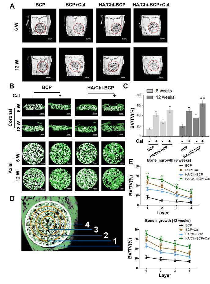
The authors regret that the image of BCP group without Cal in 12 W in the Fig. 7B was wrongly attached due to their carelessness in integrating figures. The correct version is shown below. This image substitution would not affect any results presented in the originally-published version, nor the corresponding text description and the conclusion of the paper. The authors apologize for any inconvenience or misunderstanding that this error may have caused.
Micro-CT analysis of the effects of various BCP scaffolds on new bone formation in the critical-size bone defect model of OVX rats. (A) Three-dimensional reconstruction of micro-CT images of the various scaffolds implanted in the rat calvarium at 6 and 12 weeks (scale bars: 5 mm). (B) Two-dimensional reconstruction of micro-CT images of various scaffolds implanted in the rat calvarium at 6 and 12 weeks (the white color component shows the remaining scaffold, bone that grew around and into the scaffolds is labelled in green) (scale bars: 2 mm for coronal images and 1 mm for axial images). (C) Regenerated bone volumes on the various scaffolds were quantified as bone volume divided by total volume (BV/TV). (D) General sketch of the scaffold, which was divided into four layers. (E) Percentages of bone volume regenerated into the scaffold in different layers from the edge to the center. Data are presented as the mean ± S.D. Significant differences among scaffold groups are indicated as ** P < 0.01, * P < 0.05, compared with BCP; ## P < 0.01, # P < 0.05 compared with HA/Chi-BCP, and && P < 0.01, & P < 0.05 compared with BCP+Cal; for each group, n = 5.

References
1. Tang Q, Hu Z, Jin H, Zheng G, Yu X, Wu G, Liu H, Zhu Z, Xu H, Zhang C, Shen L. Microporous polysaccharide multilayer coated BCP composite scaffolds with immobilised calcitriol promote osteoporotic bone regeneration both in vitro and in vivo. Theranostics. 2019;9(4):1125-1143 doi:10.7150/thno.29566
Author contact
![]() Corresponding authors: Liyan Shen, Tel:+86-577-88002760 Email: shenliyanedu.cn; Changqing Zhang, Tel: +86-21-64369181 Email: zhangcqedu.cn; Huazi Xu, Tel: +86-13616632111Email: spinexucom
Corresponding authors: Liyan Shen, Tel:+86-577-88002760 Email: shenliyanedu.cn; Changqing Zhang, Tel: +86-21-64369181 Email: zhangcqedu.cn; Huazi Xu, Tel: +86-13616632111Email: spinexucom
 Global reach, higher impact