Impact Factor
- Issue 14; 2026
- Issue 13; 2026
- Issue 12; 2026
- Issue 11; 2026
- Issue 10; 2026
- Volume 16; 2026
- Advance Articles
- Past Issues
- Cover Images
- Cover Suggestion
- Index & Coverage
- Special Issues
1. Introduction
2. Experimental Section
3. Results and Discussion
4. Conclusion
Acknowledgements
References
International Journal of Biological Sciences
International Journal of Medical Sciences
 Global reach, higher impact

Theranostics 2013; 3(4):275-281. doi:10.7150/thno.4983 This issue Cite
Research Paper
Facile Synthesis and Potential Bioimaging Applications of Hybrid Upconverting and Plasmonic NaGdF4: Yb3+, Er3+/Silica/Gold Nanoparticles
Sha Liu1,2, Guanying Chen1, Tymish Y. Ohulchanskyy1, Mark T. Swihart1,2 ![]() , Paras N. Prasad1
, Paras N. Prasad1 ![]()
1. Institute for Lasers, Photonics and Biophotonics, University at Buffalo, The State University of New York, Buffalo, New York 14260, United States;
2. Department of Chemical and Biological Engineering, University at Buffalo, The State University of New York, Buffalo, New York 14260, United States.
Received 2012-8-4; Accepted 2012-9-23; Published 2013-3-21
Abstract
We present a simple method for preparing water dispersible NaGdF4: Yb3+, Er3+/silica/gold nanoparticles. The emission intensity and color of the upconverting cores are modulated by the plasmonic absorbance and field enhancement from the gold nanoparticles. The applicability of hybrid NPs for multi-modal bioimaging probes is illustrated by in vitro confocal microscopy of living cancer cells.
Keywords: Upconversion, Gold, Nanoparticles, Photoluminescence, Bioimaging.
1. Introduction
Hybrid nanoparticles are of great interest because of their potential to combine desirable properties of different nanostructures in a single construct[1]. They have potential applications in multi-modal bioimaging[2, 3], diagnostics, and therapy[4, 5] as well as in photonics and optoelectronics. Lanthanide-doped upconversion (UC) nanophosphors are promising optical contrast agents for biomedical applications due to their photostability, sharp emission peaks, and long emission lifetime[6, 7]. Upon near infrared (NIR) excitation, UC nanoparticles exhibit intense visible emission via multiphoton processes involving the lanthanide ions within them[8-10]. For in vitro or in vivo imaging, the use of NIR excitation minimizes absorbance, scattering, and fluorescence from cells and tissues, allowing imaging against a dark background[11]. In contrast, commercially available labels, such as organic dyes and quantum dots, typically must be imaged against a background of Stokes-shifted tissue auto-fluorescence induced by UV, blue, or green excitation[12]. In addition, because of the existence of real intermediate energy levels in lanthanide ions, this upconversion process can be much more efficient than conventional multiphoton-absorption-induced fluorescence of organic dyes or quantum dots, where the intermediate levels are virtual. This high efficiency enables the use of low-cost, CW, NIR diode lasers as excitation sources, rather than the expensive and complex femtosecond laser sources required for conventional multiphoton fluorescence of organic dyes and quantum dots[13].
Judiciously combining these UC nanoparticles (NPs) with other nanostructures may enable even greater control of their optical properties or generation of entirely new combinations of properties not available in single-component nanoparticles[14]. Au NPs have potential applications in thermal therapy[15], bioimaging[16], photosensing[17], and other imaging and therapeutic technologies based on their strong surface plasmon resonance absorption[18] and scattering. Integration of Au NPs with UC NPs could provide enhancement of UC emission, due to local field enhancement by the Au NPs, or could provide attenuation of UC emission via energy transfer from the UC NPs to the Au NPs[1, 19]. Understanding and harnessing these interactions are of both scientific and practical interest. NaGdF4 provides one of the best host lattices for creating lanthanide-doped UC NPs[20, 21]. Moreover, NaGdF4 NPs have been demonstrated as magnetic resonance imaging contrast agents, using positive-contrast enhancement[22-24] which opens the door to multimodal imaging using Gd-containing UC NPs[25]. In this communication, we report a simple method to prepare water-dispersible hybrid nanoparticles of NaGdF4: Er,Yb (UC NPs) and gold NPs. The hybrid particles combine the magnetic and optical properties of the Gd-based UC NPs and surface plasmon resonance of gold NPs, and have potential applications in the fields of bioimaging[26], MRI and thermal therapy.
Very recently, as we were drafting this manuscript, Li et al.[27] published a similar method for synthesizing hybrids of 20 nm diameter NaYF4:Yb/Er nanophosphors and gold nanoparticles for use in biosensors. However, we present here a hybrid nanostructure of 10 nm diameter NaGdF4:Yb/Er nanophosphors and gold nanoparticles for their applications in multi-modal bioimaging. Although MRI is not an explicit focus of this paper, the known effectiveness of these Gd-containing nanoparticles as MRI contrast agents provides these particles with potential for multimodal (MRI plus optical) imaging for which the NaYF4 based particles could not be used.
2. Experimental Section
2.1 Synthesis of β-NaGdF4:Yb20%Er2% Nanocrystals
0.39 mmol gadolinium(III) chloride hexahydrate (GdCl3·6H2O, Aldrich, 99.999%), 0.1 mmol ytterbium (III) chloride hexahydrate (YbCl3·6H2O, Aldrich), and 0.01 mmol erbium chloride hexahydride (ErCl3·6H2O, Aldrich, 99.9%) were added to a 100 mL three-neck round-bottom flask. Then 4 mL oleic acid (Aldrich, 90%) and 16 mL 1-octadecene (Aldrich, 90%) were added. The flask was heated to 160 °C under argon gas flow with constant stirring for 2 hours, and then cooled to room temperature. 1.8 mmol ammonium fluoride (NH4F, sigma-Aldrich) and 1.25 mmol sodium hydroxide (NaOH, Sigma-Aldrich) dissolved in methanol (Fisher) were added, and the mixture was stirred at 50 °C for 30 min. After the methanol had evaporated, the solution was heated to 300 °C as quickly as possible under argon gas flow with vigorous stirring, held at that temperature for 60 min, and then cooled to room temperature. The obtained nanoparticles were precipitated by the addition of 15 mL acetone, collected by centrifugation at 11000 rpm for 10 min, washed with ethanol several times, and finally dispersed in 10 mL cyclohexane (Sigma-Aldrich, 99%).
2.2 Synthesis of 5 nm PVP capped-gold nanoparticles
0.16 mL of gold chloride trihydrate (HAuCl4, Aldrich, 99%) aqueous solution (5 mM) was diluted with 2 mL DI water and then 2 mL of 7 mg/mL polyvinylpyrrolidone (PVP, Aldrich, M.W. 40,000) in water was added. After 30 min of stirring, 200 µL of 0.1 M NaBH4 aqueous solution was added. After several minutes, the color of the solution changed to red indicating gold NP formation. The synthesized particles were used in further experiments without washing
2.3 Coating upconverting particles with silica shell
0.35 mL Igepal CO-520 (Aldrich) was mixed with 6 mL cyclohexane, and then 0.5 mL of a 100 mM (concentration of Gd3+) dispersion of upconverting particles was added. After 30 min stirring, 200 μL ammonium hydroxide (Sigma-Aldrich, 28%-30%) was added and the stirring was maintained for another 1 hour until the solution became clear. 40 μL tetraethylorthosilicate (TEOS, Aldrich, 97%) was then added, and the solution was stirred overnight. The synthesized particles were precipitated with acetone and then collected by centrifugation at 8000 rpm for 10 min. 6 mL DI water was used to redisperse particles for further experiments.
2.4 APTES functionalized NaGdF4:Yb, Er/silica
3 mL of 12 mM 3-aminopropyltriethoxysilane (APTES, Aldrich, 99%) in ethanol was added to the NaGdF4:Yb, Er/silica nanoparticles from the previous step, and then the solution was refluxed at 80 ºC for 1 hour. Functionalized particles were centrifuged out at 8000 rpm and were further washed with a mixture of DI water and ethanol. Finally, the obtained particles were dispersed in 2 mL ethanol.
2.5 Gold nanoparticle attachment to NaGdF4: Yb, Er/silica
2 mL ethanol solution of NaGdF4: Yb, Er/silica nanoparticles was mixed with another 2 mL ethanol and then 1.5 mL water solution of synthesized gold nanoparticles was added with stirring. After 1 hour, particles were centrifuged out, and then redispersed either in ethanol or water for further characterization.
2.6 Cellular imaging
For in vitro imaging, HeLa cells were cultured in Dulbecco's minimum essential medium (DMEM) with 10% fetal bovine serum (FBS), 1% penicillin, and 1% amphotericin B. The day before nanoparticle treatment, cells were seeded in 35 mm culture dishes. On the treatment day, the cells, at a confluency of 50-60%, in serum-supplemented medium, were treated with the nanoparticles at a specific concentration (100 μL per 1 mL of medium) and incubated overnight at 37 °C. After incubation, the cells were rinsed with the medium once.
Confocal microscopy images were obtained using a Leica TCS SP2 AOBS spectral confocal microscope (Leica Microsystems Semiconductor GmbH, Wetzler, Germany) with laser excitation at 514 nm (reflection and transmission images) or 975 nm (UC luminescence image).
2.7. Instruments
The size and morphology of gold and UC nanocrystals were characterized by transmission electron microscopy using a JEOL JEM-2010 microscope at an acceleration voltage of 200 kV. Absorption spectra were acquired using a Shimadzu 3600 UV-Visible-NIR scanning spectrophotometer. The UC emission spectra were obtained using a Fluorolog-3.11 spectrofluorometer (Jobin Yvon) under excitation at 975 nm using a fiber-coupled laser diode (Q-Photonics). The decay profiles were recorded by an Infinium oscilloscope (Hewlett-Packard) coupled to the output of the photomultiplier tube (PMT) of the Fluorolog-3.11 spectrofluorimeter, using laser diode excitation at 975 nm operating in the pulsed mode (pulse width of 300 μs) at 400 Hz. Samples with and without addition of Au NPs were measured under identical conditions and at the same concentration. To achieve this, two samples were taken from a given batch of silica-coated NPs. Au NPs were added to one sample, while the other was retained for comparison. If some UC NPs were lost during centrifugation and re-dispersion after Au NP addition, then the concentration of the sample with Au NPs could be slightly lower than that without Au NP addition.
3. Results and Discussion
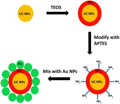
Direct growth of gold NPs on UC NPs was not successful, because homogeneous nucleation of Au NPs is favored over heterogeneous nucleation of Au on the UC NPs under all conditions we have explored. Moreover, direct, intimate contact between the UC NPs and Au NPs may not be desirable, as it could lead to complete quenching of UC NP emission. Therefore, a simple method was designed here for decorating UC NPs with gold NPs and simultaneously transfering the hydrophobic UC NPs to an aqueous environment. The overall approach is illustrated schematically in figure 1, and described in greater detail in the experimental section. Monodispersed UC NaGdF4:2% Er, 20% Yb NPs capped by oleic acid were first synthesized via a procedure adapted from our recent work[24]. The hydrophobic UC NPs were then capped with silica via a reverse-micelle method[28] to make them hydrophilic. Amino-propyltriethoxysilane (APTES) was then used to functionalize the silica-capped UC NPs with amine groups. Finally, 5 nm gold NPs capped with PVP were mixed with the silica-coated UC NPs. Amine groups on the UC NPs displaced the PVP from the AuNP surface, yielding AuNP-decorated UC NPs that were dispersible in water or ethanol.
Schematic illustration of synthetic procedures.
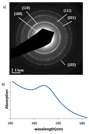
The NaGdF4: Er, Yb particles have hexagonal crystal structure, as demonstrated by selected area electron diffraction (figure 2(a)), which typically leads to higher quantum yield than the cubic phase[29]. TEM images (figure 3), show that the UC NPs have a mean diameter of about 10 nm, and the thickness of the silica shell is about 6 nm, yielding an overall diameter near 22 nm for the coated particles. Figure 3(b) shows the size of UC NPs/silica core-shell particles and their mono-dispersity. Figure 3(c) and (d) show the hybrid particles following the attachment of 5 nm Au NPs. After the UC NPs were decorated with Au NPs, the dispersion turned red. Figure 2(b) shows the extinction spectrum of the NaGdF4:Er,Yb/silica/AuNP dispersion in water, with a peak near 530 nm, attributed to the plasmon resonance absorbance and scattering by Au NPs.
(a) Selected area electron diffraction image of NaGdF4: Er3+,Yb3+ nanocrystals. (b) Absorption spectrum of NaGdF4:Er3+,Yb3+/silica/AuNP water dispersion.
TEM images of (a) UC NPs; (b) UC/silica NPs; (c) UC/silica/Au NPs; (d) higher magnification of UC/silica/Au NPs.

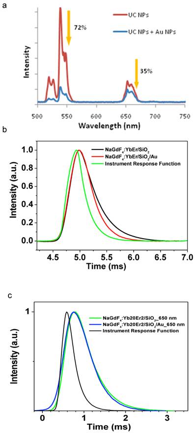
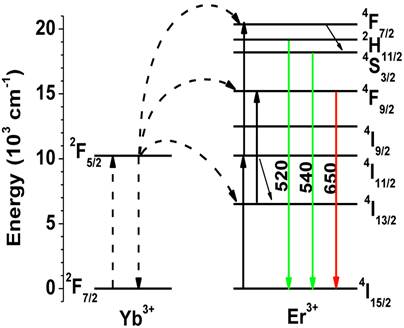
Figure 4(a) shows the emission spectra of the hybrid particles under 975 nm excitation. Two UC bands with maxima at 520/540 nm and 650 nm are seen, corresponding to the 2H11/2/4S3/2→4I15/2 and 4F9/2→4I15/2 transitions (of Er3+), respectively. The up-conversion mechanisms are shown schematically in figure 5. The 975 nm laser excites Yb3+ ions from the ground state to the 2F5/2 state. Then, two discrete energy transfers from the excited Yb3+ ions bring the Er3+ ions from the ground state to the 2H11/2/4S3/2 states, which produces UC emission at 520/540 nm by radiative decay to the ground 4I15/2 state. The red UC emission at 650 nm arises from the 4F9/2 state, which can be populated by either nonradiative decay from the 2H11/2/4S3/2 state or by exciting Er3+ ions from the 4I13/2 state to the 4F9/2 state through energy transfer from Yb3+ ions.
Photoluminescence spectra (a) and lifetime (b) of green emission for UC NPs before and after decorating with Au NPs. (c) Decay of red emission for UC NPs before and after decorating with Au NPs. Spectra presented in part (a) were measured under identical conditions and NP concentration.
Energy level diagrams of Yb3+ and Er3+ ions and the proposed upconversion mechanism in NaGdF4: Yb3+, Er3+ nanocrystals
Upon decoration with Au NPs, the relative UC emission intensities of the hybrid NPs was modulated dramatically, as shown in figure 4 (a). The intensity of the green emission peak at 520/540 nm was reduced by 72% after Au NP addition, while the red emission near 650 nm was attenuated by only 35%. The difference in attenuation for the two bands demonstrates the utility of plasmonic particles for tuning UC colors, which is potentially useful in creating bio-sensors[27]. The surface plasmon resonance of gold can also affect the emission intensities of luminescent particles by shortening the luminescence lifetime[30]. To further clarify this effect, we measured the decay of the green emission at 540 nm. As illustrated in figure 4(b), the lifetime of the green emission was decreased from ~300 µs to ~200 µs. The lifetime of the red emission at 650 nm appeared to be almost unchanged under these conditions(figure 4(c)), but it noticeably decreased for a sample with a higher concentration of gold nanoparticles. The different lifetime shortening for the green and the red emissions is apparently induced by Au NPs and associated with energy transfer to the gold nanoparticles[31]. If the lifetime shortening was due to increased radiative deacitivation rate, then we would expect enhancement of UC emission intensity[30]. However, a decreased intensity was observed for both emission bands. Energy transfer from the UC NP to the Au NP at 520/540 nm could provide a parallel, nonradiative pathway that would compete with the radiative one and decrease the emission intensity. However, a parallel nonradiative path that decreased the lifetime by only 30% would also only decrease the total emission intensity by about 30%. Thus, the direct energy transfer may contribute to the intensity decrease, but does not appear to be the dominant effect. Finally, the emission intensity can also be decreased due to absorption of the emitted light by the Au NPs, without affecting the emission lifetime. All of these effects: enhancement of radiative recombination rate, direct energy transfer (non-radiative rate), and Au reabsorption are expected to be strongest for the green emission band that overlaps with the plasmon resonance of the gold nanoparticles. Therefore, the intensity of this band decreased more than that of the band peaked at 650 nm. Some researchers have observed an increase in photoluminescence of upconverting NPs upon encapsulation with a gold shell. However, others have shown that a decrease in photoluminescence is also possible. Multiple factors may affect the interaction between gold and upconverting NPs, including the distance between gold and upconverting NPs, the ratio of gold NPs to UC NPs, and the surfactants used. These factors will affect both radiative and non-radiative pathways, such that different combinations may lead to either a net enhancement or net attenuation of luminescence.
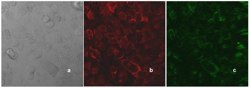
The water-dispersible hybrid nanoparticles possess both plasmonic and UC photoluminescence properties, which can be exploited for bioimaging. Figure 6 illustrates the bimodal imaging capability of the NaGdF4:Er,Yb/silica/Au NPs. HeLa cells were incubated with these nanoparticles overnight at 37 °C, rinsed, and imaged with a confocal microscope. Plasmonic properties of the nanoparticles taken up by cells produce strong contrast in confocal reflection imaging using the 514 nm laser excitation (figure 6b) because of the strong scattering from gold particles. On the other hand, due to the photoluminescence of UC nanoparticle cores, the luminescence cellular imaging can also be obtained when excited at 975 nm (figure 6c). As bioimaging probes, upconversion nanoparticles would be more valuable if their emission efficiency were higher, comparable to that of quantum dots. The doping of gold nanoparticles has been shown in this paper to tune the emission intensity of UC nanoparticles. In the future, the distance between gold and UC nanoparticles and the ratio between them can be controlled to optimize the surface plasmon enhancement effect of gold nanoparticles to increase the emission intensity of UC nanoparticles and further increase the photoluminescence efficiency. Moreover, the Gd-containing host material makes these materials relevant to MRI contrast enhancement as well. This possibility should be investigated further in the future.
Confocal microscopy images of HeLa cells treated with NaGdF4: Er,Yb/silica/Au nanoparticles: (a) transmission; (b) reflection (514 nm laser line); (c) UC photoluminescence (975 nm excitation, detection in the range of 500-700 nm).
4. Conclusion
In summary, water dispersible NaGdF4:Er3+, Yb3+/silica/Au NPs with overall diameters around 20 nm have been prepared. The color and the intensity of UC emission under NIR excitation at 975 nm were changed by the gold NPs. This can be attributed primarily to the surface plasmon resonance absorption by gold nanoparticles. The lifetime of the UC emission was found to decrease slightly as a result of interaction with the gold nanoparticles, suggesting the possibility of non-radiative energy transfer from the UC NPs to Au NPs and/or increase in the rate of radiative deactivation. UC photoluminescence and confocal reflection imaging demonstrated uptake of hybrid nanoparticles by cancer cells in vitro and the possibility of dual modality imaging exploiting both the plasmonic and the UC photoluminescent properties of the NaGdF4:Er,Yb/silica/Au NPs. Though not demonstrated here, MRI contrast enhancement by Gd is expected to enable a third imaging modality. Plasmonic effects on UC NPs depend strongly on the size of gold NPs and the distance between UC NPs and the gold NPs. Future fine tuning of these parameters might result in other interesting phenomena, such as emission enhancement, emission color change, and emission lifetime modulation.
Acknowledgements
This work was supported in part by grants from the National Institutes of Health (R01CA119358 and RO1CA104492) and by the University at Buffalo Integrated Nanostructured Systems Instrument Facility. We are grateful to Mark Kaus for assistance in synthesizing Au NPs.
Competing Interests
The authors have declared that no competing interest exists.
References
1. Wang LY, Yan RX, Hao ZY, Wang L, Zeng JH, Bao H, Wang X, Peng Q, Li YD. Fluorescence resonant energy transfer biosensor based on upconversion-luminescent nanoparticles. Angewandte Chemie-International Edition. 2005;44(37):6054-6057
2. Yi DK, Selvan ST, Lee SS, Papaefthymiou GC, Kundaliya D, Ying JY. Silica-Coated Nanocomposites of Magnetic Nanoparticles and Quantum Dots. Journal of the American Chemical Society. 2005;127(14):4990-4991
3. Chen D, Yu Y, Huang F, Yang A, Wang Y. Lanthanide activator doped NaYb1-xGdxF4 nanocrystals with tunable down-, up-conversion luminescence and paramagnetic properties. Journal of Materials Chemistry. 2011;21(17):6186-6192
4. Sokolov K, Larson TA, Bankson J, Aaron J. Hybrid plasmonic magnetic nanoparticles as molecular specific agents for MRI/optical imaging and photothermal therapy of cancer cells. Nanotechnology. 2007;18(32):325101
5. Yu MK, Park J, Jon S. Targeting Strategies for Multifunctional Nanoparticles in Cancer Imaging and Therapy. Theranostics. 2012;2(1):3-44
6. Wu SW, Han G, Milliron DJ, Aloni S, Altoe V, Talapin DV, Cohen BE, Schuck PJ. Non-blinking and photostable upconverted luminescence from single lanthanide-doped nanocrystals. Proceedings of the National Academy of Sciences of the United States of America. 2009;106(27):10917-10921
7. Mai H.X, Zhang YW, Si R, Yan ZG, Sun Ld, You LP, Yan CH. High-Quality Sodium Rare-Earth Fluoride Nanocrystals: Controlled Synthesis and Optical Properties. J. Am. Chem. Soc. 2006;128(19):6426-6436
8. Boyer JC, Cuccia LA, Capobianco JA. Synthesis of Colloidal Upconverting NaYF4: Er3+/Yb3+ and Tm3+/Yb3+ Monodisperse Nanocrystals. Nano Letters. 2007;7(3):847-852
9. Heer S, Kömpe K, Güdel HU, Haase M. Highly Efficient Multicolour Upconversion Emission in Transparent Colloids of Lanthanide-Doped NaYF4 Nanocrystals. Advanced Materials. 2004;16(23-24):2102-2105
10. Chen GY, Ohulchanskyy T, Kumar R, Agren H, Prasad PN. Ultrasmall Monodisperse NaYF4:Yb3+/Tm3+ Nanocrystals with Enhanced Near-Infrared to Near-Infrared Upconversion Photoluminescence. ACS Nano. 2010;4(6):3163-3168
11. Nyk M, Kumar R, Ohulchanskyy TY, Bergey EJ, Prasad PN. High Contrast in Vitro and in Vivo Photoluminescence Bioimaging Using Near Infrared to Near Infrared Up-Conversion in Tm3+ and Yb3+ Doped Fluoride Nanophosphors. Nano Letters. 2008;8(11):3834-3838
12. Wang M, Mi CC, Wang WX, Liu CH, Wu YF, Xu ZR, Mao CB, Xu SK. Immunolabeling and NIR-Excited Fluorescent Imaging of HeLa Cells by Using NaYF4:Yb,Er Upconversion Nanoparticles. ACS Nano. 2009;3(6):1580-1586
13. Prasad PN. Introduction to Biophotonics. New York: Wiley-Interscience. 2003
14. Chen D, Lei L, Yang A, Wang Z, Wang Y. Ultra-broadband near-infrared excitable upconversion core/shell nanocrystals. Chemical Communications. 2012;48(47):5898-5900
15. O'Neal DP, Hirsch LR, Halas NJ, Payne JD, West JL. Photo-thermal tumor ablation in mice using near infrared-absorbing nanoparticles. Cancer Letters. 2004;209(2):171-176
16. Sharrna P, Brown S, Walter G, Santra S, Moudgil B. Nanoparticles for bioimaging. Advances in Colloid and Interface Science. 2006;123:471-485
17. Jang B, Choi Y. Photosensitizer-Conjugated Gold Nanorods for Enzyme-Activatable Fluorescence Imaging and Photodynamic Therapy. Theranostics. 2012;2(2):190
18. Liu S, Chen G, Prasad PN, Swihart MT. Synthesis of Monodisperse Au, Ag, and Au-Ag Alloy Nanoparticles with Tunable Size and Surface Plasmon Resonance Frequency. Chemistry of Materials. 2011;23(18):4098
19. Kondo M, Uchikawa M, Kume S, Nishihara H. Alcohol- and acid-causing reversible switching of near-infrared absorption and luminescence in a donor-acceptor conjugated system. Chemical Communications. 2009;15:1993-1995
20. Liu YS, Tu DT, Zhu HM, Li R, Luo WQ, Chen XY. A Strategy to Achieve Efficient Dual-Mode Luminescence of Eu3+ in Lanthanides Doped Multifunctional NaGdF4 Nanocrystals. Advanced Materials. 2010;22(30):3266-3271
21. Wang F, Wang JA, Liu XG. Direct Evidence of a Surface Quenching Effect on Size-Dependent Luminescence of Upconversion Nanoparticles. Angewandte Chemie-International Edition. 2010;49(41):7456-7460
22. Abel KA, Boyer JC, Veggel FCJMv. Hard Proof of the NaYF4/NaGdF4 Nanocrystal Core/Shell Structure. Journal of the American Chemical Society. 2009;131(41):14644-14645
23. Zhen Z, Xie J. Development of Manganese-Based Nanoparticles as Contrast Probes for Magnetic Resonance Imaging. Theranostics. 2012;2(1):45-54
24. Chen GY, Ohulchanskyy TY, Law WC. Agren, H, and Prasad, P.N, Monodisperse NaYbF4: Tm3+/NaGdF4 core/shell nanocrystals with near-infrared to near-infrared upconversion photoluminescence and magnetic resonance properties. Nanoscale. 2011;3:2003-2008
25. Kumar R, Nyk M, Ohulchanskyy TY, Flask CA, Prasad PN. Combined Optical and MR Bioimaging Using Rare Earth Ion Doped NaYF4 Nanocrystals. Advanced Functional Materials. 2009;19(6):853-859
26. Xing H, Bu W, Zhang S, Zheng X, Li M, Chen F. He, Q, Zhou, L, Peng, W, Hua, Y, and Shi, J, Multifunctional nanoprobes for upconversion fluorescence, MR and CT trimodal imaging. Biomaterials. 2012;33(4):1079-1089
27. Li Z, Wang L, Wang Z, Liu X, Xiong Y. Modification of NaYF4:Yb,Er@SiO2 Nanoparticles with Gold Nanocrystals for Tunable Green-to-Red Upconversion Emissions. The Journal of Physical Chemistry C. 2011;115(8):3291-3296
28. Han Y, Jiang J, Lee SS, Ying JY. Reverse Microemulsion-Mediated Synthesis of Silica-Coated Gold and Silver Nanoparticles. Langmuir. 2008;24(11):5842-5848
29. Krämer KW, Biner D, Frei G. Güdel, H.U, Hehlen, M.P, and Lüthi, S.R, Hexagonal Sodium Yttrium Fluoride Based Green and Blue Emitting Upconversion Phosphors. Chemistry of Materials. 2004;16(7):1244-1251
30. Zhang H, Li YJ, Ivanov IA, Qu YQ, Huang Y, Duan XF. Plasmonic Modulation of the Upconversion Fluorescence in NaYF4:Yb/Tm Hexaplate Nanocrystals Using Gold Nanoparticles or Nanoshells. Angewandte Chemie-International Edition. 2010;49(16):2865-2868
31. Zhang H, Xu D, Huang Y, Duan XF. Highly spectral dependent enhancement of upconversion emission with sputtered gold island films. Chemical Communications. 2011;47(3):979-981
Author contact
![]() Corresponding author: pnprasadedu or swihartedu.
Corresponding author: pnprasadedu or swihartedu.