Impact Factor
- Issue 14; 2026
- Issue 13; 2026
- Issue 12; 2026
- Issue 11; 2026
- Issue 10; 2026
- Volume 16; 2026
- Advance Articles
- Past Issues
- Cover Images
- Cover Suggestion
- Index & Coverage
- Special Issues
1. Introduction
2. Application of...
3. Endogenous stimuli-responsive...
4. Exogenous stimuli-responsive...
5. Dual stimuli-responsive...
6. Types of wound applied with...
7. Conclusion and outlook
Abbreviations
Acknowledgements
References
International Journal of Biological Sciences
International Journal of Medical Sciences
 Global reach, higher impact

Theranostics 2026; 16(12):7029-7066. doi:10.7150/thno.133095 This issue Cite
Review
Stimuli-responsive nanomaterials for wound healing: advances and emerging directions
Yijia Dong1, Yaqing Zhang1, Junrong Wu1, Yujun Yang1, Yanli Zhang1, Zhenjun Zhu2, Rabiya Noor3, Jawad Hussain3, Longquan Shao1,4 ![]() , Yi Xing1
, Yi Xing1 ![]()
1. Stomatological Hospital, School of Stomatology, Southern Medical University, Guangzhou, 510280, PR China.
2. Department of Stomatology, Shenzhen People’s Hospital, Shenzhen, 518020, PR China.
3. Riphah International University, Lahore, Pakistan.
4. Guangdong Provincial Key Laboratory of Construction and Detection in Tissue Engineering, Southern Medical University, Guangzhou, 510280, PR China.
Received 2026-2-12; Accepted 2026-5-4; Published 2026-5-18
Abstract

In wound healing applications, conventional nanomaterials fail to precisely regulate the wound healing process because of the lack of dynamic adaptation to the wound microenvironment. Conversely, stimuli-responsive nanomaterials can respond to endogenous stimuli such as pH, enzymes, reactive oxygen species (ROS), and glucose, as well as exogenous stimuli such as light, electricity, and magnetism. Thus, stimuli-responsive nanomaterials can facilitate precise drug delivery and effectively treat wounds, and some of these materials can even achieve integrated diagnosis and treatment. Previous reviews have focused mainly on hydrogel carriers with stimuli-responsive properties. This review focuses on stimuli-responsive nanomaterials, classifies these materials on the basis of their stimulus sources, and systematically summarizes the response mechanisms, application outcomes, and design strategies of endogenous, exogenous, and dual stimuli-responsive nanomaterials applied in wound healing. Furthermore, this review explores the research gaps and future developments of stimuli-responsive nanomaterials for wound healing.
Keywords: stimuli-responsive nanomaterials, wound healing, microenvironment, drug release, integrated diagnosis and treatment
1. Introduction
Wound healing is among the most complicated processes in the human body and requires intricate spatial and temporal coordination of cells and tissues [1]. However, due to the increasing prevalence of diseases such as diabetes, cardiovascular diseases, and autoimmune disorders, this process is often disrupted, resulting in chronic nonhealing wounds and even severe consequences such as amputation and death.
Nanomaterials and hydrogels are the most common strategies for wound treatment [2, 3]. Nanomaterials refer to materials that have at least one dimension in the three-dimensional space within the nanoscale range (1–100 nm) or are composed of units of this scale as the basic building blocks [4]. Hydrogels are three-dimensional network structures formed by natural or synthetic polymers through physical or chemical cross-linking [5]. Because of their high specific surface area and small particle size, nanomaterials exhibit significant advantages over traditional hydrogels in promoting wound healing [6].
The large specific surface area of nanomaterials endows them with three major advantages: strong enzymatic catalytic activity, excellent drug-loading capacity, and the ability to regulate cellular biological functions. First, the large specific surface area of nanomaterials allows them to expose more active sites and increase the reaction area, enabling certain nanomaterials to have superior catalytic activity, achieving functions such as efficient sterilization, oxygen production, and catalytic decomposition of ROS, thereby regulating the microenvironment of the wound [7-10]. In contrast, hydrogels usually need to be combined with nanomaterials with catalytic activity to achieve the corresponding effect [11]. Second, the large specific surface area enables nanomaterials to achieve stable drug loading through various mechanisms, such as electrostatic adsorption, physical binding, and chemical bonding, thereby significantly enhancing drug-loading efficiency and accelerating wound healing through targeted drug delivery and controlled drug release [12]. While most hydrogels rely primarily on physical encapsulation for drug delivery, if the interaction between the drug and the gel matrix is weak, stable and efficient drug loading is difficult to achieve, and drug burst release is likely to occur [13]. Third, the high specific surface area of nanomaterials provides an important structural basis for regulating the biological behavior of cells. For example, the large specific surface area of nanofibers can provide a larger attachment area for skin tissue cells, effectively regulating cell proliferation and migration by mimicking the structure of the natural extracellular matrix, thereby accelerating wound healing [14].
In addition, the small particle size of nanomaterials allows them to better penetrate tissue and the bacterial biofilm barrier. Moreover, the size advantage also facilitates the arrival of nanomaterials at deep wound areas, thereby exerting corresponding biological effects [8]. In bacteria-infected wounds, pathogenic bacteria aggregate and secrete extracellular polymers, forming a dense bacterial biofilm, which leads to persistent wound infection [15]. With their small particle size, nanomaterials can effectively penetrate the dense extracellular matrix of the bacterial biofilm and kill bacteria deep within the biofilm, thereby eliminating wound infection [16]. Moreover, nanomaterials can penetrate the dermis layer and even deeper damaged tissues through intercellular spaces or capillary walls to achieve deep drug delivery and promote deep wound healing [17]. Traditional hydrogels usually cover only the wound surface and are difficult to penetrate the bacterial biofilm barrier or penetrate deep into the tissue to act on deep wounds [18].
However, the complex pathological wound microenvironment, which is characterized by hyperglycemia, inflammation, infection, and hypoxia, results in abnormal physicochemical indicators, including pH, ROS, glucose, and enzyme expression profiles [19-21]. Conventional nanomaterials cannot dynamically respond to such pathological microenvironments, and their effects are difficult to precisely control; therefore, they cannot achieve targeted intervention at the critical stage of wound healing, leading to problems such as off-target effects, pathogen resistance, and difficulty in adapting to complex wounds (Figure 1) [22,23]. Therefore, stimuli-responsive nanomaterials have garnered increasing attention and research.
Schematic diagram illustrating the major categories of stimuli-responsive nanomaterials and their working mechanisms. The figure shows how nanomaterials are designed to respond to various endogenous stimuli and exgenous stimuli. Upon exposure to a specific trigger, the nanomaterials undergo energy conversion or structural change, leading to directed pro-healing effects or controlled release of therapeutic agents. Created in https://BioRender.com.
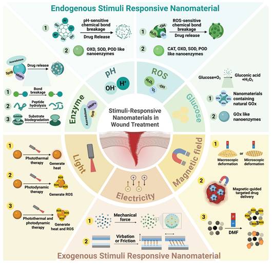
Stimuli-responsive nanomaterials refer to nanomaterials that can respond to different stimulus, thereby changing their physical and chemical structures or undergoing energy conversion [24]. On the basis of the advantages of these nanomaterials, such as dynamically adapting to the wound microenvironment, preventing the development of drug resistance, targeted drug delivery, and promoting wound healing in multiple stages, new ideas and strategies for wound treatment have been introduced [25-28]. To date, stimuli-responsive nanomaterials have demonstrated excellent antibacterial, anti-inflammatory, and immunomodulatory effects [29-32]. These findings indicate the great application potential of these materials in skin wound treatment.
In the different types of wound microenvironments and at different stages of healing, endogenous indicators such as pH, enzymes, ROS, and glucose are constantly changing. For example, in bacteria-infected wounds, the expression levels of azide reductase and serine protease are relatively high [33,34], whereas in diabetic wounds, the expression levels of matrix metalloproteinases (MMPs) and local glucose concentrations are relatively high [35,36]. Furthermore, certain indicators shift dynamically during different healing stages (including the hemostasis phase, inflammatory phase, proliferation phase and remodeling phase). For instance, the pH gradually increases from 5.5–6.0 during the inflammatory phase to 6.5–7.4 during the proliferation phase and the remodeling phase [37]. Additionally, during the inflammatory phase, ROS levels abnormally increase but gradually decrease during the proliferative and remodeling periods, then remain at a relatively low level [38]. Based on these features, researchers have designed nanomaterials that respond to endogenous stimulus. By accurately sensing microenvironmental cues (including pH, ROS, and enzymatic levels), these responsive materials can undergo programmed degradation or exhibit nanozyme-like activity, thereby synergistically promoting wound repair [39,40].
When exposed to external physical energy (such as light, mechanical force or magnetism), exogenous stimuli-responsive nanomaterials can undergo energy conversion or structural changes, thereby regulating cellular functions and accelerating wound healing. For example, light-responsive nanomaterials can generate heat energy through the photothermal effect [41], electro-responsive nanomaterials can generate electrical signals through the piezoelectric effect or the triboelectric effect [42-45], and magnetic-responsive nanomaterials can undergo macroscopic deformation or microscopic topological structure changes under the stimulation of a magnetic field, thereby generating mechanical signals for skin tissue cells [46]. By adjusting the external energy stimulation parameters, exogenous stimuli-responsive nanomaterials can prevent harm to surrounding tissues and precisely control their therapeutic effects, thereby ensuring the safety and controllability of wound treatment [47].
Additionally, dual stimuli-responsive nanomaterials can respond to two distinct stimulus sources. These mainly include dual endogenous stimuli-responsive nanomaterials, dual exogenous stimuli-responsive nanomaterials, and endogenous-exogenous stimuli-responsive nanomaterials. By integrating two distinct response mechanisms, dual stimuli-responsive nanomaterials can adapt to more complex wound microenvironments and promote wound healing in multiple healing stages [48].
This review starts by exploring the types of stimulus sources and systematically reviews the mechanism, design strategies, and modification methods of various stimuli-responsive nanomaterials in promoting wound healing. Unlike previous reviews, this review focuses on analyzing how stimuli-responsive nanomaterials, by changing their own physical and chemical properties or by utilizing their own characteristics, exert multiple mechanisms, such as bactericidal, anti-inflammatory, immune regulatory, and promote tissue regeneration effects to accelerate wound healing. In addition, this review proposes corresponding solutions to the biological safety risks and scale-up problems faced by stimuli-responsive nanomaterials during application. The aim of this review is to provide scientific guidance and theoretical support for the design and clinical translation of stimuli-responsive wound treatment nanomaterials in the future.
2. Application of stimuli-responsive nanomaterials in wound healing
Stimuli-responsive nanomaterials can be mainly classified into endogenous stimuli-responsive nanomaterials, exogenous stimuli-responsive nanomaterials, and dual stimuli-responsive nanomaterials. It can exert multiple effects in accelerating the healing of bacterial-infected wounds, diabetic wounds, burn wounds and so on. Table 1 summarizes the material composition, mechanism of action, and application types of wounds of stimuli-responsive nanomaterials (Table 1).
Summary of application of stimuli-responsive nanomaterials for wound healing
| Stimuli-responsive nanomaterials | Components | Role in wound healing | Animal model | Ref. | |
|---|---|---|---|---|---|
| Endogenous stimuli- responsive nanomaterials | pH-responsive | Allicin@ZIF-8/Ag nanoparticles | Antibacterial, Anti-inflammation, Antioxidant, Angiogenesis | S. aureus-infected full-thickness wound E. coil- infected full thickness wound | [49] |
| Chlorhexidine-Si nanoparticles | Antibacterial | E. coil-infected full-thickness wound | [50] | ||
| Enzyme- responsive | Ag nanoparticles-TA-COF nanosheets @ ebselen @ polyethylene glycol | Antibacterial, Anti-inflammation | S. aureus-infected full-thickness wound | [33] | |
| Linear poly(ethyleneimine)-immobilized nanofiber/ plasmid Human epidermal growth factor | Promote re-epithelization | Full-thickness diabetic wound | [51] | ||
| ROS- responsive | Trisulfide-derived lipid nanoparticle/ interleukin-4 mRNA | Anti-inflammation, Antioxidant | Full-thickness diabetic wound | [52] | |
| Polyvinylpyrrolidone -Ir nanoparticles /Ag nanoparticles / methacrylate gelatin | Antibacterial, Anti-inflammation | Bacteria-infected full-thickness wound | [53] | ||
| Glucose- responsive | Bacterial cellulose/ polypropylene-Fe@ mesoporous carbon nanosphere/ GOx | Hemostasis, Antibacterial | S. aureus-infected full-thickness wound | [54] | |
| Copper nanoclusters/ glucose oxidase / oxidized hyaluronic acid | Reduce blood sugar, Antibacterial | MRSA(Methicillin-Resistant Staphylococcus Aureus)-infected full-thickness diabetic wound | [55] | ||
| GSH- responsive | CuCo2O4 nanoflowers | Antibacterial | MRSA-infected full-thickness wound. MRSA-infected full-thickness burn wound | [56] | |
| ATP- responsive | Zeolitic imidazolate framework-8/ Indole-3-acetic acid /horseradish peroxidase/ polyacrylamide | Antibacterial | S. aureus-infected full-thickness diabetic wound | [57] | |
| Exogenous stimuli- responsive nanomaterials | Light- responsive | PDA-PEI-PEG-PMB nanoparticles | Antibacterial | S. aureus-infected full-thickness wound | [58] |
| UCNPs@TiO2@GO/ poly(vinylidene) fluoride | Antibacterial, Anti-inflammation | S. aureus-infected full-thickness wound | [59] | ||
| Au nanoparticle @corn stalk/chitin | Hemostasis, Antioxidant, Antibacterial | Full-thickness wound | [60] | ||
| Electro- responsive | BaTiO3 nanocrystal @MMSa | Antibacterial | S. aureus-infected full-thickness wound | [61] | |
| Poly (l-lactic acid) nanofiber | Antibacterial, Promote cell proliferation and migration | Full-thickness wound | [62] | ||
| Nano-ZnO/ Sodium alginate/ polyvinylidene fluoride | Antibacterial, Promote cell proliferation and migration | Full-thickness wound | [63] | ||
| Magnetic- responsive | Fe2O3 nanoparticle labeled MSC-Exo | Promote cell proliferation and migration, Angiogenesis | Full-thickness burn wound | [64] | |
| CoFe2O4 nanoparticle/ polyvinylidene fluoride nanofiber | Antibacterial, Promote the secretion of growth factors | S. aureus-infected full-thickness diabetic wound | [65] | ||
| Ultrasound- responsive | Platinum nanoparticle assembly/gelatin-methacryloyl | Anti-inflammation | S. aureus-infected full-thickness diabetic wound | [66] | |
| (K,Na)NbO3 nanocrystals/ reduced graphene oxide/gelatin/polyvin-yl alcohol | Promote angiogenesis and nerve regeneration | S. aureus-infected full-thickness diabetic wound | [67] | ||
| Thermal- responsive | Polyurethane /polyvinyl butyral | Promote wound contraction | Full-thickness wound | [68] | |
| Poly-(lactic acid-co-trimethylene carbonate) nanofibers /methacrylate gelatin /Epinecidin-1@chitosan nanoparticles | Antibacterial, Anti-inflammation | S. aureus-infected full-thickness diabetic wound | [69] | ||
| Dual stimuli- responsive nanomaterials | pH-ROS- responsive | GSC/PBE@Lut (Luteolin) | Antibacterial, Anti-inflammation and Promote angiogenesis | S. aureus-infected full-thickness wound | [70] |
| Light-Electro-responsive | Poly-L-lactic acid-calcium peroxide-reduced graphene oxide | Antibacterial, Promote cell proliferation and migration | E. coil-infected full-thickness wound | [71] | |
| Light-Enzyme- responsive | Hyaluronic acid-Porphyrin-based Fe covalent organic frameworks | Antibacterial and Promote angiogenesis | S. aureus-infected full-thickness wound | [72] | |
3. Endogenous stimuli-responsive nanomaterials
Endogenous stimuli-responsive nanomaterials include pH-, enzyme-, ROS-, glucose-, ATP-, and GSH-responsive nanomaterials. These nanomaterials accelerate wound healing mainly through targeted delivery of therapeutic substances and by mimicking the activity of natural enzymes.
3.1. pH-responsive nanomaterials
The pH value undergoes significant changes during wound healing. Specifically, during the inflammatory stage, the pH is acidic, whereas during the proliferation stage and remodeling stage, it is weakly acidic to neutral [73]. Therefore, researchers have designed acidic pH-responsive and alkaline pH-responsive nanomaterials to promote wound healing (Figure 2).
Diagrammatic illustration of the pH-responsive nanomaterials applied in wound treatment. The figure illustrates how these nanomaterials respond to pH changes in the wound microenvironment. Upon exposure to abnormal pH, the nanomaterials undergo structural changes to release therapeutic cargoes or mimic natural enzyme activity, thereby promoting wound healing. Created in https://BioRender.com.
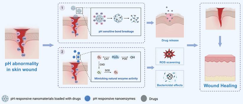
When the wound pH is acidic, nanomaterials that respond to acidic pH can act through the following two mechanisms. The first mechanism involves structural disintegration to deliver therapeutic components. The second mechanism involves mimicking natural enzyme activities and thereby accelerating wound healing.
In acidic microenvironments, acidic pH-responsive nanomaterials can undergo structural disintegration and deliver therapeutic components, thereby accelerating wound healing [26, 49, 74-77]. For instance, researchers developed an acidic pH-responsive nanosystem by modifying the surface of silver nanoparticles (Ag NPs) with ZIF-8 and then encasing allicin within the ZIF-8 framework. In the acidic wound microenvironment, H+ will react with the ZIF-8 framework through protonation interactions and cause structural disintegration of the nanosystem, thereby releasing Ag+ and allicin to exert antibacterial and antioxidant effects. In addition, ZIF-8 on the surface of Ag NPs can prevent their aggregation to lower toxicity, thereby accelerating the healing of bacterially infected wounds efficiently and safely [49, 78]. Additionally, some researchers have developed acidic pH-responsive polyserotonin (PST)/Ag nanoparticles. In the acidic microenvironment of bacterially infected wounds, the PST shell layer of these nanoparticles breaks down and enables the release of Ag+. Then by inducing oxidative stress in bacteria, disrupting the bacterial membrane and affecting their metabolic pathways to exert bactericidal effects and accelerate skin wound healing [76, 79].
Furthermore, designing a dual acidic pH-responsive nanosystem can enhance the efficacy and safety of pH-responsive nanomaterials [80, 81]. For instance, researchers have designed P-ZIF nanoparticles by loading polyhexamethylene biguanide (PHMB) into a ZIF-8 framework. Then, the dual pH-responsive HASPZ was developed by incorporating these nanoparticles via boronic ester bonds into a sodium alginate-based hydrogel. In the mildly acidic microenvironment of infected wounds, the boronic ester bonds break down [82, 83], releasing P-ZIF nanoparticles to exert a preliminary antibacterial effect. In a severely infected wound with a more acidic microenvironment, the P-ZIF nanoparticles further disintegrate to release PHMB, providing a potent antibacterial effect. Additionally, during the disintegration of P-ZIF nanoparticles, Zn2+ can be released to accelerate angiogenesis. This release pattern can prevent drug overdose to avoid side effects. Eventually, this nanohydrogel efficiently and safely promotes deep second-degree burn wound healing [80]. Additionally, researchers have constructed a dual acidic pH-responsive nanomaterial system by loading α-phase manganese sulfide nanoparticles (α-MnS NPs) into a hydrogel crosslinked by Schiff base bonds. When the wound microenvironment is acidic, it triggers the hydrolysis of Schiff base bonds, leading to the release of α-MnS NPs. These α-MnS NPs subsequently degrade in the acidic microenvironment and release H2S, thereby exerting anti-inflammatory effects by reducing the expression of proinflammatory factors (such as IL-6 and IL-1β) to accelerate wound healing [81].
In addition to delivering therapeutic components, acidic pH-responsive nanomaterials can also mimic natural enzyme activity to accelerate wound healing [84-87]. For example, researchers have used sulfated fucoidan as both a reducing agent and carrier to synthesize pH-responsive gold nanoparticles (Fuc@AuNPs). In the acidic wound microenvironment, a high concentration of H+ alters the surface charge distribution of AuNPs, thereby exhibiting oxidase (OXD)-like activity by promoting the adsorption of O2 on its surface and catalyzing the transformation of O2 into singlet oxygen (1O2), thus exerting a bactericidal effect. As the wound infection is controlled, the local pH gradually returns to the normal physiological level. At this stage, the OXD-like activity of Fuc@AuNPs is inhibited, and instead, the Fuc@AuNPs exhibit superoxide dismutase (SOD)-like activity to scavenge ROS while synergizing with the intrinsic anti-inflammatory properties of fucoidan to mitigate inflammatory responses and accelerate the healing of bacteria-infected wounds [87].
When the wound pH is alkaline, nanomaterials that respond to alkaline pH can act through the following two mechanisms. The first mechanism involves structural disintegration through covalent bond cleavage to deliver therapeutic components. The second mechanism involves mimicking natural enzyme activities and thereby accelerating wound healing.
On the one hand, in the alkaline wound microenvironment, the silicon‒oxygen bonds (Si‒O‒Si) in the silica nanoparticle (SiNP) framework can undergo nucleophilic substitution reactions with OH- and break down. This process leads to the disintegration of SiNPs due to substrate etching and increased porosity. Afterward, the encapsulated chlorhexidine (CHX) is released to exert a bactericidal effect, thereby effectively treating chronic infected wounds [50].
On the other hand, alkaline pH-responsive nanomaterials can also act by mimicking natural enzyme activities to accelerate wound healing [88, 89]. For instance, researchers developed a nanocomposite with multienzyme-like activity (Mo, Fe/Cu, I-Ag@GOx) and anchored it within a hydrogel. This nanocomposite operates through a “pH-switched glucose-initiated cascade reaction” to accelerate diabetic wound healing. In the hyperglycemic microenvironment of diabetic wounds, the nanocomposite first initiates the first cascade reaction relies on glucose oxidase (GOx), which catalyzes the decomposition of glucose and O2 into gluconic acid and H2O2 to consume excess glucose. The nanocomposite subsequently mimics peroxidase (POD)-like and OXD-like activities, converting H2O2 into superoxide anions (·O2-) and hydroxyl radicals (·OH), thereby exerting bactericidal effects. As inflammation diminishes and the local pH gradually increases to a mildly alkaline state, the activity of the nanocomposite switches to a second cascade reaction and simulates SOD-like activity to scavenge excess ROS and generate O2, thereby mitigating oxidative stress and tissue hypoxia [89].
3.2. Enzyme-responsive nanomaterials
In the wound microenvironment, the abnormal overexpression of various enzymes (such as MMPs) often prolongs inflammation, resulting in delayed wound healing [90]. To overcome this problem, researchers have designed various enzyme-responsive nanomaterials to precisely deliver therapeutic components and accelerate wound healing [91-94]. These nanomaterials respond primarily to changes in local enzyme expression levels through three mechanisms: first, by the cleavage of enzyme-responsive covalent bonds within the nanomaterials; second, by the hydrolysis of enzyme-responsive peptide sequences within the nanomaterials; and third, by the biodegradation of macromolecular substrate components within the nanomaterials that correspond to the enzymes (Figure 3).
Diagrammatic illustration of the enzyme-responsive nanomaterials applied in wound treatment. The figure shows how these nanomaterials are designed to respond to specific enzymes that are overexpressed in the wound microenvironment. Upon encountering elevated levels of enzymes, the nanomaterials undergo triggered degradation, leading to on-demand release of therapeutic cargoes precisely at the wound site. Created in https://BioRender.com.
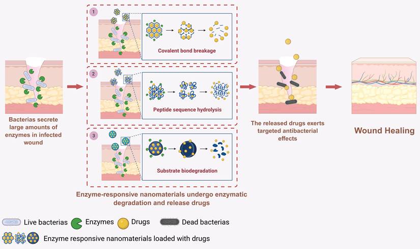
First, enzyme-responsive nanomaterials can react with the high-enzyme wound microenvironment through covalent bond cleavage. Various bacteria involved in wound infections (such as Escherichia coli and Staphylococcus aureus) secrete azoreductase, leading to elevated expression levels of azoreductase in bacterially infected wounds. Therefore, covalent organic frameworks (COFs) containing azo bonds can cleave in response to high azoreductase levels in such wound microenvironments, enabling the targeted release of loaded ebselen (EBS) and silver ions to exert bactericidal and anti-inflammatory effects and thereby accelerating the healing of bacterially infected wounds (Figure 4A-D) [33].
Enzyme-responsive nanomaterials for wound treatment. A) Diagrammatic illustration of the synthesis and antibacterial mechanism of Ag-TA-CON@EBS@PEG. B) Lethality fluorescence staining images of bacterial colonies on agar plates. C) Images of wound with different treatments at different times during the therapeutic process. D) Simulation analysis of skin wound healing process with different treatments. Adapted with permission from [33]. Copyright 2022, American Chemical Society. E) Diagrammatic illustration of enzyme responsive ANAs for infected wound. F) Description of the synthesis of enzyme-responsive ANAs. Adapted with permission from [34]. Copyright 2020, American Chemical Society.
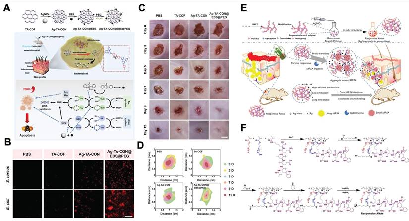
Second, enzyme-responsive nanomaterials can achieve therapeutic component delivery through the hydrolysis of enzyme-sensitive peptide sequences. For example, the bacterium secretes large amounts of serine protease-like B enzyme proteins (SplB) in MRSA-infected wounds. On this basis, researchers have designed enzyme-responsive Ag nanoparticle assemblies incorporating the WELQK peptide sequence (methacrylate-tryptophan-glutamate-leucine-glutamine-lysine-methacrylate). The WELQK sequence can specifically respond to the SplB secreted by MRSA and then be hydrolyzed to release Ag nanoparticles. Eventually, this nanoassembly can exert bactericidal effects and promote the healing of MRSA-infected wounds (Figure 4E–F) [34]. Additionally, in the diabetic wound microenvironment, the expression level of MMPs is elevated. Therefore, researchers have constructed MMP-responsive nanomaterials by incorporating MMP-cleavable peptide sequences into the nanofiber scaffold. In the high-MMP wound microenvironment, the MMP-responsive peptide sequences within the nanofiber scaffold undergo hydrolysis and deliver plasmids encoding human epidermal growth factor (hEGF), upregulate hEGF expression levels in fibroblasts and then accelerate the re-epithelialization process, thereby effectively promoting diabetic ulcer wound healing [51].
Finally, enzyme-responsive nanomaterials can also deliver therapeutic components through the enzymatic degradation of the corresponding macromolecular substrate [40, 95, 96]. For instance, gelatin nanoparticles (Cur@Gel NPs), derived from gelatin, which is a substrate of MMPs, can undergo biodegradation in response to highly expressed MMPs in wounds, thereby releasing loaded curcumin to exert anti-inflammatory effects and accelerate wound healing [40]. Moreover, hyaluronic acid (HA) can be electrostatically adsorbed onto the surface of a monolayer graphene quantum dot–carbon monoxide-releasing molecular nanocomplex (SGQDs-CORM), thereby constructing a hyaluronidase (HAase)-responsive nanomaterial, SGQDs-CORM@HA (SCH). At the site of bacterial infection, the HA on the surface of this enzyme-responsive nanomaterial can be specifically degraded by HAase secreted by MRSA, exposing the ultrathin nanosheet-structured SGQDs-CORM, which then exerts efficient bactericidal effects and accelerates the healing of bacterially infected wounds [95].
Currently, in the application of enzyme-responsive nanomaterials, the challenge of insufficient specificity of the enzyme-sensitive components still exists, which refers to the fact that the substrate recognition sequences of enzymes have some degree of overlap, such as those of proteases such as fibrinolytic enzymes and MMP. This overlap can easily lead to incorrect responses of enzyme-sensitive nanomaterials, causing cross-hydrolysis of enzyme substrates and subsequently resulting in the incorrect release of drugs in nontargeted wound microenvironments, significantly reducing therapeutic efficiency. To address this issue, future approaches can consider the use of phage display technology to design novel short peptide sequences that can specifically bind to the MMP. This peptide sequence can serve as a novel enzyme-sensitive element to effectively prevent erroneous release triggered by nontargeting enzymes.
In recent years, studies have quantitatively analyzed the targeting performance of “ultrahigh specificity” peptides obtained through “phage display technology” and in vivo validation has been completed in wound models. Common methods for quantifying affinity include isothermal titration calorimetry, surface plasmon resonance, and biolayer interferometry [97-100]. For example, Liu et al. first screened a short peptide, KG7, that specifically targeted the bacterial biofilm protein PSMα1 from a twelve-peptide phage display library. Using the ITC method, they measured the dissociation constant of KG7 and PSMα1 and confirmed that its high affinity was approximately 66 times greater than that of the control peptide. The results showed that KG7 could bind to the key functional region of PSMα1 and interfere with the assembly of the biofilm matrix. Furthermore, researchers modified KG7 onto the surface of photothermal nanomaterials to construct a targeted nanoplatform. It was confirmed that this nanocomposite could accumulate at the biofilm site and exert a photothermal effect to destroy the biofilm, thereby accelerating diabetic infected wound healing [100]. These studies have provided examples for the quantitative determination of the affinity of highly specific targeting peptides and in vivo functional validation.
3.3. ROS-responsive nanomaterials
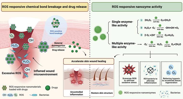
Excessive ROS (such as H2O2, ·OH, and ·O2-) in the wound microenvironment can prevent the physiological transition from the inflammation stage to the proliferation stage, thereby delaying wound healing [101]. ROS-responsive nanomaterials are nanomaterials that can detect and react to excessive ROS. They primarily accelerate wound healing through the following two mechanisms: (1) Chemical bonds within the nanomaterial react with ROS and cleavage, thereby triggering the release of loaded drugs. (2) ROS-responsive nanozymes that can detect and respond to ROS, mimicking single-enzyme or multiple-enzyme activities, thereby exerting bactericidal or anti-inflammatory effects (Figure 5).
Diagrammatic illustration of the ROS-responsive nanomaterials applied in wound treatment. The figure illustrates how these nanomaterials respond to ROS changes in the wound microenvironment. Upon exposure to elevated ROS levels, the nanomaterials undergo structural changes to release therapeutic cargoes or mimic natural enzyme activity, thereby promoting wound healing. Created in https://BioRender.com.
Firstly, ROS-responsive nanomaterials can deliver drugs through the cleavage of trisulfide bonds. For example, in the high oxidative stress microenvironment of diabetic wounds, lipid nanoparticles containing trisulfide bonds can respond to high levels of ROS, leading to the cleavage of trisulfide bonds and releasing encapsulated IL-4 mRNA, inducing macrophages polarizing toward anti-inflammatory phenotype and inhibiting excessive inflammation, thereby promoting diabetic wound healing [52]. Furthermore, ROS-responsive nanomaterials can also trigger drug release by breaking the borate ester bonds. For example, in the high level of ROS microenvironment of diabetic wounds, nanoliposomes containing borate ester bonds will undergo oxidative cleavage, triggering the sustained release of loaded antimicrobial peptides and puerarin. These nanoliposomes can promote angiogenesis, exert antibacterial, anti-inflammatory effects, thereby accelerating diabetic wound healing [32].
Secondly, ROS-responsive nanozymes can mimic single-enzyme or multiple-enzyme activities, thereby exerting bactericidal, antioxidant effects and relieving tissue hypoxia to accelerate wound healing. On the one hand, ROS-responsive nanozymes that mimic single enzyme activities (such as CAT-like, POD-like, and SOD-like) can accelerate wound healing by exerting bactericidal or antioxidant effects [102-105]. For example, researchers embedded lanthanide elements and transition metal ions into the lattice of iron-based nanozymes, reducing the free energy barrier of the catalytic reaction and facilitating electron transfer, thereby successfully constructing a high-entropy nanocrystalline structure nanozyme (C-ZnFeO@Nd NPs) with advanced POD-like catalytic ability. This nanozyme can respond to the high level of H2O2 at the infected wound site and catalyze H2O2 decomposition into ·OH. Subsequently, by disrupting the bacterial cell membrane and DNA, this nanozyme exerts bactericidal effect and accelerates bacterial-infected wound healing [103]. In addition, ROS-responsive nanozymes that can mimic CAT-like or SOD-like activities can exert antioxidant effects by decomposing high concentrations of ROS in the wound microenvironment. For example, porphyrin-based bimetallic MOF nanoparticles with Mn-N4 active sites can catalyze the decomposition of excessive H2O2 in diabetic wounds and generate oxygen through their CAT-like activity. These nanoparticles can mitigate oxidative stress and tissue hypoxia, thereby accelerating wound healing [105]. Furthermore, the Cu2Se nanosheets with SOD-like activity have been proven to be able to catalytically decompose O2·- in the wound microenvironment, thereby relieving oxidative stress and promoting acute wound healing [104].
On the other hand, ROS-responsive nanoenzymes with the ability to simulate multi-enzyme activities can exert antioxidant and hypoxia-relieving effects to accelerate wound healing [105,106]. For instance, cerium oxide-zoledronic acid nanozyme with surface-modified malic acid can accelerate diabetic wound healing by cascading the simulation of SOD-CAT-like activities. Firstly, the nanozyme responds to ROS through the redox cycle of Ce3+/Ce4+, exerting SOD-like activity and catalyzing the decomposition of ·O2- at the wound site to generate H2O2; meanwhile, the Ce3+-related oxygen vacancies can enhance the efficiency of ROS adsorption and electron transfer, further improving its SOD-like activity. Additionally, by the stable binding and activation of H2O2, Ce4+ within the nanozymes can provide CAT-like activity, which catalyzes the decomposition of H2O2 to generate oxygen. By mitigating oxidative stress and tissue hypoxia in diabetic wounds, this strategy decomposes the potentially harmful intermediate H2O2, promotes macrophage polarization to the M2 phenotype and enhances angiogenesis, thereby promoting diabetic wound healing [106].
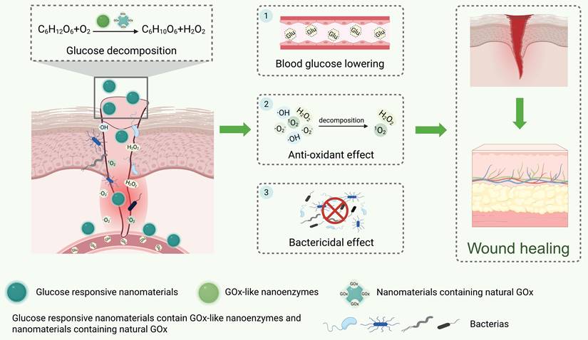
3.4. Glucose-responsive nanomaterials
Hyperglycemia in diabetic wounds leads to a wound microenvironment characterized by persistent inflammation, impaired angiogenesis, and susceptibility to infection, thereby delaying wound healing [36, 107]. GOx can convert glucose into gluconic acid and H2O2 to reduce glucose concentration [108]. Thereby, designing glucose-responsive nanomaterials can target the hyperglycemic state of diabetic wounds to exert therapeutic effects [109,110]. It is therefore the key component enabling glucose-responsive properties in nanomaterials. Currently, there are two primary strategies for designing glucose-responsive nanomaterials: the first is designing nanoenzymes that can mimic GOx activity. The second is incorporating natural GOx components into the nanomaterials. However, nanomaterials that can only mimic GOx activity will generate H2O2 and induce oxidative stress, thereby diminishing their therapeutic efficacy. Therefore, glucose-responsive nanomaterials exhibiting multi-enzyme-like activity or combined with other therapeutic agents are required for treating diabetic wounds (Figure 6).
Diagrammatic illustration of the glucose-responsive nanomaterials applied in wound treatment. The figure shows how these nanomaterials are designed to respond to high glucose levels in diabetic wound microenvironments. Upon encountering elevated glucose, the nanomaterials can mimic GOx activity and exert certain effects, thereby promoting diabetic wound healing. Created in https://BioRender.com.
Firstly, nanomaterials exhibiting multi-enzyme-like activity can trigger a cascade reaction through mimicking GOx-like activity first, thereby reversing multiple harmful factors in the diabetic wound microenvironment [111-113]. For example, it has been reported that the platinum nanoparticles (Pt NPs) in the nanocomposite PFOB@PLGA@Pt/GelMA/ODex can mimic multi-enzyme activities, including GOx, OXD, and SOD. The Pt NPs first imitate GOx-like activity to lower the concentration of glucose. Then, it can mimic OXD and SOD activities to exert bactericidal and antioxidant effects, respectively, thus promoting diabetic wound healing (Figure 7A-C) [113].
Glucose-responsive nanomaterials for wound healing. A) Diagrammatic illustration of PFOB@PLGA@Pt/GelMA/ODex nanohybrid hydrogel for diabetic infected wound. B) Images of bacteria colonies on agar plates incubated with nanohybrid hydrogels containing different Pt concentrations for different times. C) Images of wounds with different treatments at different times during the therapeutic process. Adapted with permission from [113]. Copyright 2023, American Chemical Society. D) Transmission electron microscope (TEM) image of GOx@MnS nanoparticles. E) 3D CLSM and SEM images of GOx@MnS nanoparticles against MRSA. F) Angiogenesis and degree of inflammation in diabetic infected wound model with different treatments. Adapted with permission from [115]. Copyright 2023, American Chemical Society.
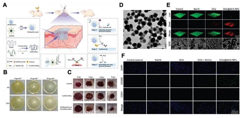
Secondly, nanomaterials containing natural GOx components combined with gas therapy or drug delivery can also promote diabetic wound healing [110, 114, 115]. For example, the GOx component of GOx@MnS nanoparticles can detect high glucose levels at diabetic wound sites and break them down to generate H2O2. It then initiates the reaction between MnS and H2O2, which generates H2S and ·OH to exert anti-inflammatory effects and bactericidal activity, thereby accelerating diabetic infected wound healing (Figure 7D-F) [115].
Based on the characteristics of natural enzymes, nanomaterials containing natural GOx have higher catalytic specificity and enzyme activity. However, their activity is easily influenced by factors such as pH and temperature in the wound microenvironment [116, 117]. While the catalytic activity of the nanoenzyme mimicking GOx is weaker than that of natural GOx, it is less susceptible to other factors. Consequently, considering the features of these two kinds of glucose-responsive nanomaterials, we suggest that future material selection should consider blood glucose levels and the duration of treatment. For treating wounds with excessively high blood glucose levels that require rapid glucose reduction, the use of nanomaterials containing natural GOx components may be more appropriate. In contrast, for wounds with mildly raised blood glucose levels requiring sustained normal glucose levels, the durability of materials should be prioritized. Thus, nanoenzymes that mimic GOx activities may be the optimal selection.
Alongside GOx, introducing specific glucose-sensitive groups is another strategy to construct glucose-responsive nanomaterials. For example, introducing phenylboronic acid groups into nanoparticles and loading specific molecules or drugs within them can construct novel glucose-responsive nanomaterials. This kind of nanomaterial can sense the high concentration of glucose in diabetic wounds, and then initiate the react between the phenylboronic acid group and glucose, thereby causing the material to disintegrate, then release the loaded drugs and accelerate diabetic wound healing.
3.5. Other endogenous stimuli-responsive nanomaterials
Besides the commonly used pH, enzyme, ROS, and glucose-responsive nanomaterials mentioned previously, other endogenous stimuli-responsive (such as GSH and ATP) nanomaterials can also promote wound healing and are gaining more attention. However, overall, glutathione (GSH) and adenosine triphosphate (ATP)-responsive nanomaterials still present significant research potential.
GSH-responsive nanomaterials can respond to GSH through redox reactions, thereby exerting bactericidal or immunomodulatory effects to accelerate wound healing [56, 118-122]. When bacterial infections occur, widespread cell necrosis will release intracellular GSH and accumulate at the wound site. As a natural antioxidant, GSH can increase bacteria’s resistance to oxidative bactericidal agents (such as ROS and RNS), which reduces the effectiveness of these agents [123]. To solve this problem, researchers have designed nanocomposites that can produce ROS and decompose GSH simultaneously. For example, CuCo2O4 nanoflowers can mimic OXD and POD activities, which enable them to react with surrounding H2O2 and O2 to generate ROS. At the same time, this nanomaterial also exhibits glutathione peroxidase (GPx)-like activity to deplete GSH in the wound microenvironment through redox reaction. These nanoflowers can weaken bacteria’s ROS resistance to exert potent bactericidal effects, thereby promoting bacteria-infected wound healing [56]. Additionally, GSH-responsive nanomaterials can also promote wound healing through regulating immune response. For instance, the nitro-quantum dots (N-CDs) in the MACNL (melanin@AuNPs@CPPO@N-CDs@L-menthol) nanocomposite can undergo a redox reaction with GSH to release NO, thereby increasing macrophage polarization to the M2 phenotype and exert immunomodulatory effects. Eventually, this nanomaterial can accelerate diabetic infected wounds healing through anti-inflammatory and angiogenesis-promoting effects [120].
ATP-responsive nanomaterials can accelerate wound healing through targeted drug delivery [124-126]. In bacterial-infected wounds, a notable feature is the continuous release of ATP by pathogens, which enables ATP-responsive nanomaterials to target areas of bacterial accumulation [124]. For example, ZIF-8 (a nanomaterial composed of Zn2+ and dimethylimidazole linked by coordinate bonds) will degrade in high-level ATP environments. This occurs because ATP exhibits a higher affinity for Zn2+ than for 2-methylimidazole, enabling ATP to replace the 2-methylimidazole units within ZIF-8 and causing its framework to collapse [127]. Researchers designed ZIF-8 nanocomposites that encapsulate indole-3-acetic acid (IAA) based on this principle. In bacterially infected wounds, the ZIF-8 nanocomposites will degrade to release IAA, which accelerates wound healing through generating ROS to exert bactericidal effect [125]. However, the ATP-responsive nanomaterials designed using the above methods have the drawback of inaccurate response, as they may also respond to other stimulus sources (for instance, ZIF-8 also demonstrates pH-responsiveness). This can cause ZIF-8 to degrade before reaching the desired site, thereby restricting the therapeutic effect. To address this issue, future research should try to construct nanomaterials incorporating ATP-specific aptamers. Aptamers are single-stranded nucleic acids that have distinct secondary and tertiary structures, and they can selectively bind to target molecules [128,129]. In the future, one possible approach is that we can utilize the principle of base complementary pairing to integrate ATP aptamers and their complementary DNA into a three-dimensional nanostructure and then load drugs onto it. In wound microenvironments with high ATP concentrations, ATP can establish a more stable three-dimensional conformational alignment with the aptamer due to spatial structural complementarity, hydrogen bonding, base stacking, and electrostatic interactions. This interaction will disrupt the original three-dimensional nanostructure formed by the ATP aptamer and its complementary DNA, ultimately facilitating drug release and wound treatment.
4. Exogenous stimuli-responsive nanomaterials
4.1. Light-responsive nanomaterials
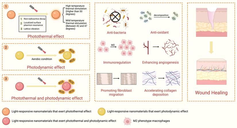
Light-responsive nanomaterials can detect and respond to external light stimuli and, in turn, exert photothermal effects, photodynamic effects, or photothermal-photodynamic synergistic effects (PTDT). This kind of nanomaterial can accelerate wound healing through bactericidal effect, regulating wound microenvironment, and promoting tissue regeneration (Figure 8).
Diagrammatic illustration of the light-responsive nanomaterials applied in wound treatment. The figure illustrates the working principle by which these nanomaterials respond to external light stimuli. Upon light irradiation, the nanomaterials undergo photothermal conversion, photodynamic ROS generation, or exert photothermal-photodynamic synergistic effects to promote wound healing. Created in https://BioRender.com.
4.1.1. Photothermal effect
Light-responsive nanomaterials can absorb specific wavelengths of light (such as visible and near-infrared light) and transform them into thermal energy through nonradiative decay [130]. These nanomaterials can enhance wound healing through the following mechanisms: (1) exerting bactericidal effects; (2) modulating the wound microenvironment; and (3) promoting tissue regeneration.
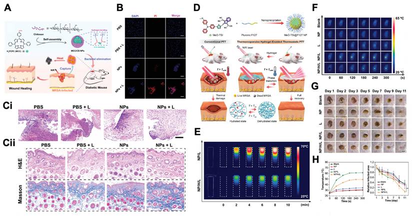
Light-responsive nanomaterials can exert bactericidal effects in two ways to promote wound healing. For one, the generation of high-temperature photothermal stimulation at temperatures higher than 50 °C directly causes thermal damage to bacterial structure [131,132]. For instance, monocarboxylic phenol/chitosan nanoparticles (MCC/CS NPs) with positively charged surfaces can adsorb bacteria through electrostatic interactions. Under 606 nm near-infrared laser irradiation, the temperature of the MCC/CS NPs can subsequently reach 55 °C and further damage the phospholipid bilayer of the bacterial cell membrane, causing membrane rupture and the release of intracellular substances. Eventually, MCC/CS NPs can exert bactericidal effects and accelerate infected wound healing (Figure 9A–C) [132]. As another example, molybdenum disulfide/chemically modified chitosan (MoS2@CSH) exhibits excellent photothermal properties and can exert POD-like activity. Under 808 nm near-infrared laser irradiation, the temperature of MoS2@CSH can increase to 51.9 °C, thereby exerting photothermal bactericidal effects. Additionally, it can simulate enzymatic activity similar to POD. Through a Fenton-like reaction, excessive H2O2 is broken down in the wound microenvironment and ·OH is generated, which has synergistic antibacterial effects and thereby accelerates infected wound healing by reshaping the wound microenvironment. In this example, the MoS2 nanosheets can stably combine with CSH because of their large specific surface area, thereby improving their dispersibility. This, in turn, enhances the photothermal conversion efficiency, enzyme-like activity, and biocompatibility [133].
Light-responsive nanomaterials that exert photothermal effects for wound treatment. A) Diagrammatic illustration of MCC/CS nanoparticles for diabetic infected wound. B) Fluorescence images of MRSA with different treatments. C) H&E and Masson staining of the wound section with different treatment (Ci: Day 4, Cii: Day 14). Adapted with permission from [132]. Copyright 2022, John Wiley and Sons. D) Diagrammatic illustration of the thermo-responsive hydrogel containing MeO-TSI@F127 nanoparticles for bacteria-infected wound. E) Thermal images of hydrogel containing MeO-TSI@F127 nanoparticles upon NIR irradiation. F) Thermal images of the bacteria-infected wounds with different treatment. G) Photographs of the bacteria-infected wounds with different treatment. H) Temperature profiles extracted from the corresponding thermal images and relative infected areas with different treatment. Adapted with permission from [146]. Copyright 2023, John Wiley and Sons.
For another, Light-responsive nanomaterials can also exert bactericidal effects by generating mild photothermal stimulation between 42 °C and 47 °C and combined with other mechanisms (such as antimicrobial ion release, chemodynamic therapy, etc.) to synergistically kill bacteria. This combined approach is necessary because mild thermal stimulation usually has only an inhibitory effect on bacteria rather than directly killing them. Therefore, it needs to be combined with other mechanisms to exert a bactericidal effect [134-136]. For instance, a composite QPQH hydrogel dressing can be developed by loading polydopamine-coated zinc oxide nanoparticles (PDA@ZnO NPs) into a hydrogel containing quercetin (QT) and quaternary ammonium chitosan (QCS). Under 808 nm near-infrared laser irradiation, the PDA@ZnO NPs can generate mild thermal stimulation at approximately 45 °C, which can synergize with the slowly released Zn2+ to disrupt bacterial membrane integrity and metabolic balance. Simultaneously, the cationic groups of QCS exert electrostatic attraction and membrane-disruptive effects on bacteria, resulting in a combined bactericidal effect to accelerate infected wound healing [134]. Additionally, light-responsive nanomaterials can also cooperate with chemodynamic therapy (CDT) to achieve bactericidal effects. For instance, researchers have fabricated CuXO@PDA nanoparticles (CP NPs) capable of promoting wound healing through mild-temperature photothermal stimulation combined with POD-like activity. Under 808 nm near-infrared laser irradiation, CP NPs can generate mild thermal stimulation of approximately 45 °C that can disrupt bacterial metabolism. Moreover, mild thermal stimulation can also increase POD-like activity through reducing the energy required for the catalytic reaction, thereby accelerating ROS production to exert a chemodynamic bactericidal effect. Eventually, CP NPs can efficiently accelerate infected wound healing [135].
Second, light-responsive nanomaterials can accelerate wound healing through modulating wound inflammatory microenvironment [137,138]. Under 808 nm near-infrared laser irradiation, the graphene oxide nanoparticles can generate mild thermal stimulation of 45 °C to increase the expression levels of the anti-inflammatory cytokines IL-4 and IL-10 in macrophages. This promotes their polarization toward the M2 phenotype to exert anti-inflammatory effects and accelerate wound healing [137].
Finally, light-responsive nanomaterials can accelerate wound healing by promoting tissue regeneration [139,140]. For instance, under 808 nm laser irradiation, Prussian blue nanoparticles can generate a mild photothermal stimulus of 41 °C, upregulating hypoxia inducible factor-1α (HIF-1α) expression in human umbilical vein endothelial cells (HUVECs) and further promoting VEGF secretion, thereby enhancing cellular angiogenesis to accelerate diabetic wound healing [139]. Additionally, under 908 nm laser irradiation, CuS@BSA nanoparticles can generate a mild thermal stimulus of 47 °C and upregulate the expression level of vimentin in mesenchymal stem cells (MSCs), thereby promoting MSC proliferation and increasing the fibroblast differentiation rate, accelerating collagen fiber deposition, and facilitating wound healing [140].
The photothermal temperature threshold includes the bactericidal temperature threshold and the tissue damage threshold. Specifically, the temperature generated by the photothermal effect can exert a bactericidal effect when it exceeds 50 °C, and the temperature threshold that can cause damage to healthy skin tissue is approximately 44 °C [141,142]. High-temperature photothermal effects are often used to treat bacteria-infected wounds. These effects can rapidly sterilize by destroying bacterial structure through heat, but the heat generated can cause thermal damage to surrounding healthy tissues. Studies have shown that the damage to the skin caused by a temperature of 44 °C for 6 h is essentially equivalent to that caused by a temperature of 55 °C for 30 s [143]. These findings suggest that in addition to temperature, the duration of action is another important factor for determining the degree of tissue damage. Therefore, when high-temperature photothermal effects are used to promote skin wound healing, the duration of action should be precisely controlled to prevent thermal damage to the surrounding tissues.
To address the aforementioned issue, in addition to controlling the duration of action, the “photothermal temperature wall” theory can also be utilized to precisely control the temperature [144]. Specifically, when the temperature reaches a preset threshold, the color or transparency of the light-responsive nanomaterial can change reversibly, thereby reducing or blocking light absorption. This process will terminate the photothermal conversion and prevent excessive temperature increase. One approach is to combine light-responsive nanomaterials with thermochromic materials. For example, by using spiro lactone, myristic acid, bisphenol A (BPA), and a hollow-structured silica nanocarrier, researchers have constructed a temperature-controlled smart nanoparticle (TCSN). In the solid myristic acid without light irradiation, SL receives a proton from BPA to form SL-H+ and adopt an NIR-absorbing colored state. Under NIR light irradiation, when the temperature reaches the melting point of the myristic acid, SL-H+ loses a proton from BPA to adopt an NIR-transparent colorless state. Therefore, this nanoplatform can precisely control the temperature around the melting point (49 °C) of myristic acid and safely accelerate the healing of bacteria-infected wounds without damaging surrounding skin tissue [145]. Another approach is to utilize the phase transition property of the hydrogel. When the temperature reaches the phase transition temperature of the hydrogel, it dehydrates and transforms into a white solid state, creating numerous light-scattering centers, thereby inhibiting further heating. For instance, the thermosensitive hydrogel P (NIPAM-AM) loaded with light-responsive MeO-TSI@F127 nanoparticles can be used to accelerate the healing of bacterially infected wounds without damaging surrounding skin tissue. Under 808 nm laser irradiation, MeO-TSI@F127 nanoparticles can generate thermal stimulation to kill bacteria. When the temperature reaches the phase transition temperature of P (NIPAM-AM), the PNIPAM undergoes a phase change to control the temperature, maintaining the photothermal equilibrium temperature between 45 and 50 °C (Figure 9D-H) [146].
High-temperature photothermal effects and mild-temperature photothermal effects can be switched by changing the incident light wavelength, frequency, and duration (Table 2). Therefore, in the future, on the basis of the different types of wounds and different healing stages, an optimal photothermal effect mode can be selected to treat wounds in a personalized manner. The following factors may be important to consider for the selection of different photothermal modes. From the perspective of wound types, the high-temperature mode, owing to its antibacterial properties, may be more suitable for infected wounds. Considering that diabetic wounds are characterized by both susceptibility to infection and impaired vascularization, the combined photothermal mode of high- and mild-temperature photothermal therapy may result in better therapeutic effects. During the wound healing stage, the high-temperature mode affects mainly the inflammatory stage through its antibacterial effect, whereas the mild-temperature mode can promote cell proliferation, differentiation and vascular regeneration, which is more suitable for the proliferative and remodeling stages. Therefore, the high-temperature mode is more suitable for the early stage of wound healing, whereas the mild-temperature mode is more appropriate for the later stage.
Light-responsive nanomaterial for wound healing: lighting parameters, photothermal performance, and therapeutic outcomes
| Light-responsive nanomaterial | Lighting parameters | Photothermal conversion efficiency | Surface Temperature | Animal model | Pro-healing effect | Ref. | |
|---|---|---|---|---|---|---|---|
| High temperature photothermal stimulation | CuO@AgO/ZnO nanoparticle | 808 nm laser, 2 W/cm2, 7min | 23% | 55 °C | S. aureus-infected full-thickness wound | Antibacterial | [147] |
| Highly graphitic- N-doped GQDs | 1064 nm laser, 1 W/cm2, 5 min | 50.4% | 50.11 °C | MRSA-infected full-thickness wound | Antibacterial | [148] | |
| Fe3O4 nanoparticle | 808 nm laser, 1 W/cm2, 10 min | 28.5% | 50.5 °C | S. aureus-infected full-thickness wound | Antibacterial | [149] | |
| Polypropylene/ polyacrylonitrile x%polydopamine | 808 nm laser, 0.2 W/cm2, 3 min | - | 60 °C | S. aureus-infected full-thickness diabetic wound | Antibacterial, Promote volatilization of the exudate | [150] | |
| MoS2/Silk Sericin | 808 nm laser, 1 W/cm2, 5 min | - | > 50 °C | MRSA-infected full-thickness wound | Antibacterial | [151] | |
| Mild temperature photothermal stimulation | mPDA@deferoxamine@chitosan-graft-third generation poly(amidoamine) polymer with terminal S-nitrosothiol groups | 808 nm laser, 2.5 W/cm2, 10 min | 32.3% | 45 °C | S. aureus-infected full-thickness wound | Synergistic antibacterial, Anti-inflammation, Angiogenesis | [30] |
| Curcumin-based metal- organic framework | 808 nm laser, 1.5 W/cm2, 5 min | - | 43.9 °C | S. aureus-infected full-thickness wound | Synergistic antibacterial, Anti-inflammation, Angiogenesis, Promote nerve regeneration | [152] | |
| CuSi nanowires | 808 nm laser, 1 W/cm2, 15 min | - | 45 °C | S. aureus-infected full-thickness wound | Synergistic antibacterial, Angiogenesis | [153] | |
| CuS@BSA nanoparticle | 980 nm laser, 0.8 W/cm2, 4 min | 42% | 42 °C | Full-thickness wound | Inducing MSCs differentiation to fibroblasts | [140] | |
| Poly-l-lactic acid/quaternized chitosan/black phosphorus/Hemoglobin | 808 nm laser, 1.5 W/cm2, 3 min | - | 40 °C | S. aureus-infected full-thickness diabetic wound | Synergistic antibacterial, Angiogenesis | [154] |
4.1.2. Photodynamic effect
When activated by light in aerobic environments, light-responsive nanomaterials (nanophotosensitizers) can generate high levels of ROS through photodynamic effects [155], thereby exerting bactericidal effects and promoting wound healing.
Nanophotosensitizers can undergo energy level transitions and transfer electrons and energy to nearby molecules, thereby generating high concentrations of ROS to exert bactericidal effects. Research has shown that MOF-based nanomaterials, black phosphorus nanosheets, and graphene quantum dots doped with halogens and nitrogen atoms can generate ROS through the photodynamic effect, thereby promoting wound healing through bactericidal and anti-inflammatory effects. For instance, researchers have embedded 4-octyl itaconate (4OI)-modified black phosphorus nanosheets into hydrogels. Under 808 nm laser irradiation, black phosphorus nanosheets can generate ROS to exert bactericidal effects. Moreover, the large specific surface area of black phosphorus nanosheets effectively increased the loading amount of 4OI. In the absence of laser irradiation, black phosphorus nanosheets can act as carriers to control the release of 4OI and exert anti-inflammatory effects, thereby accelerating the healing of diabetic ulcer wounds. In addition, as a degradable two-dimensional material, black phosphorus can gradually be oxidized and degraded into nontoxic phosphate ions under physiological conditions, avoiding long-term material residue. This significantly reduces the risks of chronic toxicity and foreign body reactions. In combination with its excellent light-responsive properties and antibacterial performance, it has unique application advantages in the field of wound healing (Figure 10A-C) [156-159].
Light-responsive nanomaterials that exert photodynamic effects for wound treatment. A) SEM images of the GelMA, 4OI@Gel and 4OI-BP@Gel hydrogel. B) Giemsa staining of bacteria infected wound tissues after 1 week treatment. Red arrows show the bacteria. C) H&E staining of the wound section at the DAY7 and DAY14. Adapted with permission from [157]. Copyright 2023, Elsevier. D) Description of the preparation of UCNPs@ZrMOF-Pt composite hydrogel and its antibacterial mechanism. E) Fluorescence images of live/dead bacteria after different treatment (1. PBS, 2. UCNPs@ZrMOF-Pt hydrogel, 3. UCNPs@ZrMOF + Laser, 4. UCNPs@ZrMOF-Pt + Laser, 5. UCNPs@ZrMOF + H2O2 + Laser, 6. UCNPs@ZrMOF-Pt + H2O2 + Laser). F) Fluorescence images of live/dead bacteria after different treatment (1. PBS, 2. Laser, 3. UCNPs@ZrMOF-Pt hydrogel, 4. UCNPs@ZrMOF hydrogel+ Laser, 5. UCNPs@ZrMOF-Pt hydrogel + Laser). G) Fluorescence images of bacteria-infected wound tissue with different treatment [169]. Adapted with permission from Copyright 2022, John Wiley and Sons.
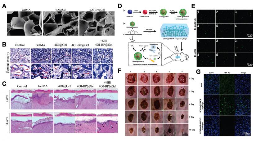
The hypoxic microenvironment of chronic wounds and the fact that certain nanophotosensitizers can only be activated by visible light are two major factors that limit the production of ROS. To increase the efficiency of ROS generation by nanophotosensitizers and promote their bactericidal function [160-163], given the high H2O2 concentration in chronic wound microenvironments [164], researchers have developed nanophotosensitizers that simultaneously exert photodynamic effects and exhibit CAT-like activity, such as atomically dispersed Fe-doped oxygen-deficient molybdenum oxide MoO3-X (ADFM) and Se@CeO2 nanoparticles. Such nanomaterials can decompose H2O2 to generate O2, thereby alleviating the hypoxic state and improving the efficacy of photodynamic reactions [165,166]. Additionally, some nanophotosensitizers can be activated only by visible light, which has limited tissue penetration, thus limiting their application in deep wounds [167,168]. To overcome this problem, researchers have combined nanophotosensitizers with upconversion nanoparticles (UCNPs) that can convert penetrating near-infrared light into visible light [169,170]. For example, incorporating UCNPs with MOF-based nanophotosensitizers and modifying with platinum nanoparticles can simultaneously increase the photodynamic efficacy and address the problem of penetration depth. Under NIR irradiation, the UCNPs can transform the penetrating NIR laser into visible light and, in turn, activate the nanophotosensitizers, producing 1O2 to exert bactericidal effects through a photodynamic reaction. Moreover, the CAT-like activity of platinum nanoparticles can transform high levels of H2O2 into O2, thereby increasing the photodynamic antibacterial efficacy and accelerating wound healing (Figure 10D-G) [169].
However, most nanophotosensitizers cannot specifically target pathogens. Therefore, the high levels of ROS they generate spread throughout the entire illuminated area, damaging normal tissue cells at the wound edge and negatively impacting wound healing. To address this issue, a possible strategy can involve providing nanophotosensitizers with the ability to target pathogens. For example, nanophotosensitizers can be gathered around bacteria using biomimetic strategies, such as modifying the surface of nanomaterials with aptamers that specifically recognize bacteria (e.g., aptamer Apc for Escherichia coli [171] and aptamer SA31 for Staphylococcus aureus [172]) or applying the bacterial cell membrane coating technique. This will enable the generated ROS to target bacteria more accurately, increasing both the bactericidal efficacy and the protection of surrounding tissue, thereby effectively and safely promoting wound healing.
4.1.3. Photothermal-photodynamic synergistic effect
Nanomaterials that generate PTDT effects can accelerate wound healing through enhanced bactericidal activity and the activation of repair-related mechanisms. On the one hand, through photodynamic processes, light-responsive nanomaterials can facilitate the production of large amounts of ROS and oxidatively damage bacterial cell membranes. On the other hand, through photothermal processes, light-responsive nanomaterials can generate localized hyperthermia that directly increases bacterial cell membrane permeability and destroys bacterial biofilms. This synergistic effect makes it easier for ROS to pass through cell membranes and exert an oxidizing bactericidal effect. Additionally, PTDT effects lower the temperature and amount of ROS needed for bactericidal activity, thereby protecting skin tissue around the wound [170, 173-176]. For instance, in bacteria-infected wounds, under 400 nm visible light irradiation, PAM-PDA/Ag@AgCl nanomaterials can simultaneously release thermal energy and generate ROS through the PTDT effect, thereby efficiently promoting the healing of infected wounds while avoiding damage to surrounding healthy tissue. Additionally, light-responsive nanomaterials can activate repair-related mechanisms to accelerate tissue regeneration. For example, under 808 nm laser irradiation, the CaSiO3-ClO2 @PDA-ICG nanoparticles (CCPI NPs) can exert a PTDT effect to activate AMPK signaling in macrophages and promote their polarization toward the M2 phenotype, thereby increasing transforming growth factor beta 1 (TGF-β1) secretion. Subsequently, it can activate epithelial‒mesenchymal transition (EMT) in tissue cells near the periphery of the wound, which significantly increases fibroblast migratory ability and accelerating granulation tissue formation and wound closure [177].
4.2. Electro-responsive nanomaterials
Electro-responsive nanomaterials refer to nanomaterials that can generate specific electrical responses to external mechanical stimuli (such as friction, physiological motion, ultrasound, magnetic force, etc.) and transform mechanical energy into electrical signals. Electro-responsive nanomaterials mainly include piezoelectric nanomaterials and triboelectric nanomaterials [178-181].
4.2.1. Piezoelectric nanomaterials
Piezoelectric nanomaterials have noncentrosymmetric crystal structures. Under external mechanical force stimulation, these materials will undergo internal polarization and generate electrical signals. They can accelerate wound healing by modulating the wound microenvironment; promoting cell proliferation, migration, and differentiation; and exerting bactericidal effects (Figure 11).
Diagrammatic illustration of the piezoelectric nanomaterials applied in wound treatment. The figure illustrates how these nanomaterials convert mechanical energy into electrical signals via the piezoelectric effect, thereby accelerating wound healing. Created in https://BioRender.com.
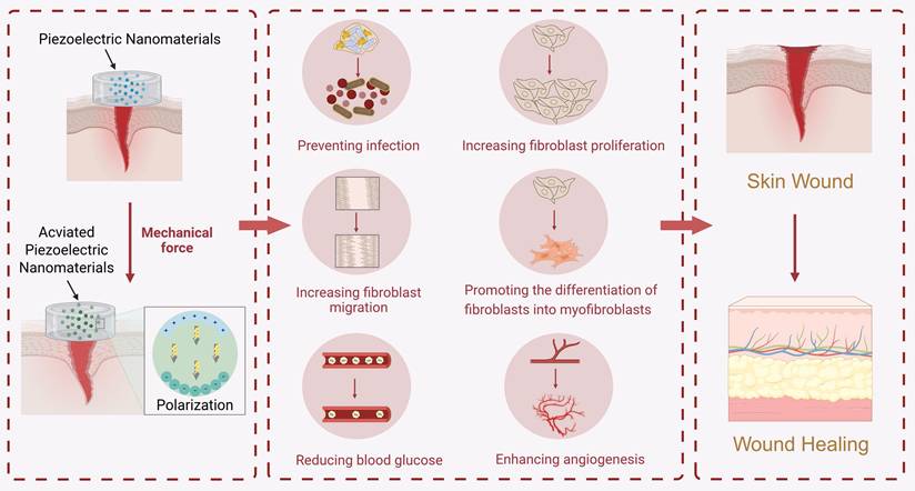
Piezoelectric nanomaterials can mimic natural enzyme activity to modulate the wound microenvironment [11,182-184]. For instance, in diabetic wounds, by integrating hyaluronic acid-encapsulated L-arginine, ultrasmall gold nanoparticles, and Cu1.6O nanoparticles loaded with phosphorus-doped graphitic carbon nitride nanosheets, researchers have constructed a piezoelectric ACPCAH nanocomposite, which can be activated by ultrasound and alkaline wound microenvironment. By regulating the surface electron transfer process, it can cascade mimics of the enzymatic activities of SOD-CAT-GOx-POD/nitric oxide synthase (NOS), thereby reducing blood sugar levels, exerting anti-inflammatory and bactericidal effects, alleviating hypoxia, and promoting vascular regeneration. Eventually, this piezoelectric nanocomposite can accelerate wound healing by modulating the diabetic wound microenvironment [183].
Furthermore, piezoelectric nanomaterials can generate electrical signals to promote cell proliferation, migration, and differentiation [63,185,186]. For instance, when the exudate from a wound is absorbed, a ZnO nanoparticle-modified polyvinylidene fluoride/sodium alginate piezoelectric hydrogel (ZPFSA) can undergo vertical swelling. During physiological movement, it generates horizontal friction with the skin. Both of the aforementioned actions can provide mechanical stress to piezoelectric hydrogels, thereby generating electrical stimuli that promote fibroblast proliferation and migration to accelerate wound healing [63]. Additionally, electrical stimulation generated by piezoelectric nanomaterials can regulate cell differentiation. For example, an electrically stimulated “Lock-ON/OFF” drug delivery system can be constructed by loading carboxylated carbon nanotubes/vancomycin hydrochloride (c-MWCNTs-VAN) onto a composite membrane composed of polyvinylidene fluoride (PVDF) and polyethylene oxide (PEO). This system can respond to mechanical stress generated by physiological movement and generate electrical stimulation through the piezoelectric effect. On the one hand, electrical stimulation can precisely control the on-demand release of VAN to exert its bactericidal effect. On the other hand, it can promote fibroblast proliferation and migration and myofibroblast differentiation, thereby accelerating wound closure [185]. As another example, under the stimulation of a rotating magnetic field, the PCL/Ti3C2Tx MXene nanofiber membrane can generate an approximately 10.8 μA microcurrent. By activating and upregulating calcium signaling, it drives calcium influx, thereby regulating the migration, proliferation and differentiation of fibroblasts and accelerating the healing of diabetic wounds [187].
Finally, piezoelectric nanomaterials can also exert bactericidal effects by generating oxidative substances to accelerate wound healing [179,188,189]. For instance, under ultrasound stimulation, piezoelectric C3N4 nanosheets can generate H2 and holes, thereby disrupting the electron transfer chain of bacteria and blocking their respiration to achieve bactericidal effects, thus promoting infected diabetic wound healing [189].
Improving the mechanical-electrical energy conversion efficiency of piezoelectric nanomaterials or their interactions with cells/bacteria is one possible way to enhance the efficacy of piezoelectric nanomaterials in treating skin wounds. This process involves three primary strategies: (1) modulating the internal structure of nanomaterials; (2) adjusting exogenous mechanical forces; and (3) combining other therapeutic approaches.
First, constructing a specific “Schottky junction” structure in piezoelectric nanomaterials can enhance their ability to produce oxidizing substances for bactericidal effects [190]. For instance, the deposition of Au nanoparticles onto the surface of barium titanate (BT) nanocubes can form a “Schottky barrier” structure. By inhibiting electron‒hole recombination and promoting electrochemical reactions, compared with single-component piezoelectric nanomaterials, composite piezoelectric nanomaterials have demonstrated superior oxidizing substance generation and have exhibited enhanced efficacy in promoting infected wound healing [191].
Second, different mechanical stress parameters result in different levels of electrical stimulation from piezoelectric nanomaterials [192]. Therefore, modifying mechanical stress conditions can regulate the effects of piezoelectric nanomaterials. For example, altering the intensity and duration of ultrasound exposure can produce distinct effects, such as antibacterial activity and promotion of tissue regeneration. These strategies can be applied during different phases of wound healing. For example, after 5 min of exposure to 1.5 W/cm2 ultrasound, barium titanate@macrophage membranes preactivated by Staphylococcus aureus (BTO@MMSa) can generate oxidative substances and exert bactericidal effects, thereby acting on the inflammatory phase of wound healing. While following 1 min of 0.8 W/cm2 ultrasound exposure, BTO@MMSa can act during the tissue regeneration phase of wound healing. It can upregulate the expression levels of repair-related genes such as VEGF, COL-I, and COL-III in fibroblasts, thereby promoting cell migration. The sequential application of high- and low-power ultrasound to infected wounds significantly accelerates the healing process [61].
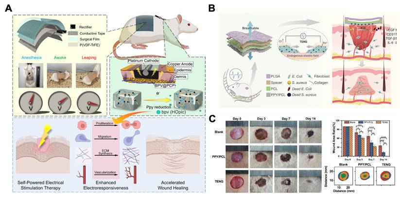
Finally, combining piezoelectric nanomaterials with other therapeutic approaches can synergistically promote wound healing. First, piezoelectric nanomaterials can be combined with drug delivery. For example, phosphatase and tensin homolog (PTEN) has been proved to suppress cellular responsiveness to electrical stimuli (Figure 12A) [193]. Therefore, combining piezoelectric nanomaterials with BPV, an inhibitor of PTEN, can amplify the cellular response to electrical stimulation. On this basis, researchers have developed a piezoelectric nanoplatform loaded with BPV (BPV@PCP). This nanoplatform can respond to physiological activities and convert them into electrical stimuli to control BPV release, thereby enhancing cell responsiveness to electrical stimulation. Compared with piezoelectric nanomaterials without BPV, this material effectively accelerates wound healing [194]. Second, combining piezoelectric nanomaterials with cell membrane coating technology can also increase their efficacy. For instance, the use of macrophage membranes preactivated by Staphylococcus aureus to coat piezoelectric nanomaterials can increase their bactericidal activity. Compared with conventional macrophage membranes, piezoelectric nanomaterials preactivated by Staphylococcus aureus express higher levels of pathogen-associated molecular patterns (PAMPs), thereby enabling more precise bacterial recognition. Therefore, this piezoelectric nanomaterial can target infected areas and precisely exert bactericidal effects, thereby promoting wound healing. In addition to the above two approaches, other methods, such as modifying the surface charge properties and hydrophilicity/hydrophobicity of piezoelectric nanomaterials, can also enhance the interaction between them and bacteria or cells, thereby improving their efficacy in promoting wound healing [61].
Electro-responsive nanomaterials for wound healing. A) Diagrammatic illustration of nanogenerator accelerates wound healing. Adapted with permission from [194]. Copyright 2023, American Chemical Society. B) Diagrammatic illustration of triboelectric nanogenerator accelerates diabetic infected wound healing. C) Effect of TENG patch on diabetic infected wound healing in different treatments. Adapted with permission from [196]. Copyright 2023, Elsevier.
4.2.2. Triboelectric nanomaterials
Triboelectric nanomaterials are mainly used in the form of triboelectric nanogenerators (TENGs), which consist of electrode layers and triboelectric layers with opposite charges. They primarily utilize the principles of electrostatic induction and contact electrification to convert mechanical force into electrical energy. TENGs mainly accelerate wound healing by exerting bactericidal effects and promoting cell proliferation and migration (Figure 13).
Diagrammatic illustration of the triboelectric nanomaterials applied in wound treatment. The figure shows how triboelectric nanomaterials convert mechanical friction into electrical signals and thereby promoting wound healing. Created in https://BioRender.com.
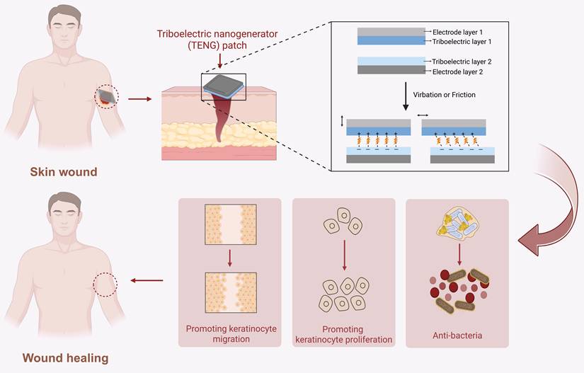
Firstly, TENGs can exert bactericidal effect by mediating transient electroporation to disrupt bacterial membranes and promote H2O2 formation, thereby accelerating wound healing [180,195,196]. For instance, researchers constructed a single-electrode TENG composed of Polypyrrole/ Polycaprolactone (PPY/PCL), PCL, and poly(lactic-co-glycolic acid) (PLGA) layers. Under physiological movement stimulation, two tribo layers (PCL and PLGA) vibrate according to the “contacted-separated-contacted” cycles. Because of their different electron affinities, electrons will transfer and cause regular changes in the open-circuit voltage and the short-circuit current. By the time TENG contacts with infected wound, the positively charged surface of TENG can effectively adsorb bacteria through electrostatic induction. Simultaneously, the electrical stimulation produced by TENG can produce H2O2 and induce electroporation of bacterial membranes, resulting in bacterial contents leaking, which achieves precise bactericidal effects and accelerates diabetic infected wound healing (Figure 12B-C) [196].
Secondly, TENGs can generate electric signals to regulate cellular functions, thereby promoting wound healing [197-200]. For example, a TENG device (Electro-Generating Dressing, EGD) combined with negative pressure wound therapy (NPWT) can promote wound healing through regulating immune responses and accelerating the re-epithelialization process. Specifically, negative pressure therapy can induce the periodic contact-separation cycles within the TENG’s friction layer. Based on the different electron affinities of two tribo layers, the mechanical deformation can be converted into a stable electric field. On the one hand, this electric field can promote macrophages to polarize towards M2 phenotype and mitigate inflammation. On the other hand, it can activate the PI3K/Akt and MAPK/ERK signaling pathways to enhance the directional migration and proliferation of epidermal cells [197].
When electro-responsive nanomaterials are used to treat wounds, physical wear, wound exudate corrosion, and mechanical fatigue may gradually decrease their effectiveness [201]. To address this issue, there are three ways to optimize material design. First, electro-responsive nanomaterials resistant to mechanical fatigue can be selected as the core components. Second, biocompatible coatings (such as polyethylene glycol) can be used to modify material surfaces, thereby protecting against the corrosive effects of wound exudate. Third, sensors can be integrated within the electro-responsive nanomaterials, and wireless communication technology can be integrated to monitor the material’s electrical output performance and wound healing status in real time, which can enable precise indication of the material replacement time, ensuring its treatment efficiency.
Most electro-responsive nanomaterials currently in use tend to overemphasize therapeutic efficacy while neglecting real-time feedback on wound status. This limitation may lead to issues such as the ability to monitor material effectiveness decreasing in real time (such as reduced electrical signal generation efficiency), which can cause treatment failure. Moreover, it is difficult to precisely adjust the electrostimulation intensity on the basis of the wound status, potentially leading to either excessive stimulation or insufficient efficacy. To overcome this challenge, in the future, electro-responsive nanomaterials can be integrated with wireless communication technology and artificial intelligence algorithms, thereby establishing a closed-loop therapeutic system featuring “sensing-analysis-regulation”. This system would rely on sensors to capture real-time signals of the electrical properties of the material and the wound microenvironment (such as pH and temperature). After analysis by AI algorithms, it would dynamically optimize electrical stimulation parameters, ultimately achieving more precise and adaptive wound interventions.
4.3. Magnetic-responsive nanomaterials
Magnetic-responsive nanomaterials refer to nanomaterials that exhibit specific physical or chemical responses to stimulation by an external magnetic field [202]. They can accelerate wound healing through three mechanisms: (1) providing mechanical cues to skin tissue cells; (2) exerting bactericidal effects; and (3) enhancing the targeting ability of pro-healing substances (Figure 14).
Diagrammatic illustration of the magnetic-responsive nanomaterials applied in wound treatment. The figure shows how magnetic nanomaterials (e.g., iron oxide nanoparticles) respond to an external magnetic field to accelerate wound healing. Upon magnetic field application, these nanomaterials can exert mechanical forces on skin tissue cells, generate local heat, produce ROS, or achieve targeted release of therapeutic agents, thereby promoting wound healing. Created in https://BioRender.com.
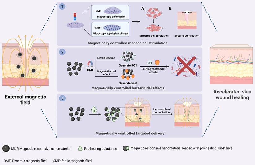
First, under static or dynamic magnetic field stimulation, magnetic-responsive nanomaterials can respond to and undergo macroscopic deformation or microscopic topological changes, thereby providing directional mechanical stimulation to skin tissue cells and promoting wound healing [46, 203-205]. On the one hand, under the stimulation of external dynamic magnetic fields, membranes containing Fe3O4 nanoparticles will undergo periodic macroscopic deformation, which will activate mechanosensitive channels on fibroblast and keratinocyte membranes and upregulate the expression of the mechanosensitive proteins YAP and Lamin A/C. Consequently, this membrane can promote wound contraction and re-epithelialization to accelerate skin wound healing (Figure 15A-D) [46]. On the other hand, under a static magnetic field, magnetic-responsive nanomaterials can undergo orientation according to the direction of the magnetic field, inducing changes in their microscopic topological structure and thereby promoting wound regeneration. For instance, under static magnetic field stimulation, magnetic-responsive cellulose nanocrystals within the Alg-SF-CNC composite nanoscaffold can align along the magnetic field direction, which will create an ordered topological structure within the scaffold, thereby providing migration guidance for fibroblasts and keratinocytes. Consequently, these cells migrate in an organized manner along the scaffold orientation to accelerate wound healing [204].
Magnetic-responsive nanomaterials for wound treatment. A) Diagrammatic illustration of the magnetic-responsive massage membrane accelerates wound healing. B) Effect of magnetic field duration on the expression level of mechanical transduction-related protein LaminA/C. C) Effect of magnetic field duration on the expression level of mechanical transduction-related protein YAP D) Relative expression level of two proteins under different magnetic field duration. Adapted with permission from [46]. Copyright 2024, John Wiley and Sons. E) Description of the synthesis of nanoparticle-incorporated exosomes (Exo + nanoparticles) and its pro-healing effect in injured skin. F) Expression level of proteins associated with HUVEC proliferation, migration, and angiogenesis with different treatment. G) H&E staining of wound sections under different treatment with magnetic guidance 5 weeks post-wounding. Arrows indicate scar edges. Adapted with permission from [64]. Copyright 2020, Springer Nature.
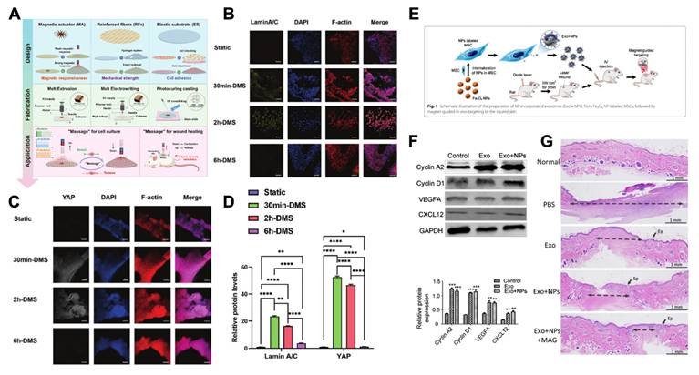
Second, magnetic-responsive nanomaterials can exert bactericidal effects under the influence of a magnetic field by generating ROS or through the magnetic heating effect, thereby accelerating the healing of infected wounds. For instance, AIron NPs can accelerate the release of Fe2+ in an alternating magnetic field, increase the amount of ·OH, and exert a bactericidal effect by oxidizing and destroying the bacterial cell membrane and nucleic acids, thereby promoting the healing of infected wounds. Additionally, under the stimulation of an alternating magnetic field, Fe3O4 NPs can convert magnetic energy into thermal energy and increase the local temperature at the wound to 50 °C, thereby exerting antibacterial and anti-inflammatory effects and accelerating the healing of diabetic infected wounds [206, 207].
Finally, magnetic fields, magnetic-responsive nanomaterials and pro-healing substances (such as growth factors, stem cells, and extracellular vesicles) can form a “magnetic targeting system” together [202]. By modulating the magnetic field, the targeted accumulation of these substances at the wound site can be enhanced. This is because when the magnetic force exceeds the blood flow velocity, the pro-healing substances labeled with magnetic-responsive nanomaterials can be retained in the affected area, thereby increasing their local concentration [208]. For example, in burn wound treatment, when magnetic-responsive Fe3O4 nanoparticles are internalized by MSCs, the secreted exosomes (MSC-Exos) are labeled with Fe3O4 nanoparticles. Under magnetic field stimulation, the homing of MSC-Exos to the wound site significantly increases, thereby rapidly promoting angiogenesis, collagen deposition, and re-epithelialization to accelerate burn wound healing (Fig. 15E-G) [64].
Currently, the “magnetic targeting system” still faces the challenge of limited depth of targeted delivery. This limitation is influenced mainly by the distance between the magnet and the magnetic-responsive nanomaterials, the blood flow at the wound site, and the circulation time of the nanoparticles.
The targeted aggregation effect of magnetic-responsive nanomaterials depends on the magnetic field strength, and the magnetic field decreases as the distance between the magnet and the magnetic-responsive nanomaterials increases [209], which results in magnetic-responsive nanomaterials having a targeted depth limited to less than 5 mm of the body surface [210]. This restriction makes it difficult to meet the therapeutic component delivery requirements for deep wounds. Several possible strategies can be considered to overcome this limitation. First, single magnets can be replaced with array or multipole magnets to optimize the magnet architecture. This method mitigates the decrease in the magnetic field due to distance by using field superposition to increase the magnetic field strength and uniformity in deep wounds. Moreover, the sensitivity of magnetic-responsive nanomaterials to magnetic fields can be enhanced. The magnetic moment of magnetic-responsive nanomaterials can be increased by modification techniques such as adding metal dopants (including Zn, Co, and Mn), developing specific-shaped magnetic-responsive nanomaterials (such as cubic-shaped), and decreasing their size. These modifications will enable sufficient magnetic force even under distant weak magnetic fields, thereby achieving targeted aggregation. Through the above methods, the targeted effect of magnetic-responsive nanomaterials may be enhanced, making them more suitable for treating deep wounds.
Hemodynamics is also a key factor in determining whether magnetic nanomaterials can successfully remain at the wound site [211, 212]. In the treatment of deep wounds, due to the weakened magnetic field gradient, the magnetic force often fails to fully resist the flushing effect caused by blood flow or a large amount of exudate, making it difficult for magnetic nanomaterials to stably remain in the wound area. To solve this problem, the surface of the nanoparticles can be modified with ligands (such as antibodies and peptides) that specifically bind to the surface of target cells in the target area [214]. By utilizing the high affinity between the receptor and ligand, combined with magnetic force, magnetic nanomaterials can enhance their adhesion to the wound tissue, jointly resisting the shear forces of local blood flow and exudate, thereby facilitating the stable retention of the materials at deep wound sites.
Finally, the in vivo circulation time of magnetic nanomaterials is another factor for their effectiveness [215]. The mononuclear phagocytic system is the main obstacle for rapid clearance of nanoparticles from the body. Researchers have designed various strategies to enable nanoparticles to evade recognition by the MPS, thereby prolonging their in vivo circulation time. These strategies include coating the surface of the nanoparticles with hydrophilic polymers (such as PEGylation) to form a hydration layer to inhibit plasma protein absorption [216]; designing biomimetic nanoparticles (such as red blood cell membranes and platelet membranes) [217]; and using specific cells (such as neutrophils and monocytes) as carriers to implement strategies such as “cell hitchhiking”. These strategies could extend the circulation half-life of the nanoparticles in the blood, providing the necessary circulation time basis for their targeted enrichment in deep tissues under the influence of the magnetic field [218].
4.4. Other exogenous stimuli-responsive nanomaterials
Besides the commonly used light-, electro-, and magnetic-responsive nanomaterials mentioned earlier, other exogenous stimuli-responsive (such as ultrasound and temperature) nanomaterials can also promote wound healing and have achieved significant research breakthroughs.
Ultrasound-responsive nanomaterials can accelerate wound healing through three mechanisms: (1) promoting nerve and vascular regeneration; (2) modulating immune responses and (3) exerting bactericidal effects.
First, ultrasound-responsive nanomaterials can accelerate wound healing by converting ultrasonic waves into electrical signals, thereby promoting nerve and vascular regeneration [67, 219]. Under the stimulation of ultrasound waves, ultrasound-responsive nanomaterials with non-centrosymmetric structures can undergo internal charge separation between positive and negative charges, thereby generating an internal electric field and producing electrical signals. For example, hydrogels containing ultrasound-responsive nanoparticles (K,Na)NbO3 can convert ultrasonic waves into electrical signals to activate the AMPK pathway, increasing the transmembrane transport of nerve growth factor-containing vesicles by promoting ATP synthesis. Consequently, this nanohydrogel can enhance the neurotrophic factors secreted by Schwann cells (such as NGF, BDNF, and VEGF), thereby inducing nerve and vascular regeneration. This process enhances local oxygen levels and healing-related growth factor secretion to accelerate diabetic wound healing [67].
Second, ultrasound-responsive nanomaterials can accelerate wound healing by modulating immune response [66, 220]. Under the effect of ultrasound, platinum nanoparticle assemblies (PNA) can promote electron polarization through the plasma resonance effect, thereby generating GSH to scavenge high concentrations of oxidative substances in the wound microenvironment. This subsequently accelerates diabetic wound healing by regulating immune responses and restoring the proliferation and migration capabilities of fibroblasts and keratinocytes [66].
Finally, ultrasound-responsive nanomaterials can promote wound healing by generating ROS to exert bactericidal effect [221, 222]. Under the stimulation of ultrasound, electrons within ultrasound-responsive nanomaterials will undergo transitions. When these transitioned electrons return to their ground state, the released energy can be transferred to substances such as O2 to generate ROS [223]. For example, under ultrasound stimulation, oxygen-rich vacancy porous titanium dioxide nanosheets/ultrafine copper peroxide nanoclusters (OV-TiO2/CuO2) can generate ROS to kill MRSA and suppress excessive inflammatory responses, thereby accelerating MRSA-infected wound healing [222]. For another example, under ultrasound stimulation, the two-dimensional Bi2WO6 nanosheets can generate ROS, which induces lipid oxidation and membrane rupture of bacterial cell membranes, thereby exerting bactericidal and accelerating the healing of MRSA-infected wounds [224].
In response to temperature changes at the wound site, thermo-responsive nanomaterials can undergo structural transformation and drive early wound closure, thereby accelerating wound healing. During the healing process, wound temperature undergoes dynamic changes. During the inflammatory stage, the wound temperature exceeds the normal surface skin temperature [225,226]. Nanomaterials can respond to this change and accelerate wound healing [68, 227, 228]. For example, when wound temperature is above 37 °C, the polyurethane/polyvinyl butyral (PU/PVB) nanofiber membrane can release its stored energy, undergo centripetal contraction, and generate “micro-deforming forces” along the wound edges. This can increase fibroblast proliferation and migration, promote early wound closure and shield the healing process from external adverse factors (such as bacteria or foreign bodies), thereby providing a stable microenvironment that expedites wound healing [68].
However, there is currently limited application of temperature-responsive nanomaterials in wound treatment. This may be because their effect of accelerating wound closure only provides a physical barrier to the healing process, and their effective duration is relatively constrained. To enable temperature-responsive nanomaterials to more comprehensive effects in promoting wound healing, firstly, it is advisable to develop temperature-responsive nanomaterials with specific morphologies. For instance, nanofibers with porous structures, aligned structures, or directional orientation have been proven to provide directional guidance for the migration of skin tissue cells, thereby accelerating skin tissue regeneration. Secondly, it is recommended to combine temperature-responsive nanomaterials with antibiotics, growth factors, etc., thus promoting wound healing through multiple effects.
5. Dual stimuli-responsive nanomaterials
The previous text has already elaborated on the role of single stimuli-responsive nanomaterials in wound treatments. To further enhance the functionality of stimuli-responsive nanomaterials, researchers have designed dual stimuli-responsive nanomaterials. They can respond to two distinct stimulus sources, thereby exerting multiple functions to promote wound healing. These mainly include (1) dual endogenous stimuli-responsive nanomaterials; (2) dual exogenous stimuli-responsive nanomaterials and (3) endogenous-exogenous stimuli-responsive nanomaterials (Figure 16).
Diagrammatic illustration of dual stimuli-responsive nanomaterials applied in wound treatment. The figure shows how these nanomaterials are designed to respond to two complementary triggers. Upon sequential or simultaneous exposure to both stimuli, the nanomaterials undergo synergistic structural changes or energy conversion, leading to on-demand release of therapeutics or enhanced physical effects, thereby promoting wound healing. Created in https://BioRender.com.
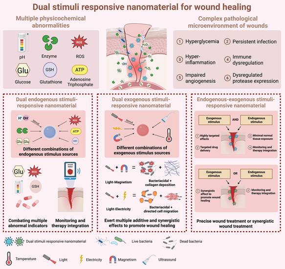
5.1. Dual endogenous stimuli-responsive nanomaterials
Dual endogenous stimuli-responsive nanomaterials refer to nanomaterials that can respond to two kinds of endogenous stimulus sources. For example, pH-glucose-responsive nanomaterials, pH-enzyme-responsive nanomaterials, and pH-GSH-responsive nanomaterials [55, 229, 230]. Such stimuli-responsive nanomaterial can confront the complex wound microenvironment with multiple abnormal indicators, such as diabetic wounds and bacterially infected wounds. Besides, they can also monitor wound physicochemical indicators during wound therapy. For instance, researchers designed glucose-pH-responsive nanohydrogel copper nanoclusters/GOx/oxidized hyaluronic acid (CGH) to deal with the hyperglycemic and acidic pH microenvironment of diabetic wounds. Firstly, the GOx incorporated in the nanohydrogel can reduce glucose levels. Subsequently, the acidic pH at the wound site triggers the release of copper nanoclusters (CuNCs), which then exert bactericidal and anti-inflammatory effects through the Fenton reaction. This nanohydrogel can promote re-epithelialization, collagen deposition, and vascular regeneration to accelerate diabetic wound healing (Figure 17A) [55].
Dual stimuli-responsive nanomaterials for wound treatment. A) Diagrammatic illustration of CGH hydrogel for diabetic wound. Adapted with permission from [55]. Copyright 2024, Elsevier. B) Diagrammatic illustration of PGCC for infected wound. Adapted with permission from [71]. Copyright 2023, Elsevier. C) Diagrammatic illustration of the preparation process of HA-Fe-COF and its pro-healing effect for infected chronic wound. Adapted with permission from [72]. Copyright 2024, American Chemical Society. D) Diagrammatic illustration of LAMC/CD-C@M@P hydrogel for diabetic wound. Adapted with permission from [237]. Copyright 2024, John Wiley and Sons.
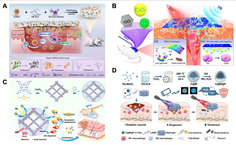
5.2. Dual exogenous stimuli-responsive nanomaterials
Dual exogenous stimuli-responsive nanomaterials refer to nanomaterials that can respond to two kinds of exogenous stimulus sources. Such as light-electro and light-magnetic-responsive nanomaterials. This type of stimuli-responsive nanomaterial can synergistically promote wound healing through combined effects [71, 231-233]. For instance, light-electro-responsive nanomaterials (PGCC) containing poly-L-lactic acid (PLLA), reduced graphene oxide (rGO), calcium peroxide (CaO2) and CAT that exert photothermal and piezoelectric effects can accelerate infected wound healing by exerting bactericidal effect and promoting tissue regeneration. Under ultrasound stimulation, PLLA generates oxidative substances through piezoelectric effect to exert bactericidal and anti-inflammatory effects. Meanwhile, under 808 nm near-infrared laser irradiation, rGO can facilitate the photothermal effect and produce mild thermal stimulus, thereby up-regulating the secretion of heat shock protein 90 (Hsp90) to maintain protein homeostasis and accelerate tissue regeneration (Figure 17B) [71]. In addition, by employing a “dual-drive” strategy, researchers developed a magnetic nanoparticle-protein fiber complex (GMPF0.1) with oriented fibrillar structures containing magnetic nanoparticles and coated with polydopamine (PDA). Specifically, under 808 nm near-infrared laser irradiation, the PDA shell generates photothermal effects and produces thermal stimulation to disrupt bacterial cell membranes, thereby achieving bactericidal effect. Subsequently, magnetic nanoparticles undergo directional alignment under magnetic field stimulation, further inducing the formation of highly oriented fiber structures within the magnetic nanoparticle-protein fiber complex. This oriented structure can provide growth guidance for fibroblasts and regulates their ordered proliferation and migration, thereby accelerating tissue regeneration and ultimately promoting bacterial-infected wound healing [233].
5.3. Endogenous-exogenous stimuli-responsive nanomaterials
Endogenous-exogenous stimuli-responsive nanomaterials refer to nanomaterials that can respond endogenous and exogenous stimulus. Such as pH-light-responsive, ROS-light-responsive, enzyme-light-responsive, and pH-electro-responsive nanomaterials [72, 173, 234-236]. The characteristics of such nanomaterials are as follows: Firstly, they can initiate a response triggered by endogenous stimuli within the wound microenvironment, while simultaneously combining with exogenous stimuli to enhance therapeutic efficacy. Secondly, these nanomaterials can monitor wound physicochemical indicators during wound therapy enabled by endogenous stimuli, enabling real-time assessment of wound state and achieving integrated “monitoring-treatment” capabilities. For instance, researchers designed an enzyme-light-responsive HA-Fe-COF nanoplatform to promote bacterially infected wound healing. Only when the hyaluronidase secreted by bacteria degrades the HA encapsulated on the surface of the nanoplatform, Fe-COF can be released. Subsequently, under the irradiation of NIR light, it can generate the PTDT effect to exert advanced bactericidal effect, thereby ensuring that the material only functions in the presence of bacteria, improving antibacterial efficiency, and preventing the generation of heat and ROS from damaging the surrounding normal skin tissue (Figure 17C) [72]. Additionally, by incorporating ceria-molybdenum disulfide nanoparticles (C@M@P) and carbon quantum dots (CDs) encapsulated in a polydopamine shell into a hydrogel, researchers developed a pH-light-responsive hydrogel that can both monitor and treat diabetic infected wounds. First, CDs exhibit photoluminescence properties. Specifically, when excited by light at specific wavelengths, they exhibit pH-sensitive fluorescence emission based on protonation/deprotonation reactions of surface functional groups and defect-state electron transitions. Consequently, under UV irradiation, the fluorescence intensity emitted by CDs linearly increases with rising pH values. Based on this property, fluorescent images of the hydrogel can be captured by smartphone and converted into RGB signals to rapidly estimate wound pH levels, enabling early warning of bacterial infections (typically an alkaline pH environment). When monitored pH signals indicate bacterial infection, under 808 nm near-infrared irradiation, C@M@P can generate high-temperature thermal stimulation through photothermal effects to exert bactericidal effects. Simultaneously, the triggered photothermal effects can also accelerate the valence transition of Ce3+ to Ce4+ in cerium dioxide through thermal energy input. This enhances C@M@P’s ROS scavenging capacity, ultimately accelerating diabetic wound healing through synergistic bactericidal, anti-inflammatory, and antioxidant effects (Figure 17D) [237].
6. Types of wound applied with stimuli-responsive nanomaterials
6.1. Acute wound healing
Acute wounds are usually caused by accidental injuries or cuts and generally heal within a few weeks. They are characterized by rapid injury occurrence, fast repair processes, and relatively simple healing. The core goal is to prevent infection and achieve rapid closure of the wound [3, 238]. On the basis of the acidic pH characteristics during the inflammatory stage of acute wounds, acidic pH-responsive nanomaterials can release Ag⁺ in a low-pH environment, exerting bactericidal effects and accelerating the healing of acute wounds [239]. Currently, the application of stimuli-responsive nanomaterials for acute wounds is relatively limited. On the basis of these findings, temperature-responsive, electro-responsive, and magnetic-responsive nanomaterials can promote the migration of fibroblasts and keratinocytes and accelerate wound contraction; therefore, these materials may have considerable application prospects for acute wounds.
6.2. Chronic wound healing
6.2.1. Chronic infected wound healing
When a wound becomes infected, bacteria colonize the wound site and trigger an uncontrolled inflammatory response, resulting in delayed healing. The pathological microenvironment of infected wounds is characterized by the accumulation of bacterial metabolites, pH imbalance, and elevated oxidative stress levels [240, 241]. For these wounds, pH, enzymes, ROS, and light- and magnetic-responsive nanomaterials can all have therapeutic effects, but the choice of treatment depends on the pathological characteristics.
In infected wounds where biofilms have not yet formed or where only a thin layer of biofilm exists, the acidic pH and elevated ROS microenvironment can enable pH- and ROS-responsive nanomaterials to exert antibacterial and anti-inflammatory effects through drug delivery or mimicking enzyme activity, thereby accelerating wound healing [242, 243]. This is because the dense extracellular polymer (EPS) matrix of the biofilm can hinder the penetration of pH- and ROS-responsive nanomaterials, making it difficult for them to act on bacteria deep within the biofilm [244]. Additionally, for wounds where specific bacteria need to be eliminated and the biofilm has not yet fully formed, enzyme-responsive nanomaterials, which incorporate chemical bonds recognized by specific bacterial enzymes (such as the V8 protease secreted by Staphylococcus aureus), are specifically degraded by these enzymes at the site of infection, thereby precisely releasing antimicrobial drugs. This enables the selective elimination of target pathogens and accelerates wound healing [245].
In infected wounds where biofilms have formed, light-responsive nanomaterials can generate local high-temperature or reactive oxygen species under external light irradiation, effectively destroying the EPS matrix of the biofilm and promoting the penetration of the material into the deeper layers of the biofilm to exert bactericidal effects [246]. Magnetic-responsive nanomaterials are suitable for infected wounds with deep biofilm colonization because of their advantages, such as strong tissue penetration force and noninvasive and controllable characteristics. They can rapidly lyse the biofilm under the influence of a magnetic field, effectively eliminating internal bacteria and accelerating wound healing [247].
6.2.2. Chronic diabetic wound healing
In diabetic wounds without bacterial infection, the main pathological features include hyperglycemia, elevated levels of oxidative stress, excessive degradation of the extracellular matrix (ECM), and impaired angiogenesis [107]. In response to these pathological features, various stimuli-responsive nanomaterials can exert corresponding therapeutic effects. Specifically, in the alkaline microenvironment of such wounds, pH-responsive nanomaterials can respond to the alkaline environment by releasing therapeutic substances or exerting nanoenzymatic activity, thereby accelerating wound healing. In response to excessive ECM degradation and poor formation of granulation tissue, the expression levels of specific host enzymes (such as MMPs and elastase) in the local microenvironment are relatively high. At this time, enzyme-responsive nanomaterials can exert therapeutic effects through drug delivery [51]. In terms of the characteristics of persistent inflammation and high oxidative stress levels, ROS-responsive nanomaterials can respond to ROS in the microenvironment and exert therapeutic effects by releasing bioactive substances and eliminating excessive ROS [52]. In response to the significant fluctuations in glucose concentration in the wound microenvironment, glucose-responsive nanomaterials can specifically respond to the high-glucose microenvironment of the wound and break down glucose to accelerate wound healing [55]. In terms of the poor formation of microenvironmental blood vessels and the extracellular matrix, nanomaterials that can generate mild-temperature photothermal effects can promote granulation tissue regeneration and re-epithelialization [248], and magnetic-responsive nanomaterials can also play a similar role by generating mechanical stimulation to accelerate tissue repair [249]. In addition, in terms of persistent oxidative stress and poor vascular formation, electro-responsive nanomaterials can simulate the activity of natural enzymes, generate electrical signals, eliminate excessive ROS, and accelerate tissue regeneration [67].
With respect to diabetic wounds with bacterial infection, in addition to the abovementioned microenvironmental characteristics of simple diabetic wounds, infections in this type of wound often persist and are prone to forming biofilms. For such complex wounds, all kinds of stimuli-responsive nanomaterials have good therapeutic effects. On the basis of the above pathological characteristics, light-responsive nanomaterials that generate high-temperature photothermal effects and magnetic-responsive nanomaterials can not only exert rapid and efficient bactericidal effects but also destroy bacterial biofilms under the action of external physical fields, thereby effectively accelerating the healing of such wounds [58, 250]. Furthermore, glucose-responsive nanomaterials can respond to the high-glucose environment of wounds, break down glucose and produce ROS, working together to lower blood sugar levels and kill bacteria, thereby accelerating wound healing [115].
6.2.3. Chronic burn wound healing
The pathological characteristics of the burn wound microenvironment primarily include an excessive inflammatory response, high levels of oxidative stress, susceptibility to infection, and impaired angiogenesis [3]. pH, ROS, enzymes, light, and electro-responsive nanomaterials all have great application prospects in burn wounds, but must be reasonably selected on the basis of the pathological characteristics of the wounds.
Changes in the pH of burn wounds are complex. Initially, wounds are alkaline, and as infection or inflammation progresses, they can become acidic, accompanied by high levels of ROS [251]. At this time, pH-responsive and ROS-responsive nanomaterials can accelerate wound healing by delivering drugs and removing excessive amounts of ROS [252, 253]. In terms of poor vascular regeneration, electro-responsive nanomaterials can accelerate wound healing by accelerating vascular regeneration and collagen fiber deposition [253]. Because burn wounds are prone to infection, light-responsive nanomaterials can exert bactericidal effects through high-temperature photothermal effects, photodynamic effects and PTDT effects to accelerate wound healing [254].
7. Conclusion and outlook
Despite their significant application potential in promoting skin wound healing, stimuli-responsive nanomaterials still face several challenges that urgently require resolution. In each section, we have identified the specific challenges faced by different types of these nanomaterials in wound healing applications. Below, we will systematically summarize these challenges and discuss future research directions.
7.1. Retention capability and adaptability
Stimuli-responsive nanomaterials exhibit general challenges in terms of their wound retention ability and adaptability to exudative environments. In wounds with abundant exudate, stimuli-responsive nanomaterials can hardly maintain stable adhesion to the wound surface and are easily removed by exudate, resulting in a short effective duration and hindering long-term therapeutic effects. To overcome this problem, future studies should concentrate on developing in situ curing and active anchoring stimuli-responsive nanosystems. On the one hand, in situ gelation or self-assembling nanomaterials can form a stable three-dimensional network structure when in contact with wound exudate or specific enzyme environments, thereby physically blocking the flushing of wound exudate. On the other hand, dynamic covalent chemistry can be utilized to engineer the surface of the materials; for instance, modifying stimuli-responsive nanomaterials with phenylboronic acid groups can enable the formation of reversible covalent bonds between the abundant salivary acid residues in the skin tissue and the materials, resulting in an increase from “passive adhesion” to “active anchoring”, thereby significantly prolonging the retention time and treatment window of the stimuli-responsive nanomaterials at the wound site.
7.2. Protein corona effect
Upon contact with wounds, endogenous stimuli-responsive nanomaterials can rapidly adsorb proteins from wound exudate to form protein coronas. These coronas primarily consist of opsonin (such as coagulation proteins and complement proteins) and anti-opsonin (such as apolipoproteins and albumin). Because of their high specific surface area and irregular morphology, endogenous stimuli-responsive nanomaterials readily adsorb opsonin-based protein coronas. The formation of such protein coronas obscures the response sites on the material surface that perceive and respond to microenvironmental stimuli, preventing the release of therapeutic components; moreover, it can alter the inherent physicochemical properties of the material, compromising its “stealth” characteristics and thereby mediating the recognition of immune molecules such as complement proteins as signaling molecules by immune cells, prompting the material to be identified as a foreign object and subsequently phagocytosed and cleared. Consequently, this significantly decreases the circulation and functional time of endogenous stimulus-responsive nanomaterials within the wound site. Several possible strategies can be considered to overcome this limitation, such as suppressing conformation-regulating protein adsorption. Grafting polyethylene glycol or zwitterionic polymers onto the surface of stimuli-responsive nanomaterials can result in the formation of a dense hydration layer, thereby reducing protein binding through steric hindrance and electrostatic repulsion; alternatively, the construction of nanoscale topological structures (e.g., nanocolumns) on the stimuli-responsive nanomaterial surface to minimize the protein-surface contact area can also lower the adsorption energy and thereby suppress conformation-regulating protein adsorption.
7.3. Disordered response sequence
Although dual-stimuli-responsive nanomaterials display promising application potential in complex wound environments because of their ability to integrate the ability to perceive both endogenous and exogenous signals, it is worth noting that most current dual-stimuli-responsive nanomaterials possess only the physical capacity to respond to dual stimuli but lack intrinsic logical control mechanisms. This lack of control results in the risk of mutual interference between functional modules. When exposed to multiple stimulus signals, dual stimuli-responsive nanomaterials may experience competing or complex response pathways, leading to uncontrolled drug release and therapeutic effects. To achieve precise regulation and orderly activation of dual-response units, future studies could concentrate on the following two directions.
The first solution is to develop a spatiotemporal decoupled response mechanism. Through precise structural design, the physical isolation of different response modules of stimuli-responsive nanomaterials in space and sequential activation in time can be achieved. For example, in a light- and pH-responsive nanosystem, placing the light-sensitive unit and the pH-responsive unit at different spatial levels of the core-shell structure could prevent functional interference.
The second solution is to design dual stimuli-responsive nanomaterials on the basis of the NOT, YES, OR, and AND logic gates of Boolean logic operations in computer science. For example, “AND”-type dual stimuli-responsive nanomaterials, which must simultaneously meet the condition restrictions of two independent parameters, should be developed to ensure that the materials respond only under specific conditions, thereby significantly improving the accuracy and spatiotemporal specificity of wound treatment.
Currently, Boolean logic gates include mainly DNA logic gates and logic gates constructed on the basis of enzyme cascade reactions. The former relies on the specific recognition of DNA molecules to guide the reaction pathways and sequences, thereby generating corresponding logical functions. However, its application is primarily limited by the following factors: DNA degradation by nucleases in physiological environments, which causes logic gates to fail; potential immunogenicity; signal leakage; and complex synthesis methods. The latter uses the substrates and products of enzymes as input and output signals and performs logical operations such as AND, OR, NOT, and XOR through the design of single-enzyme catalytic reactions or multienzyme cascade reactions. However, its stability is easily disrupted by the microenvironment of the wound (such as temperature and pH), and there are problems such as logical errors caused by cross-reactions during application, difficulty in system reset caused by irreversible enzymatic reactions, and poor biocompatibility. Due to the high heterogeneity of the wound microenvironment and the incomplete evaluation system for the biocompatibility of related nanomaterials, the transformation and application of this stimuli-responsive platform based on Boolean logic gates in the field of wound healing still face significant challenges. Therefore, in the future, more in-depth research should be conducted on the application of this nanotechnology platform in skin wound healing to accelerate its development and transformation.
7.4. AI assisted material design
Due to the diversity and complexity of pathological wound microenvironments, traditional methods struggle to rapidly screen and design stimulus sources and nanomaterial combinations that match specific pathological microenvironments. Future applications of AI, with its unique modeling and predictive capabilities, may resolve this challenge. For instance, after the wound type and specific requirements for AI are described, vast datasets can be analyzed to recommend suitable stimulus sources, nanomaterials, and their combination methods. Furthermore, AI can also establish calculation models that predict variables such as a nanomaterial’s reaction time to particular stimuli, which can help determine the best nanomaterial ratios, combinations, and structures for wound healing. Additionally, dual stimuli-responsive nanomaterials remain the main focus of wound healing applications. However, in intricate wound microenvironments with several aberrant indicators, it is difficult for these materials to meet therapeutic requirements. Thus, AI-guided development of multistimuli-responsive nanomaterials should be the focus of future research. AI can suggest multistimulus combination techniques by examining the ways in which nanomaterials respond to various stimuli and their interactions. Furthermore, AI can intelligently control the order of responses to different stimuli after creates predictive models and simulate the reactions of nanomaterials to multiple stimuli. This enables the precise design of multistimuli-responsive nanomaterials with optimal performance. Consequently, this approach reduces trial-and-error costs and advances materials toward “intelligent adaptation”. This finding further supports the development goals of intelligent and precision-driven clinical treatment.
7.5. Biocompatibility
Cytotoxicity is the basis for evaluating the in vitro biocompatibility of materials. Ideal stimuli-responsive nanomaterials should not only exert therapeutic effects but also avoid damaging the surrounding tissue cells of the wound, thereby achieving more effective biological effects for wound healing. However, some stimuli-responsive nanomaterials still have potential cytotoxic effects. For instance, some nanoenzymes that can respond to endogenous stimuli have poor degradation performance because of their metal components and may have potential cytotoxicity. To avoid this issue, it is advisable to introduce biocompatible natural polymer materials or synthetic biodegradable polymers into the material system to enhance the biological safety of such materials.
In terms of in vivo biological safety, when stimuli-responsive nanomaterials enter the circulation system, they can be recognized by the immune system, leading to immune activation, immune suppression or immune disorders. Existing studies have not yet systematically clarified the influence of the physicochemical properties of nanomaterials on the immune response of the body. Moreover, most studies focus only on the induction of immune responses in the wound area by stimuli-responsive nanomaterials and their therapeutic effects and lack a comprehensive assessment of the systemic immune effects and potential negative effects they induce. Therefore, future research should focus on the precise regulation of the material’s physicochemical properties and the resulting immune responses and systematically evaluate the systemic immune responses it triggers. While maintaining therapeutic efficacy, the material should reduce nonspecific immune toxicity and achieve a balance between immune safety and efficacy. In addition, degradation characteristics and long-term biocompatibility are key factors affecting the in vivo biocompatibility of stimuli-responsive nanomaterials. When these materials enter the body, they can be internalized by the mononuclear phagocytic system and accumulate in organs such as the liver and spleen. Low degradability will lead to long-term material deposition, causing chronic inflammation and tissue damage through the induction of oxidative stress, inflammatory activation, and immune responses. However, current research focuses mostly on short-term biocompatibility assessments of stimuli-responsive nanomaterials and neglects long-term safety. Therefore, future design should prioritize the degradability of the materials and strengthen the systematic assessment of degradation products and long-term in vivo biocompatibility.
7.6. Regulation and scalability
In terms of regulatory approval, due to characteristics such as requiring external stimulation and ability to load bioactive substances, stimuli-responsive nanomaterials often possess multiple attributes, including drugs, biological products, and medical devices, resulting in ambiguous regulatory attributes and making it difficult for researchers to clearly identify the applicable laws and regulations. These multiple attributes also mean that the material must simultaneously meet the regulatory requirements for different products, significantly prolonging the research and clinical translation cycles. Moreover, there is currently a lack of efficacy and safety standards for such materials, restricting cross-study comparisons. Therefore, in the future, their regulatory attributes should be strictly defined, and unified standards should be established to accelerate clinical transformation.
In terms of large-scale production, the complex pathological microenvironment of wounds poses high requirements for the design and fabrication of stimuli-responsive materials. Researchers need to precisely control the physical and chemical parameters of materials to achieve precise drug release or a continuous response to multiple stimuli. Small-scale stable synthesis under laboratory conditions can be achieved. However, in industrial-scale large-scale production, process scaling often leads to unstable product efficacy and inconsistent quality between batches. In the future, advanced technologies such as microfluidic continuous flow synthesis technology and ultrasonic-enhanced continuous flow technology can be utilized to improve quality uniformity and assist in the large-scale production of stimulus-responsive nanomaterials.
The lack of long-term biological safety data is another key factor limiting the clinical application of such materials. Current research focuses mainly on evaluating the safety of stimuli-responsive nanomaterials during wound treatment but lacks in-depth exploration of their metabolic pathways and the toxicity of degradation products, resulting in unclear in vivo metabolic kinetic characteristics of the materials and hindering clinical transformation. In the future, comprehensive pharmacokinetic analyses and long-term toxicity assessments of such materials should be performed to improve their safety.
In summary, future research should focus on the precise regulation of material interface properties, the synergistic integration of multistimuli response modules, and the AI-assisted design and screening of stimuli-responsive nanomaterials to overcome existing technical limitations. The advances of these studies will provide theoretical support and technical pathways for developing smart stimuli-responsive nanomaterials tailored to the complex pathological wound microenvironment.
Abbreviations
ROS: Reactive oxygen species
MMPs: Matrix metalloproteinases
MRSA: Methicillin-Resistant Staphylococcus Aureus
Ag NPs: Silver nanoparticles
PST: Polyserotonin
PHMB: Polyhexamethylene biguanide
α-MnS NPs: α-phase manganese sulfide nanoparticles
OXD: Oxidase
1O2: Singlet oxygen
SOD: Superoxide dismutase
CAT: Catalase
SiNPs: Silica nanoparticles
CHX: Chlorhexidine
GOx: Glucose oxidase
O2·⁻: Superoxide anions
OH: Hydroxyl radicals
COFs: Covalent organic frameworks
EBS: Ebselen
SplB: Serine protease-like B enzyme proteins
hEGF: Human epidermal growth factor
HA: Hyaluronic acid
HAnase: Hyaluronidase
Pt NPs: Platinum nanoparticles
TEM: Transmission electron microscope
GSH: Glutathione
ATP: Adenosine triphosphate
GPx: Glutathione peroxidase
N-CDs: Nitro-quantum dots
IAA: Indole-3-acetic acid
PTDT: Photothermal-photodynamic synergistic effects
MCC/CS NPs: Monocarboxylic phenol/chitosan nanoparticles
PDA@ZnO NPs: Polydopamine-coated zinc oxide nanoparticles
QT: Quercetin
QCS: Quaternary ammonium chitosan
CP NPs: CuxO@PDA nanoparticles
HIF-1α: Hypoxia inducible factor-1α
HUVECS: Human umbilical vein endothelial cells
MSCs: Mesenchymal stem cells
BPA: Bisphenol A
TCSN: Temperature controlled smart nanoparticle
UCNPs: Upconversion nanoparticles
CCPI NPs: CaSiO3-ClO2 @PDA-ICG nanoparticles
TGF-β1: Transforming Growth Factor Beta 1
EMT: Epithelial-mesenchymal transition
NOS: Nitric oxide synthase
ZPFSA: ZnO nanoparticle-modified polyvinylidene fluoride/sodium alginate piezoelectric hydrogel
c-MWCNTs-VAN: Carboxylated carbon nanotubes/vancomycin hydrochloride
PVDF: Polyvinylidene fluoride
PEO: Polyethylene oxide
BT: Barium titanate
BTO@MMSa: Barium titanate@macrophage membranes pre-activated by Staphylococcus aureus
PTEN: Phosphatase and tensin homolog
PAMPs: Pathogen-associated molecular patterns
TENGs: Triboelectric nanogenerators
PPY/PCL: Polypyrrole/Polycaprolactone
PLGA: Poly(lactic-co-glycolic acid)
EGD: Electro-Generating Dressing
PNA: Platinum nanoparticle assemblies
OV-TiO2/CuO2: Oxygen-rich vacancy porous titanium dioxide nanosheets/ultrafine copper peroxide nanoclusters
PU/PVB: Polyurethane/polyvinyl butyral
CGH: Copper nanoclusters/Gox/oxidized hyaluronic acid
CuNCs: Copper nanoclusters
PLLA: Poly-L-lactic acid
rGO: Reduced graphene oxide
CaO2: Calcium peroxide
Hsp 90: Heat shock protein 90
PDA: Polydopamine
C@M@P: Ceria-molybdenum disulfide nanoparticles
CDs: Carbon quantum dots
Acknowledgements
During the preparation of this manuscript, no AI tools were used for conceptualization, manuscript writing, content generation, scientific interpretation, figure creation, image generation. All authors assume full responsibility for the accuracy and integrity of all content presented in this manuscript.
Funding
This work was supported by the National Natural Science Foundation of China (No. 82571055 and 82271025).
Authorship contribution statement
Yijia Dong: Writing – review & editing, Writing – original draft, Conceptualization. Yaqing Zhang: Writing – review & editing, Investigation. Junrong Wu: Writing – review & editing, Investigation. Yujun Yang: Investigation. Yanli Zhang: Writing – review & editing, Investigation. Zhenjun Zhu: Investigation. Rabiya Noor: Investigation. Jawad Hussain:Investigation. Longquan Shao: Supervision, Funding acquisition, Conceptualization. Yi Xing: Supervision, Conceptualization, Writing – review & editing.
Data availability
Data will be made available on request.
Competing Interests
The authors have declared that no competing interest exists.
References
1. Peña OA, Martin P. Cellular and molecular mechanisms of skin wound healing. Nat Rev Mol Cell Biol. 2024;25:599-616
2. Zeng Q, Qi X, Shi G, Zhang M, Haick H. Wound dressing: From nanomaterials to diagnostic dressings and healing evaluations. ACS Nano. 2022;16:1708-33
3. Liang Y, He J, Guo B. Functional hydrogels as wound dressing to enhance wound healing. ACS nano. 2021;15:12687-722
4. Jeevanandam J, Barhoum A, Chan YS, Dufresne A, Danquah MK. Review on nanoparticles and nanostructured materials: History, sources, toxicity and regulations. Beilstein J Nanotechnol. 2018;9:1050-74
5. Roca-Arroyo AF, Gutierrez-Rivera JA, Morton LD, Castilla-Casadiego DA. Hydrogel network architecture design space: Impact on mechanical and viscoelastic properties. Gels. 2025 11
6. Wang M, Huang X, Zheng H, Tang Y, Zeng K, Shao L. et al. Nanomaterials applied in wound healing: Mechanisms, limitations and perspectives. J Control Release. 2021;337:236-47
7. Xu W, Liu F, Wang L, Qin L, Guo J, Tan F. et al. Ultrasmall batio(3) nanoparticle-based piezoelectric adhesive bandage for ultrasound-driven, drug-free antibacterial wound healing. Nano Lett. 2025;25:16894-904
8. Pan B, Jin Y, Chen Y, Wang J, Wang K, Chen S. et al. Nir light-controlled cascade regulation of mixed-valence copper (i)/(ii) nanozyme (cu2-xse) spray for infectious wound healing. Adv Funct Mater. 2026;36:e07799
9. Liu Y, Xu B, Lu M, Li S, Guo J, Chen F. et al. Ultrasmall fe-doped carbon dots nanozymes for photoenhanced antibacterial therapy and wound healing. Bioact Mater. 2022;12:246-56
10. Xu S, Yuan Y, Pan Y, Zhang H, Yin S, Zhang X. et al. A highly efficient and biocompatible cupper-based single-atom nanocatalyst with unsaturated coordination structure for efficient diabetic wound healing. Small. 2025;21:e2502896
11. Yu M, Guo Y, Zhou S, Li Y, Deng Z, Zhao X. et al. Spreadable thermosensitive nanocomposite hydrogel dressing with ultrasound-responsive bactericidal/repair-promoting regulation and cascade antioxidantion for infected burn wound repair. J Mater Sci Technol. 2025;219:189-204
12. Pourshahrestani S, Unalan I, Zeimaran E, Xu Z, Roether JA, Kerpes A. et al. Tannic acid-loaded zinc-and copper-doped mesoporous bioactive glass nanoparticles: Potential antioxidant nanocarriers for wound healing. Bioact Mater. 2025;54:71-85
13. Daly AC, Riley L, Segura T, Burdick JA. Hydrogel microparticles for biomedical applications. Nat Rev Mater. 2020;5:20-43
14. Shariatzadeh FJ, Currie S, Logsetty S, Spiwak R, Liu S. Enhancing wound healing and minimizing scarring: A comprehensive review of nanofiber technology in wound dressings. Prog Mater Sci. 2025;147:101350
15. Tan X, Lin N, Yang S, Gong H, Wang M, Li N. et al. Aucu@ cuo2 aerogels with h2o2/o2 self-supplying and quadruple enzyme-like activity for mrsa-infected diabetic wound management. Adv Sci (Weinh). 2025;12:2502391
16. Chi H, Zhang C, Pang X, Han M, Zhang F, Liu J. et al. Near-infrared-driven self-propelled janus nanomotors penetrate persistent biofilms to deliver anti-inflammatory and photothermal therapies for accelerated wound healing. Biomaterials. 2026: 124140.
17. Yang X, Zhang C, Deng D, Gu Y, Wang H, Zhong Q. Multiple stimuli-responsive mxene-based hydrogel as intelligent drug delivery carriers for deep chronic wound healing. Small. 2022;18:2104368
18. Mo D, Pan M, Wei Y, Yang Y, Chen W, Liu Q. et al. Near-infrared light-driven photocatalytic reactive oxygen species-generating antibacterial and self-shrinking hybrid hydrogels for combating drug-resistant bacterial biofilm infection and accelerating wound healing. Bioact Mater. 2026;59:396-420
19. Sun Y, Li Y, Ding X, Xu P, Jing X, Cong H. et al. An nir-responsive hydrogel loaded with polydeoxyribonucleotide nano-vectors for enhanced chronic wound healing. Biomaterials. 2025;314:122789
20. Guo J, Cao Y, Wu QY, Zhou YM, Cao YH, Cen LS. Implications of ph and ionic environment in chronic diabetic wounds: An overlooked perspective. Clin Cosmet Investig Dermatol. 2024;17:2669-86
21. Zhao Y, Zhao Y, Xu B, Liu H, Chang Q. Microenvironmental dynamics of diabetic wounds and insights for hydrogel-based therapeutics. J Tissue Eng. 2024;15:20417314241253290
22. Wei J, Zhang X, Sui B, Ding X, Li Y, Liu B. et al. Potassium-doped mno(2) nanoparticles reprogram neutrophil calcium signaling to accelerate healing of methicillin-resistant staphylococcus aureus-infected diabetic wounds. ACS Nano. 2025;19:11807-22
23. Hu J, Xu L, Cao J, Lin J, Lian C, Guan J. Glucose-fueled cationic nanomotors for promoting the healing of infected diabetic wounds. J Colloid Interface Sci. 2025;679:747-59
24. Chen X, Wu D, Chen Z. Biomedical applications of stimuli-responsive nanomaterials. MedComm (2020). 2024;5:e643
25. Wu X, Sheng S, Xu Y, Shi J, Ning X, Zhou A. Electric eel-inspired bioelectromechanical bandage with biochemical-photothermal-piezoelectric synergy for promoting postoperative recovery in diabetes. ACS Nano. 2025;19:27665-91
26. Fu S, Yi X, Li Y, Li Y, Qu X, Miao P. et al. Berberine and chlorogenic acid-assembled nanoparticles for highly efficient inhibition of multidrug-resistant staphylococcus aureus. J Hazard Mater. 2024;473:134680
27. Li J, Zhao M, Liang J, Geng Z, Fan Y, Sun Y. et al. Hollow copper sulfide photothermal nanodelivery platform boosts angiogenesis of diabetic wound by scavenging reactive oxygen species. ACS Appl Mater Interfaces. 2024;16:4395-407
28. Li L, Xie Y, Wang J, Sun Q, Gao M, Li C. Biofilm microenvironment-activated multimodal therapy nanoplatform for effective anti-bacterial treatment and wound healing. Acta Biomater. 2024;183:221-34
29. Wang X, Sun X, Bu T, Wang Q, Jia P, Dong M. et al. In situ fabrication of metal-organic framework derived hybrid nanozymes for enhanced nanozyme-photothermal therapy of bacteria-infected wounds. Compos B Eng. 2022;229:109465
30. Liu W, Song X, Liu W, Lin Q, Xu R, Li H. et al. Mild-temperature photothermal effect triggers simultaneous nitric oxide- and deferoxamine-releasing mesoporous polydopamine-based nanoplatform for robust antibacterial, anti-inflammation, and wound-healing activity. Chem Mater. 2022;34:10606-22
31. Huang X, Zheng L, Zhou Y, Hu S, Ning W, Li S. et al. Controllable adaptive molybdate-oligosaccharide nanoparticles regulate m2 macrophage mitochondrial function and promote angiogenesis via pi3k/hif-1α/vegf pathway to accelerate diabetic wound healing. Adv Healthc Mater. 2024;13:e2302256
32. Yang H, Lv D, Qu S, Xu H, Li S, Wang Z. et al. A ros-responsive lipid nanoparticles release multifunctional hydrogel based on microenvironment regulation promotes infected diabetic wound healing. Adv Sci (Weinh). 2024;11:e2403219
33. Wang X, Sun B, Ye Z, Zhang W, Xu W, Gao S. et al. Enzyme-responsive cof-based thiol-targeting nanoinhibitor for curing bacterial infections. ACS Appl Mater Interfaces. 2022;14:38483-96
34. Zuo YM, Yan X, Xue J, Guo LY, Fang WW, Sun TC. et al. Enzyme-responsive ag nanoparticle assemblies in targeting antibacterial against methicillin-resistant staphylococcus aureus. ACS Appl Mater Interfaces. 2020;12:4333-42
35. Meng H, Su J, Shen Q, Hu W, Li P, Guo K. et al. A smart mmp-9-responsive hydrogel releasing m2 macrophage-derived exosomes for diabetic wound healing. Adv Healthc Mater. 2025;14:e2404966
36. Huang F, Lu X, Yang Y, Yang Y, Li Y, Kuai L. et al. Microenvironment-based diabetic foot ulcer nanomedicine. Adv Sci (Weinh). 2023;10:e2203308
37. Sharpe JR, Booth S, Jubin K, Jordan NR, Lawrence-Watt DJ, Dheansa BS. Progression of wound ph during the course of healing in burns. J Burn Care Res. 2013;34:e201-8
38. Khalid KA, Nawi AFM, Zulkifli N, Barkat MA, Hadi H. Aging and wound healing of the skin: A review of clinical and pathophysiological hallmarks. Life (Basel). 2022 12
39. Zhang X, Li H, Liu Y, Yu J, Zhang P, Yu P. et al. Acid-responsive cst@nps enhanced diabetic wound healing through rescuing mitochondrial dysfunction. Bioact Mater. 2025;44:269-82
40. Zhang L, Luo B, An Z, Zheng P, Liu Y, Zhao H. et al. Mmp-responsive nanoparticle-loaded, injectable, adhesive, self-healing hydrogel wound dressing based on dynamic covalent bonds. Biomacromolecules. 2023;24:5769-79
41. Du J, Rybak D, Su Q, Wang Q, Yuan Q, Gao J. et al. Sprayable photothermal fiber-embedded hydrogels to engineer microenvironment for infected wound healing. Adv Funct Mater. 2025;35:2501242
42. Luo J, Zheng D, Lu B, Ding X, Wang M, Fang B. et al. Piezoelectric copper-doped bioactive glass composite dressing for antibacterial-immune synergistic regulation and wound healing. Mater Des. 2025;256:114247
43. Meng X, Xiao X, Jeon S, Kim D, Park BJ, Kim YJ. et al. An ultrasound-driven bioadhesive triboelectric nanogenerator for instant wound sealing and electrically accelerated healing in emergencies. Adv Mater. 2023;35:e2209054
44. Wei Y, Yu Q, Zhan Y, Wu H, Sun Q. Piezoelectric hydrogels for accelerating healing of diverse wound types. Biomater Sci. 2025;13:568-86
45. Yang Q, Yu M, Zhang H, Li N, Du J, Xu L. et al. Triboelectric nanogenerator based on well-dispersed and oxide-free liquid metal-doped conductive hydrogel as self-powered wearable sensor for respiratory and thyroid cartilage signal monitoring. Nano Energy. 2025;134:110530
46. Zhu M, Hu Z, Liu N, Yao K, Hong G, Li Y. et al. A cyclical magneto-responsive massage dressing for wound healing. Small. 2024;20:e2400644
47. Wang F, Wu Q, Jia G, Kong L, Zuo R, Feng K. et al. Black phosphorus/mno2 nanocomposite disrupting bacterial thermotolerance for efficient mild-temperature photothermal therapy. Adv Sci (Weinh). 2023;10:2303911
48. Li S, Li R, Zhang X, Zhu Y, Wang D, Cui W. et al. Core-shell structure-based smart hydrogel wound dressing with endogenous and exogenous dual adhesion dynamic response to promote wound healing. Adv Funct Mater. 2025;35:2500329
49. Pang Y, Zhao M, Xie Y, Wang Y, You Y, Ke Y. et al. Multifunctional ac@zif-8/agnps nanoplatform with ph-responsive and ros scavenging antibacterial properties promotes infected wound healing. Chem Eng J. 2024;489:151485
50. Pan F, Giovannini G, Zhang S, Altenried S, Zuber F, Chen Q. et al. Ph-responsive silica nanoparticles for the treatment of skin wound infections. Acta Biomater. 2022;145:172-84
51. Kim HS, Yoo HS. In vitro and in vivo epidermal growth factor gene therapy for diabetic ulcers with electrospun fibrous meshes. Acta Biomater. 2013;9:7371-80
52. Wang S, Zhang Y, Zhong Y, Xue Y, Liu Z, Wang C. et al. Accelerating diabetic wound healing by ros-scavenging lipid nanoparticle-mrna formulation. Proc Natl Acad Sci U S A. 2024;121:e2322935121
53. Wu B, Yang J, Zu Y, Chi J, Shi K. Aligned electrospun fiber film loaded with multi-enzyme mimetic iridium nanozymes for wound healing. J Nanobiotechnology. 2022;20:478
54. Zhang S, Yang Z, Hao J, Ding F, Li Z, Ren X. Hollow nanosphere-doped bacterial cellulose and polypropylene wound dressings: Biomimetic nanocatalyst mediated antibacterial therapy. Chem Eng J. 2022;432:134309
55. Li X, Meng Z, Guan L, Liu A, Li L, Nešić MD. et al. Glucose-responsive hydrogel optimizing fenton reaction to eradicate multidrug-resistant bacteria for infected diabetic wound healing. Chem Eng J. 2024;487:150545
56. Wang W, Cui Y, Wei X, Zang Y, Chen X, Cheng L. et al. Cuco2o4 nanoflowers with multiple enzyme activities for treating bacterium-infected wounds via cuproptosis-like death. ACS Nano. 2024;18:15845-63
57. Weng Y, Chen H, Chen X, Yang H, Chen C-H, Tan H. Adenosine triphosphate-activated prodrug system for on-demand bacterial inactivation and wound disinfection. Nat Commun. 2022;13:4712
58. Shen H, Xie P, Yi R, Zhou J, Huang S, Zhang L. et al. Polydopamine-polyethylenimine nanoparticles with photothermal-antimicrobial synergy for enhanced wound healing. J Colloid Interface Sci. 2025;694:137713
59. Sun J, Song L, Fan Y, Tian L, Luan S, Niu S. et al. Synergistic photodynamic and photothermal antibacterial nanocomposite membrane triggered by single nir light source. ACS Appl Mater Interfaces. 2019;11:26581-9
60. Zheng L, Gu B, Li S, Luo B, Wen Y, Chen M. et al. An antibacterial hemostatic aunps@corn stalk/chitin composite sponge with shape recovery for promoting wound healing. Carbohydr Polym. 2022;296:119924
61. Qian X, Lu T, Huang C, Zheng D, Gong G, Chu X. et al. Bioinspired sonodynamic nano spray accelerates infected wound healing via targeting and disturbing bacterial metabolism. Adv Funct Mater. 2024;34:2315576
62. Das R, Le TT, Schiff B, Chorsi MT, Park J, Lam P. et al. Biodegradable piezoelectric skin-wound scaffold. Biomaterials. 2023;301:122270
63. Liang J, Zeng H, Qiao L, Jiang H, Ye Q, Wang Z. et al. 3d printed piezoelectric wound dressing with dual piezoelectric response models for scar-prevention wound healing. ACS Appl Mater Interfaces. 2022;14:30507-22
64. Li X, Wang Y, Shi L, Li B, Li J, Wei Z. et al. Magnetic targeting enhances the cutaneous wound healing effects of human mesenchymal stem cell-derived iron oxide exosomes. J Nanobiotechnology. 2020;18:113
65. Ke Q, Zhang X, Yang Y, Chen Q, Su J, Tang Y. et al. Wearable magnetoelectric stimulation for chronic wound healing by electrospun cofe(2)o(4)@ctab/pvdf dressings. ACS Appl Mater Interfaces. 2024;16:9839-53
66. Zhang F, Kang Y, Feng L, Xi G, Chen W, Kong N. et al. Infected wound repair with an ultrasound-enhanced nanozyme hydrogel scaffold. Mater Horiz. 2023;10:5474-83
67. Wang F, Qiu J, Guan S, Chen S, Nie X, Fu Z. et al. An ultrasound-responsive hydrogel with piezoelectric-enhanced electrokinetic effect accelerates neurovascular regeneration for diabetic wound healing. Mater Today. 2025;84:48-64
68. Dong Y, Zhang X, Chen Y, Yu J, Li X, Ding B. “Stiff-elastic” binary synergistic fibrous tape with thermal-triggered shrinkable and shape recoverable performances for wound closure. Adv Funct Mater. 2024;34:2402252
69. Huang Y, Song M, Li X, Du Y, Gao Z, Zhao Y-Q. et al. Temperature-responsive self-contraction nanofiber/hydrogel composite dressing facilitates the healing of diabetic-infected wounds. Mater Today Bio. 2024;28:101214
70. Li Y, Wang Y, Ding Y, Fan X, Ye L, Pan Q. et al. A double network composite hydrogel with self-regulating cu2+/luteolin release and mechanical modulation for enhanced wound healing. ACS Nano. 2024;18:17251-66
71. Lai Y-H, Roy Barman S, Ganguly A, Pal A, Yu J-H, Chou S-H. et al. Oxygen-producing composite dressing activated by photothermal and piezoelectric effects for accelerated healing of infected wounds. Chem Eng J. 2023;476:146744
72. Chen Y, Feng T, Zhu X, Tang Y, Xiao Y, Zhang X. et al. Ambient synthesis of porphyrin-based fe-covalent organic frameworks for efficient infected skin wound healing. Biomacromolecules. 2024;25:3671-84
73. Schneider LA, Korber A, Grabbe S, Dissemond J. Influence of ph on wound-healing: A new perspective for wound-therapy? Arch Dermatol Res. 2007;298:413-20
74. Huang T, Sun Z, Heath DE, O'Brien-Simpson N, O'Connor AJ. 3d printed and smart alginate wound dressings with ph-responsive drug and nanoparticle release. Chem Eng J. 2024;492:152117
75. Casillas-Popova SN, Lokuge ND, Andrade-Gagnon B, Chowdhury FR, Skinner CD, Findlay BL. et al. Ph-responsive degradable electro-spun nanofibers crosslinked via boronic ester chemistry for smart wound dressings. Macromol Biosci. 2024;24:e2400217
76. Zhu J, Zeng Q, Liu Y, Lin Y, Ma D, Wen T. et al. Smart nanosilver strikes twice: Precision bacteria killing meets autophagy-boosted healing for infected wounds. Adv Funct Mater. 2025;35:2507797
77. Jung S, Chang S, Kim N-E, Song Y-J, Kim J. Zif-8 based active wound dressing for ph modulation and ph-responsive delivery of curcumin and zn2+: Effect of acidity on the drug release for antibacterial and antioxidant efficacy. Macromol Mater Eng. 2023;308:2300141
78. Tan L, Yuan G, Wang P, Feng S, Tong Y, Wang C. Ph-responsive ag-phy@zif-8 nanoparticles modified by hyaluronate for efficient synergistic bacteria disinfection. Int J Biol Macromol. 2022;206:605-13
79. Meng Y, Zhu J, Ding J, Zhou W. Polyserotonin as a versatile coating with ph-responsive degradation for anti-tumor therapy. Chem Commun (Camb). 2022;58:6713-6
80. Li N, Liu W, Zheng X, Wang Q, Shen L, Hui J. et al. Antimicrobial hydrogel with multiple ph-responsiveness for infected burn wound healing. Nano Res. 2023;16:11139-48
81. Sun L, Zhang Z, Cao Y, Lu Z, Zhang Y, He X. et al. Ph-responsive and on-demand hydrogen sulfide-releasing injectable hydrogel for wound healing promotion. Chem Eng J. 2025;509:160904
82. Xiong YH, Zhang L, Xiu Z, Yu B, Duan S, Xu FJ. Derma-like antibacterial polysaccharide gel dressings for wound care. Acta Biomater. 2022;148:119-32
83. Huang Y, Mu L, Zhao X, Han Y, Guo B. Bacterial growth-induced tobramycin smart release self-healing hydrogel for pseudomonas aeruginosa-infected burn wound healing. ACS Nano. 2022;16:13022-36
84. Chen Z, Shan J, Niu Q, Chen H, Zhang W, Cao D. et al. Ph-responsive double-enzyme active metal-organic framework for promoting the healing of infected wounds. J Colloid Interface Sci. 2024;657:250-62
85. Yan Y, Wang M, Zhao M, Zhang J, Liu Y, Gao X. Ph switchable nanozyme platform for healing skin tumor wound infected with drug-resistant bacteria. Adv Healthc Mater. 2023;12:2301375
86. Zhou X, Zhou Q, He Z, Xiao Y, Liu Y, Huang Z. et al. Ros balance autoregulating core-shell ceo(2)@zif-8/au nanoplatform for wound repair. Nanomicro Lett. 2024;16:156
87. Peng J, Liu C, Mo M, Huang Y, Lu Y, Xiao M. et al. Construction of multifunctional hydrogel containing ph-responsive gold nanozyme for bacteria-infected wound healing. Int J Biol Macromol. 2024;283:137746
88. Feng Y, Su L, Zhang Z, Chen Y, Younis MR, Chen D. et al. Ph-responsive wound dressing based on biodegradable cup nanozymes for treating infected and diabetic wounds. ACS Appl Mater Interfaces. 2024;16:95-110
89. Li Q, Dong M, Han Q, Zhang Y, Yang D, Wei D. et al. Enhancing diabetic wound healing with a ph-responsive nanozyme hydrogel featuring multi-enzyme-like activities and oxygen self-supply. J Control Release. 2024;365:905-18
90. Ryan A. Azoreductases in drug metabolism. Br J Pharmacol. 2017;174:2161-73
91. Zhang Y, Sun P, Zhang L, Wang Z, Wang F, Dong K. et al. Silver-infused porphyrinic metal-organic framework: Surface-adaptive, on-demand nanoplatform for synergistic bacteria killing and wound disinfection. Adv Funct Mater. 2019;29:1808594
92. Chen L, Xing S, Lei Y, Chen Q, Zou Z, Quan K. et al. A glucose-powered activatable nanozyme breaking ph and h2o2 limitations for treating diabetic infections. Angewandte Chemie. 2021;133:23726-31
93. Kamaraj M, Moghimi N, McCarthy A, Chen J, Cao S, Chethikkattuveli Salih AR. et al. Granular porous nanofibrous microspheres enhance cellular infiltration for diabetic wound healing. ACS Nano. 2024;18:28335-48
94. Mir M, Ahmed N, Permana AD, Rodgers AM, Donnelly RF, Rehman AU. Enhancement in site-specific delivery of carvacrol against methicillin resistant staphylococcus aureus induced skin infections using enzyme responsive nanoparticles: A proof of concept study. Pharmaceutics. 2019 11
95. Liu J, Li RS, He M, Xu Z, Xu LQ, Kang Y. et al. Multifunctional sgqds-corm@ha nanosheets for bacterial eradication through cascade-activated "nanoknife" effect and photodynamic/co gas therapy. Biomaterials. 2021;277:121084
96. Wu X, Shi T, Wang D, Huang Z, Li B, Liang C. et al. Enzyme-responsive 3d porous nanofiber sponge for antibiotic-free wound healing. J Drug Deliv Sci Technol. 2025;105:106627
97. Luo Z, Luo P, Gu H, Hu X, Xiao S, Lu W. et al. A novel long-acting c5a-blocking cyclic peptide prevents sepsis-induced organ dysfunction via effective blockade of the inflammatory cascade. Signal Transduct Target Ther. 2025;10:362
98. Zhang Q, Xuan Q, Wang C, Shi C, Wang X, Ma T. et al. Bioengineered “molecular glue”-mediated tumor-specific cascade nanoreactors with self-destruction ability for enhanced precise starvation/chemosynergistic tumor therapy. ACS Appl Mater Interfaces. 2023;15:41271-86
99. Zhao Y, Wang Q, Xie C, Cai Y, Chen X, Hou Y. et al. Peptide ligands targeting fgf receptors promote recovery from dorsal root crush injury via akt/mtor signaling. Theranostics. 2021;11:10125
100. Chen C, Gao Y, Qiao X, Feng Y, Yu X, Cai J. et al. Functional amyloid phenol-soluble modulin α1-targeting photothermal nanoplatform for effective elimination of biofilm-associated infections. ACS nano. 2025;19:20613-32
101. Kumar S, Theis T, Tschang M, Nagaraj V, Berthiaume F. Reactive oxygen species and pressure ulcer formation after traumatic injury to spinal cord and brain. Antioxidants (Basel). 2021 10
102. Huang Y, Liao W, Wang W, Zhang T, Zhang Y, Lu L. Facile synthesis of nanoparticles-stacked co(3)o(4) nanoflakes with catalase-like activity for accelerating wound healing. Regen Biomater. 2024;11:rbae006
103. Wang J-Y, Shen G-X, Zeng X-D, Lu J-P, Wu C-W, Zhou R-P. et al. High-entropy nanozymes with dual antibiofilm and pro-regenerative functions for wound healing. Rare Metals. 2025;44:8701-19
104. He J, Zhang W, Cui Y, Cheng L, Chen XL, Wang X. Multifunctional cu(2)se/f127 hydrogel with sod-like enzyme activity for efficient wound healing. Adv Healthc Mater. 2024;13:e2303599
105. Zhang X, Wen S, Liu Q, Cai W, Ning K, Liu H. et al. Multi-functional nanozyme-integrated astragalus polysaccharide hydrogel for targeted phased therapy in diabetic wound healing. Nano Today. 2025;62:102739
106. Li H, Wei S, Ling Q, Wang R, Liu T, Yu H. et al. Nanozyme-reinforced hydrogel spray as a reactive oxygen species-driven oxygenator to accelerate diabetic wound healing. Adv Mater. 2025;37:e2504829
107. Lan CC, Wu CS, Huang SM, Wu IH, Chen GS. High-glucose environment enhanced oxidative stress and increased interleukin-8 secretion from keratinocytes: New insights into impaired diabetic wound healing. Diabetes. 2013;62:2530-8
108. Wu F, Liu Y, Cheng H, Meng Y, Shi J, Chen Y. et al. Enhanced cancer starvation therapy based on glucose oxidase/3-methyladenine-loaded dendritic mesoporous organosilicon nanoparticles. Biomolecules. 2021 11
109. Yang Y, Fang Q, Wang J, Li M, Li Z, Xu H. et al. Glucose-activated programmed hydrogel with self-switchable enzyme-like activity for infected diabetic wound self-adaptive treatment. Adv Mater. 2025;37:e2419158
110. Zhou Q, Li X, Gao N, Ling G, Zhang P. A multimodal therapy for infected diabetic wounds based on glucose-responsive nanocomposite-integrated microneedles. J Mater Chem B. 2024;12:1007-21
111. He S, Lin M, Zheng Q, Liang B, He X, Zhang Y. et al. Glucose oxidase energized osmium with dual-active centers and triple enzyme activities for infected diabetic wound management. Adv Healthc Mater. 2024;13:e2303548
112. Li Y, Fu R, Duan Z, Zhu C, Fan D. Injectable hydrogel based on defect-rich multi-nanozymes for diabetic wound healing via an oxygen self-supplying cascade reaction. Small. 2022;18:e2200165
113. Zhou Z, Mei X, Hu K, Ma M, Zhang Y. Nanohybrid double network hydrogels based on a platinum nanozyme composite for antimicrobial and diabetic wound healing. ACS Appl Mater Interfaces. 2023;15:17612-26
114. Chen L, Chen Y, Zhang R, Yu Q, Liu Y, Liu Y. Glucose-activated nanoconfinement supramolecular cascade reaction in situ for diabetic wound healing. ACS Nano. 2022;16:9929-37
115. Ge Y, Rong F, Lu Y, Wang Z, Liu J, Xu F. et al. Glucose oxidase driven hydrogen sulfide-releasing nanocascade for diabetic infection treatment. Nano Lett. 2023;23:6610-8
116. Liao Y, Zhang Z, Zhao Y, Zhang S, Zha K, Ouyang L. et al. Glucose oxidase: An emerging multidimensional treatment option for diabetic wound healing. Bioact Mater. 2025;44:131-51
117. Fan H, Zheng J, Xie J, Liu J, Gao X, Yan X. et al. Surface ligand engineering ruthenium nanozyme superior to horseradish peroxidase for enhanced immunoassay. Adv Mater. 2024;36:e2300387
118. Kang X, Yang X, Bu F, Feng W, Liu F, Xie W. et al. Gsh/ph cascade-responsive nanoparticles eliminate methicillin-resistant staphylococcus aureus biofilm via synergistic photo-chemo therapy. ACS Appl Mater Interfaces. 2024;16:3202-14
119. Xu Y, Jiang J, Lv X, Li H, Yang D, Wang W. et al. Environment-triggered nanoagent with programmed gas release performance for accelerating diabetic infected wound healing. Chem Eng J. 2024;479:147645
120. Zhao X, Xue C, Liu Y, Wang T, Shuai Q. Glu and gsh double-unlocked immunomodulatory tri-cascade nanoreactor for biofilm-infected diabetic wounds healing. Chem Eng J. 2024;485:150002
121. Wang J, Zhao S, Chen J, Liu X, Chen H, Lu T. et al. Phage-ce6-manganese dioxide nanocomposite-mediated photodynamic, photothermal, and chemodynamic therapies to eliminate biofilms and improve wound healing. ACS Appl Mater Interfaces. 2023;15:21904-16
122. He M, Wang Z, Xiang D, Sun D, Chan YK, Ren H. et al. A h₂s-evolving alternately-catalytic enzyme bio-heterojunction with antibacterial and macrophage-reprogramming activity for all-stage infectious wound regeneration. Adv Mater. 2024;36:e2405659
123. Xu M, Hu Y, Xiao Y, Zhang Y, Sun K, Wu T. et al. Near-infrared-controlled nanoplatform exploiting photothermal promotion of peroxidase-like and oxd-like activities for potent antibacterial and anti-biofilm therapies. ACS Appl Mater Interfaces. 2020;12:50260-74
124. Qi X, Xiang Y, Li Y, Wang J, Chen Y, Lan Y. et al. An atp-activated spatiotemporally controlled hydrogel prodrug system for treating multidrug-resistant bacteria-infected pressure ulcers. Bioact Mater. 2025;45:301-21
125. Weng Y, Chen H, Chen X, Yang H, Chen CH, Tan H. Adenosine triphosphate-activated prodrug system for on-demand bacterial inactivation and wound disinfection. Nat Commun. 2022;13:4712
126. Wang Z, Hao Y, Li H, Liang H, Xue X, Chen T. et al. Bacterial microenvironment-responsive fe-ce6 nanoparticles accelerate infected wound healing via in situ generation of nanozyme and photodynamic antibacterial activity. Sci China Mater. 2025;68:2962-72
127. Deng J, Wang K, Wang M, Yu P, Mao L. Mitochondria targeted nanoscale zeolitic imidazole framework-90 for atp imaging in live cells. J Am Chem Soc. 2017;139:5877-82
128. Chopra A, Shukla R, Sharma T. Aptamers as an emerging player in biology. Aptamers Syn Antibodies. 2014;1:1-11
129. Strehlitz B, Reinemann C, Linkorn S, Stoltenburg R. Aptamers for pharmaceuticals and their application in environmental analytics. Bioanal Rev. 2012;4:1-30
130. Su M, Wu Z, Yan T, Li N, Li X, Hou T. et al. Atomic-thin 2d copper sulfide nanocrystals with over 94% photothermal conversion efficiency as superior nir-ii photoacoustic agents. Adv Funct Mater. 2024;34:2409580
131. Xu Z, Deng B, Wang X, Yu J, Xu Z, Liu P. et al. Nanofiber-mediated sequential photothermal antibacteria and macrophage polarization for healing mrsa-infected diabetic wounds. J Nanobiotechnology. 2021;19:404
132. Yu Y, Tian R, Zhao Y, Qin X, Hu L, Zou JJ. et al. Self-assembled corrole/chitosan photothermal nanoparticles for accelerating infected diabetic wound healing. Adv Healthc Mater. 2023;12:e2201651
133. Wang P, Wu J, Xiao X, Fan Y, Han X, Sun Y. Engineering injectable coassembled hydrogel by photothermal driven chitosan-stabilized mos2 nanosheets for infected wound healing. ACS nano. 2024;18:26961-74
134. Liu Y, Ma Q, Tang L, Shen Y, Zhao H, Liu X. et al. A multifunctional hydrogel with mild photothermal antibacterial and antioxidant properties based on quercetin and dopamine-coated zinc oxide nanoparticles for healing bacteria-infected wound. Chem Eng J. 2024;497:154518
135. Luo L, Zhang W, Su W, Zhuo J, Zhang L, Xie X. et al. A robust photothermal-mediated nanozyme engineering with efficient synergistic antibacterial therapy for wound healing. ACS Mater Lett. 2024;6:2487-96
136. Chen F, Liu L, Tang D, Zhang H, Wu N, Wang L. et al. Treatment of acute wound infections by degradable polymer nanoparticle with a synergistic photothermal and chemodynamic strategy. Adv Sci (Weinh). 2024;11:e2309624
137. Zhang R, Feng J, Chen H, Zhang G, Liang X, Xu C. et al. Hybrid hydrogel with photothermal stimulation elicits immunomodulation-mediated wound healing. Adv Funct Mater. 2025;35:2419170
138. Liu L, Wang L, Zhang C, Hu L, Wu N, Zheng X. et al. Biodegradable polymeric nanoparticle employing a photodynamic/photothermal/thermodynamic combination strategy for treatment of acute maxillofacial wound infections. Adv Funct Mater. 2026;36:e13316
139. Guo Q, Yin T, Huang W, Nan R, Xiang T, Zhou S. Hybrid hydrogels for immunoregulation and proangiogenesis through mild heat stimulation to accelerate whole-process diabetic wound healing. Adv Healthc Mater. 2024;13:e2304536
140. Xiao Y, Peng J, Liu Q, Chen L, Shi K, Han R. et al. Ultrasmall cus@bsa nanoparticles with mild photothermal conversion synergistically induce mscs-differentiated fibroblast and improve skin regeneration. Theranostics. 2020;10:1500-13
141. Martin N, Falder S. A review of the evidence for threshold of burn injury. Burns. 2017;43:1624-39
142. Xu X, Liu X, Tan L, Cui Z, Yang X, Zhu S. et al. Controlled-temperature photothermal and oxidative bacteria killing and acceleration of wound healing by polydopamine-assisted au-hydroxyapatite nanorods. Acta Biomater. 2018;77:352-64
143. Acton A. When we leave hospital: A patient's perspective of burn injury. Br Med J. 2004;329:504-6
144. Wang H, Lyu C, Zhang C, Yang D, Wang X, Nie J. et al. An intelligent hyperthermia system with photothermal temperature wall effect for programmable gated drug release in wound healing. Adv Funct Mater. 2025;35:2422554
145. Wang J, Hao B, Xue K, Fu H, Xiao M, Zhang Y. et al. A smart photothermal nanosystem with an intrinsic temperature-control mechanism for thermostatic treatment of bacterial infections. Adv Mater. 2022;34:e2205653
146. Fu H, Xue K, Zhang Y, Xiao M, Wu K, Shi L. et al. Thermoresponsive hydrogel-enabled thermostatic photothermal therapy for enhanced healing of bacteria-infected wounds. Adv Sci (Weinh). 2023;10:e2206865
147. Ye L, He X, Obeng E, Wang D, Zheng D, Shen T. et al. The cuo and ago co-modified zno nanocomposites for promoting wound healing in staphylococcus aureus infection. Mater Today Bio. 2023;18:100552
148. Geng B, Li Y, Hu J, Chen Y, Huang J, Shen L. et al. Graphitic-n-doped graphene quantum dots for photothermal eradication of multidrug-resistant bacteria in the second near-infrared window. J Mater Chem B. 2022;10:3357-65
149. Guo J, Wei W, Zhao Y, Dai H. Iron oxide nanoparticles with photothermal performance and enhanced nanozyme activity for bacteria-infected wound therapy. Regen Biomater. 2022;9:rbac041
150. Zhang X, Yu N, Ren Q, Niu S, Zhu L, Hong L. et al. Janus nanofiber membranes with photothermal-enhanced biofluid drainage and sterilization for diabetic wounds. Adv Funct Mater. 2024;34:2315020
151. Qiu L, Duan L, Lin H, Wang M, Liang H, Peng G. et al. Multifunctional and sprayable 2d mos2/silk sericin bio-nanocomposite dressings with enhanced photothermal effect for infected wound healing. Adv Fiber Mater. 2024;6:1074-91
152. Huang K, Liu W, Wei W, Zhao Y, Zhuang P, Wang X. et al. Photothermal hydrogel encapsulating intelligently bacteria-capturing bio-mof for infectious wound healing. ACS Nano. 2022;16:19491-508
153. Feng Y, Wu M, Zhang H, Xu H, Li H, Chen D. et al. Mild-temperature photothermal assisted cusi nanowires for promoting infected wound healing. Front Bioeng Biotechnol. 2023;11:1158007
154. Zhao Y, Tian C, Liu Y, Liu Z, Li J, Wang Z. et al. All-in-one bioactive properties of photothermal nanofibers for accelerating diabetic wound healing. Biomaterials. 2023;295:122029
155. Overchuk M, Weersink RA, Wilson BC, Zheng G. Photodynamic and photothermal therapies: Synergy opportunities for nanomedicine. ACS Nano. 2023;17:7979-8003
156. Zhang W, Wang B, Xiang G, Jiang T, Zhao X. Photodynamic alginate zn-mof thermosensitive hydrogel for accelerated healing of infected wounds. ACS Appl Mater Interfaces. 2023;15:22830-42
157. Ding Q, Sun T, Su W, Jing X, Ye B, Su Y. et al. Bioinspired multifunctional black phosphorus hydrogel with antibacterial and antioxidant properties: A stepwise countermeasure for diabetic skin wound healing. Adv Healthc Mater. 2022;11:e2102791
158. Huang H-H, Anand A, Lin C-J, Lin H-J, Lin Y-W, Harroun SG. et al. Led irradiation of halogen/nitrogen-doped polymeric graphene quantum dots triggers the photodynamic inactivation of bacteria in infected wounds. Carbon. 2021;174:710-22
159. Bigham A, Mariano A, Boccaccini AR, Ambrosio L, Raucci MG. Black phosphorus in theragenerative medicine: A multi-organ perspective on disease modulation and tissue repair. Bioact Mater. 2026;58:422-71
160. Li M, Zhou S, Yu Q, Wang C, Chen H, Ma Y. et al. A sprayable tq/ce6@sab/f-gel for accelerating wound healing via hypoxia-tolerant photodynamic therapy and immune-metabolic pathway. Biomaterials. 2026;325:123602
161. Wang Y, Shi X, Fang H, Han Z, Yuan H, Zhu Z. et al. Platinum-based two-photon photosensitizer responsive to nir light in tumor hypoxia microenvironment. J Med Chem. 2022;65:7786-98
162. Lan M, Zhao S, Liu W, Lee C-S, Zhang W, Wang P. Photosensitizers for photodynamic therapy. Adv Healthc Mater. 2019;8:1900132
163. Bai R, Yang L, Jia M, Chen R, Zhang H, Pan Y. et al. Cascade production of in situ oxygen and singlet oxygen from self-assembled nanophotosensitizer for anti-hypoxic photodynamic therapy. Nano Lett. 2025;25:9631-8
164. Kang Y, Xu L, Dong J, Yuan X, Ye J, Fan Y. et al. Programmed microalgae-gel promotes chronic wound healing in diabetes. Nat Commun. 2024;15:1042
165. Wang M, Liu Y, Yang S, Wang X, Duan Q, Liu J. et al. Collaboration in contradiction: Self-adaptive synergistic ros generation and scavenge balancing strategies used for the infected wounds treatment. Adv Healthc Mater. 2025;14:e2402579
166. Yu L, Sun Y, Niu Y, Zhang P, Hu J, Chen Z. et al. Microenvironment-adaptive nanozyme for accelerating drug-resistant bacteria-infected wound healing. Adv Healthc Mater. 2023;12:e2202596
167. Tsang CY, Zhang Y. Nanomaterials for light-mediated therapeutics in deep tissue. Chem Soc Rev. 2024;53:2898-931
168. Zhou K, Yu Y, Xu L, Wang S, Li Z, Liu Y. et al. Aggregation-induced emission luminogen based wearable visible-light penetrator for deep photodynamic therapy. ACS Nano. 2024;18:29930-41
169. Chen Y, Li D, Zhong Y, Lu Z, Wang D. Nir regulated upconversion nanoparticles@metal-organic framework composite hydrogel dressing with catalase-like performance and enhanced antibacterial efficacy for accelerating wound healing. Int J Biol Macromol. 2023;235:123683
170. Liu CL, Yang J, Bai XH, Cao ZK, Yang C, Ramakrishna S. et al. Dual antibacterial effect of in situ electrospun curcumin composite nanofibers to sterilize drug-resistant bacteria. Nanoscale Res Lett. 2021;16:54
171. Song MY, Jurng J, Park YK, Kim BC. An aptamer cocktail-functionalized photocatalyst with enhanced antibacterial efficiency towards target bacteria. J Hazard Mater. 2016;318:247-54
172. Cui A, Dong L, Hou Y, Mu X, Sun Y, Wang H. et al. Nir-driven multifunctional pec biosensor based on aptamer-modified pda/mno(2) photoelectrode for bacterial detection and inactivation. Biosens Bioelectron. 2024;257:116320
173. He X, Xu Z, Wu H, Sathishkumar G, Zhang K, Rao X. et al. A multimodal antibacterial platform for visualizing reactive oxygen species generation and promoting wound healing. Adv Funct Mater. 2024;34:2404708
174. Sun B, Ye Z, Zhang M, Song Q, Chu X, Gao S. et al. Light-activated biodegradable covalent organic framework-integrated heterojunction for photodynamic, photothermal, and gaseous therapy of chronic wound infection. ACS Appl Mater Interfaces. 2021;13:42396-410
175. Xu K, Zhang P, Zhang Y, Zhang Y, Li L, Shi Y. et al. Moo(x)nws with mechanical damage - oriented synergistic photothermal / photodynamic therapy for highly effective treating wound infections. J Colloid Interface Sci. 2024;660:235-45
176. Li J, Chu Z, Wang S, Liu H, Dong B. Study on synergistic mechanism of cu-tcpp@mno2 composites for photothermal/photodynamic/chemodynamic treatment of infected wound healing. Chem Eng J. 2025;512:162334
177. Chen H, Wu L, Wang T, Zhang F, Song J, Fu J. et al. Ptt/ pdt-induced microbial apoptosis and wound healing depend on immune activation and macrophage phenotype transformation. Acta Biomater. 2023;167:489-505
178. Dai B, Xie C, Zhang S, Li X, He A, Mou Y. et al. Synergistic photodynamic and piezocatalytic therapies for enhanced infected wound healing via piezo-phototronic effect. Adv Funct Mater. 2025;35:2505084
179. Liu D, Li L, Shi BL, Shi B, Li MD, Qiu Y. et al. Ultrasound-triggered piezocatalytic composite hydrogels for promoting bacterial-infected wound healing. Bioact Mater. 2023;24:96-111
180. Wang W, Sun W, Du Y, Zhao W, Liu L, Sun Y. et al. Triboelectric nanogenerators-based therapeutic electrical stimulation on skin: From fundamentals to advanced applications. ACS Nano. 2023;17:9793-825
181. Mao M, Kong J, Ge X, Sun Y, Yu H, Liu J. et al. Mxene-based wearable self-powered and photothermal triboelectric nanogenerator patches for wound healing acceleration and tactile sensing. Chem Eng J. 2024;482:148949
182. Yu X, Wang L, Zhu Z, Han X, Zhang J, Wang A. et al. Piezoelectric effect modulates nanozyme activity: Underlying mechanism and practical application. Small. 2023;19:e2304818
183. Shang L, Yu Y, Jiang Y, Liu X, Sui N, Yang D. et al. Ultrasound-augmented multienzyme-like nanozyme hydrogel spray for promoting diabetic wound healing. ACS Nano. 2023;17:15962-77
184. Shi S, Jiang Y, Yu Y, Liang M, Bai Q, Wang L. et al. Piezo-augmented and photocatalytic nanozyme integrated microneedles for antibacterial and anti-inflammatory combination therapy. Adv Funct Mater. 2023;33:2210850
185. Sun Y, Tang Y, He Y, Chen L, Wu C, Zhang B. et al. A self-powered wound dressing based on “lock-on/off” drug release combined electric stimulus therapy for accelerated infected wound healing. Adv Funct Mater. 2024;34:2315086
186. Li W, Tan X, Liang Y, Chen N, Feng D, Tao Y. et al. Multifunctional microneedle patch synergized with electrical stimulation to regulate microenvironment for enhanced healing of infected diabetic wounds. Chem Eng J. 2025;505:159255
187. Zhou F, Wei L, Tang L, Shan M, Wang Z, Peng J. et al. Nano-interlocking enhanced electroactive dressing: Electromagnetic induction for accelerated diabetic wound healing and wound microenvironment monitoring. Adv Funct Mater. 2025;35:e08829
188. Sun X, Chen X, Wang S, Gu H, Bao H, Ning Z. et al. Oxygenous and biofilm-targeted nanosonosensitizer anchored with pt nanozyme and antimicrobial peptide in the gelatin/sodium alginate hydrogel for infected diabetic wound healing. Int J Biol Macromol. 2025;293:139356
189. Xu Q, Chen S, Jiang L, Xia C, Zeng L, Cai X. et al. Sonocatalytic hydrogen/hole-combined therapy for anti-biofilm and infected diabetic wound healing. Natl Sci Rev. 2023;10:nwad063
190. Zhang YS, Ke S, Hu X, Wang SY, Peng WQ, Qian XH. et al. Enhancing wound healing through sonodynamic silver/barium titanate heterostructures-loading gelatin/pcl nanodressings. Int J Biol Macromol. 2024;283:137648
191. Wu M, Zhang Z, Liu Z, Zhang J, Zhang Y, Ding Y. et al. Piezoelectric nanocomposites for sonodynamic bacterial elimination and wound healing. Nano Today. 2021;37:101104
192. Shi X, Chen Y, Zhao Y, Ye M, Zhang S, Gong S. Ultrasound-activable piezoelectric membranes for accelerating wound healing. Biomater Sci. 2022;10:692-701
193. Zhao M, Song B, Pu J, Wada T, Reid B, Tai G. et al. Electrical signals control wound healing through phosphatidylinositol-3-oh kinase-gamma and pten. Nature. 2006;442:457-60
194. Fu S, Yi S, Ke Q, Liu K, Xu H. A self-powered hydrogel/nanogenerator system accelerates wound healing by electricity-triggered on-demand phosphatase and tensin homologue (pten) inhibition. ACS Nano. 2023;17:19652-66
195. Du S, Zhou N, Xie G, Chen Y, Suo H, Xu J. et al. Surface-engineered triboelectric nanogenerator patches with drug loading and electrical stimulation capabilities: Toward promoting infected wounds healing. Nano Energy. 2021;85:106004
196. Tang Q, Ke Q, Chen Q, Zhang X, Su J, Ning C. et al. Flexible, breathable, and self-powered patch assembled of electrospun polymer triboelectric layers and polypyrrole-coated electrode for infected chronic wound healing. ACS Appl Mater Interfaces. 2023;15:17641-52
197. Luo R, Liang Y, Yang J, Feng H, Chen Y, Jiang X. et al. Reshaping the endogenous electric field to boost wound repair via electrogenerative dressing. Adv Mater. 2023;35:e2208395
198. Lv X, Guo C, Luo S, Qi L, Shi J, Zhao J. et al. Self-adhesive ultrasound-mediated triboelectric nanogenerator device for subcutaneous antibacterial treatment and accelerated wound healing. Sci China Mater. 2025;68:261-9
199. Venkatesan M, Chandrasekar J, Hsu YC, Sun TW, Li PY, King XT. et al. Rationally improved surface charge density of triboelectric nanogenerator with tio(2)-mxene/polystyrene nanofiber charge trapping layer for biomechanical sensing and wound healing application. Adv Sci (Weinh). 2024;11:e2404019
200. Zhang S, Jiang T, Han F, Cao L, Li M, Ge Z. et al. A wearable self-powered microneedle system based on conductive drugs for infected wound healing: A new electrical stimulation delivery strategy. Chem Eng J. 2024;480:148347
201. Peng G, Li W, Peng L, Li R, Wang Z, Zhou Y. et al. Multifunctional DNA-based hydrogel promotes diabetic alveolar bone defect reconstruction. Small. 2024;20:e2305594
202. Li J, Yang Y, Zhang G, Sun J, Li Y, Song B. Therapeutic advances of magnetic nanomaterials in chronic wound healing. Nano Today. 2025;60:102554
203. Shou Y, Le Z, Cheng HS, Liu Q, Ng YZ, Becker DL. et al. Mechano-activated cell therapy for accelerated diabetic wound healing. Adv Mater. 2023;35:e2304638
204. Ganguly K, Jin H, Dutta SD, Patel DK, Patil TV, Lim KT. Magnetic field-assisted aligned patterning in an alginate-silk fibroin/nanocellulose composite for guided wound healing. Carbohydr Polym. 2022;287:119321
205. Wang P, Lv C, Zhou X, Wu Z, Wang Z, Wang Y. et al. Tannin-bridged magnetic responsive multifunctional hydrogel for enhanced wound healing by mechanical stimulation-induced early vascularization. J Mater Chem B. 2022;10:7808-26
206. Gao F, Li X, Zhang T, Ghosal A, Zhang G, Fan HM. et al. Iron nanoparticles augmented chemodynamic effect by alternative magnetic field for wound disinfection and healing. J Control Release. 2020;324:598-609
207. Zhang R, Lai R, Qu C, Wu J, Yan Y, Zheng X. et al. Magnetically programmable hydrogel enables sequential thermo-mechano therapy for infected diabetic wound healing. Small. 2026;22:e10260
208. Fan CH, Cheng YH, Ting CY, Ho YJ, Hsu PH, Liu HL. et al. Ultrasound/magnetic targeting with spio-dox-microbubble complex for image-guided drug delivery in brain tumors. Theranostics. 2016;6:1542-56
209. Zhou Z, Shen Z, Chen X. Tale of two magnets: An advanced magnetic targeting system. ACS Nano. 2020;14:7-11
210. Liu JF, Jang B, Issadore D, Tsourkas A. Use of magnetic fields and nanoparticles to trigger drug release and improve tumor targeting. Wiley Interdiscip Rev Nanomed Nanobiotechnol. 2019;11:e1571
211. Almosawy W, Alizadeh Aa, Koosha N, Najafi N, Abdi N, Najafi M. et al. Magnetically guided nanoparticles in cancer treatment: Impact of non-newtonian behavior, interaction force, and wall shear stress. J Drug Deliv Sci Technol. 2024;93:105410
212. Zhu Y, Chen C, Chen J, Su M, Huang W, Liu S. et al. In vivo generation and manipulation of car-t-mimicking cells via magnetic bispecific nano-antibody for solid tumor therapy. Adv Mater. 2026: e20493.
213. Jiang J, Liang H, Ye Y, Huang W, Miao J, Tan H. et al. Stem cell secretome armed magneto-actuated micromotors as spatio-temporal manipulators for wound healing acceleration. Nat Commun. 2025;16:6754
214. Chen J, Ren T, Xie L, Hu H, Li X, Maitusong M. et al. Enhancing aortic valve drug delivery with par2-targeting magnetic nano-cargoes for calcification alleviation. Nat Commun. 2024;15:557
215. Tsoi KM, MacParland SA, Ma XZ, Spetzler VN, Echeverri J, Ouyang B. et al. Mechanism of hard-nanomaterial clearance by the liver. Nat Mater. 2016;15:1212-21
216. Zhu X, Li L, Tang J, Yang C, Yu H, Liu K. et al. Cascade-responsive nano-assembly for efficient photothermal-chemo synergistic inhibition of tumor metastasis by targeting cancer stem cells. Biomaterials. 2022;280:121305
217. Wang Y, Xu X, Chen X, Li J. Multifunctional biomedical materials derived from biological membranes. Adv Mater. 2022;34:e2107406
218. Xia Y, Rao L, Yao H, Wang Z, Ning P, Chen X. Engineering macrophages for cancer immunotherapy and drug delivery. Adv Mater. 2020;32:e2002054
219. Wang F, Guan S, Chen S, Qiu J, Liu X. An ultrasound-responsive hydrogel for diabetic wound electrotherapy based on interfacial ion/electron transfer. Adv Funct Mater. 2025;35:2510243
220. Zhang J, Gong H, Chen C, Huang Z, Xu J, Tian M. et al. Ultrasound-propelled penetration of swimming nanobowls for effectively healing deep diabetic wounds. Adv Funct Mater. 2026;36:e10537
221. Zhao H, Zhou W, Cao S, Guo R, Zhou L, Qiu L. Dual-enzymatic nanocatalyst for optimizing hypoxia wound healing via linkage-enhanced sonodynamic antimicrobial therapy. Small. 2025;21:e2504524
222. Liang M, Shang L, Yu Y, Jiang Y, Bai Q, Ma J. et al. Ultrasound activatable microneedles for bilaterally augmented sono-chemodynamic and sonothermal antibacterial therapy. Acta Biomater. 2023;158:811-26
223. Zhang Y, Yang Y, Feng Y, Gao X, Pei L, Li X. et al. Sonodynamic therapy for the treatment of atherosclerosis. J Pharm Anal. 2024;14:100909
224. Lao X, Bai Q, Zhao Y, Dai X, Liu Y, Han X. et al. Enhanced sonodynamic bacterial elimination and wound healing therapy based on lanthanide ion doped bi2wo6 nanosheets and hydrogel platform. Adv Funct Mater. 2025;35:2511512
225. Prasad Mahindrakar B, Goswami AG, Huda F, Naithani M, Basu S. Wound ph and surface temperature as a predictive biomarker of healing in diabetic foot ulcers. Int J Low Extrem Wounds. 2025;24:1129-39
226. Huang J, Fan C, Ma Y, Huang G. Exploring thermal dynamics in wound healing: The impact of temperature and microenvironment. Clin Cosmet Investig Dermatol. 2024;17:1251-8
227. Huang Y, Song M, Li X, Du Y, Gao Z, Zhao YQ. et al. Temperature-responsive self-contraction nanofiber/hydrogel composite dressing facilitates the healing of diabetic-infected wounds. Mater Today Bio. 2024;28:101214
228. Huang Z, An H, Guo H, Ji S, Gu Q, Gu Z. et al. An asymmetric natural nanofiber with rapid temperature responsive detachability inspired by andrias davidianus for full-thickness skin wound healing. Adv Fiber Mater. 2024;6:473-88
229. Tang L, Zhang L, Xu Z, Abedalwafa MA, Wang J, Wang F. et al. The clinic-in-a-dressing paradigm: Synchronized diagnosis and treatment in chronic wounds via hydrogel-nanofiber matrices. Acta Biomater. 2025;205:286-301
230. Zhao J, Du Y, Cai W, Zhang P, Song X, Ye W. et al. Ph and glutathione-responsive sequentially releasing hydroxyl radical and hydrogen sulfide nanozyme for effective wound infection treatment via bimodal chemodynamic-gas therapy. ACS Appl Nano Mater. 2025;8:19518-34
231. Sun Y, Tang Y, Sheng C, Zhang B, Zhang H, Zheng W. et al. Photothermally activated pyroelectric enhanced self-powered wound dressing: Breaking through the limitations of interfacial impedance to achieve efficient electrical stimulation for wound repair. Adv Healthc Mater. 2025: e03405.
232. Yang X, Zhang C, Deng D, Gu Y, Wang H, Zhong Q. Multiple stimuli-responsive mxene-based hydrogel as intelligent drug delivery carriers for deep chronic wound healing. Small. 2022;18:e2104368
233. He C, Yin M, Zhou H, Qin J, Wu S, Liu H. et al. Magnetic nanoactuator-protein fiber coated hydrogel dressing for well-balanced skin wound healing and tissue regeneration. ACS Nano. 2025;19:1713-31
234. Zha K, Xiong Y, Zhang W, Tan M, Hu W, Lin Z. et al. Waste to wealth: Near-infrared/ph dual-responsive copper-humic acid hydrogel films for bacteria-infected cutaneous wound healing. ACS Nano. 2023;17:17199-216
235. Sun Z, Xiao M, Lv S, Wang C, Fu H, Tian L. et al. A ph-responsive, surface charge-switchable nanosystem with enhanced biofilm penetration for synergistic photodynamic and antibiotic therapy of diabetic wounds. Adv Funct Mater. 2025;35:2418711
236. Zhu Z, Xie Q, Sun Y, Yan F, Kang X, Zalewski P. et al. Electrically controlled on-demand wound therapy based on real-time ph monitoring. Chem Eng J. 2025;512:162522
237. Wang Y, Liu K, Wei W, Dai H. A multifunctional hydrogel with photothermal antibacterial and antioxidant activity for smart monitoring and promotion of diabetic wound healing. Adv Funct Mater. 2024;34:2402531
238. Qi L, Zhang C, Wang B, Yin J, Yan S. Progress in hydrogels for skin wound repair. Macromol Biosci. 2022;22:2100475
239. Gui Q, Ding N, Wu H, Liu J, Geng Y, Zhu J. et al. Development of a ph-responsive antimicrobial and potent antioxidant hydrogel for accelerated wound healing: A game changer in drug delivery. Adv Biol (Weinh). 2025;9:2400358
240. Uberoi A, McCready-Vangi A, Grice EA. The wound microbiota: Microbial mechanisms of impaired wound healing and infection. Nat Rev Microbiol. 2024;22:507-21
241. Fakher S, Westenberg D. A comprehensive overview of chronic wound infections and current treatment methods. Wound Repair Regen. 2025;33:e70115
242. Zhu Y, Liu D, Zhao M, Jian X, Peng E, Zhao X. et al. Self-adaptive allantoin@ zif8 nanocomposite hydrogel with resveratrol synergy for mrsa-infected wound regeneration. Adv Sci (Weinh). 2026: e20614.
243. Che H, Xu J, Wu D, Chen S, Liu C, Zhao C. et al. Reactive oxygen species-responsive polydopamine-ptcute nanoparticle-loaded microneedle system for promoting the healing of infected skin wounds. J Control Release. 2024;376:999-1013
244. Wang G, Zhang C, Huang Z, Chen J, Chen H, Lin T. et al. Transcytosable and ultrasound-activated liposome enables deep penetration of biofilm for surgical site infection management. Adv Mater. 2025;37:2411092
245. Craig M, Schuster E, Holmberg K. Biodegradable nanofilms on microcapsules for controlled release of drugs to infected chronic wounds. Mater Today Proc. 2015;2:118-25
246. Chen M, Sun Y, Xu B, Yang Y, Wu Q, Lu M. et al. Photo-responsive nanozyme disrupts bacterial electron transport chain for enhanced anti-biofilm therapy. Adv Funct Mater. 2025;35:2417354
247. Qian Y, Zhang L, Li N, Dong F, Yang Z, Dong H. et al. A magnetic cloud bomb for effective biofilm eradication. Adv Funct Mater. 2023;33:2214330
248. Tang Z, Xu Y, Li J, Mai H, Chen R, Fu J. et al. Multifunctional hydrogel adhesive accelerates hemostasis and wound healing with combined programmatic photothermal property and reactive oxygen species-scavenging ability. ACS Appl Mater Interfaces. 2025;17:40208-25
249. Shou Y, Le Z, Cheng HS, Liu Q, Ng YZ, Becker DL. et al. Mechano-activated cell therapy for accelerated diabetic wound healing. Adv Mater. 2023;35:2304638
250. Huang Z, Li Y, Yin W, Raby RBN, Liang H, Yu B. A magnetic-guided nano-antibacterial platform for alternating magnetic field controlled vancomycin release in staphylococcus aureus biofilm eradication. Drug Deliv Transl Res. 2025;15:1249-64
251. Osti E. Skin ph variations from the acute phase to re-epithelialization in burn patients treated with new materials (burnshield®, semipermeable adhesive film, dermasilk®, and hyalomatrix®). Non-invasive preliminary experimental clinical trial. Ann Burns Fire Disasters. 2008;21:73-7
252. Xu S, Zhang Y, Dai B, Rao J, Deng F, Zhang Sa. et al. Green-prepared magnesium silicate sprays enhance the repair of burn-skin wound and appendages regeneration in rats and minipigs. Adv Funct Mater. 2024;34:2307439
253. Liu Z, Chen X, Xu Z, Li S, Ma W, Wang Y. et al. Diamond-inspired DNA hydrogel based on tetrahedral framework nucleic acids for burn wound healing. Adv Mater. 2025;37:e09727
254. Zhao M, Kang M, Wang J, Yang R, Zhong X, Xie Q. et al. Stem cell-derived nanovesicles embedded in dual-layered hydrogel for programmed ros regulation and comprehensive tissue regeneration in burn wound healing. Adv Mater. 2024;36:2401369
Author contact
![]() Corresponding authors: Longquan Shao. E-mail(s): shaolongquanedu.cn. Yi Xing. E-mail(s): 767267217com.
Corresponding authors: Longquan Shao. E-mail(s): shaolongquanedu.cn. Yi Xing. E-mail(s): 767267217com.