Impact Factor
 Global reach, higher impact
Theranostics 2025; 15(12):5566-5567. doi:10.7150/thno.114260 This issue Cite
Erratum
Targeting the STING pathway in tumor-associated macrophages regulates innate immune sensing of gastric cancer cells: Erratum
Lei Miao1,2*, Jingjing Qi1*, Qi Zhao1*, Qi-Nian Wu1,3, Da-Liang Wei1, Xiao-Li Wei1,4, Jia Liu1, Jun Chen5, Zhao-Lei Zeng1, Huai-Qiang Ju1, Hui-yan Luo1 ![]() , Rui-Hua Xu1
, Rui-Hua Xu1 ![]()
1. State Key Laboratory of Oncology in South China, Sun Yat-sen University Cancer Center, Collaborative Innovation Center for Cancer Medicine, Guangzhou 510060, China.
2. Department of Pediatric Surgery, Guangzhou Women and Children's Medical Center, Guangzhou Medical University, Guangzhou, Guangdong, China.
3. Department of Pathology, Sun Yat-sen University Cancer Center, State Key Laboratory of Oncology in South China, Collaborative Innovation Center for Cancer Medicine, Guangzhou 510060, China.
4. Department of Medical Oncology, Sun Yat-sen University Cancer Center, State Key Laboratory of Oncology in South China, Collaborative Innovation Center for Cancer Medicine, Guangzhou 510060, China.
5. Zhongshan School of Medicine, Sun Yat-sen University, Guangzhou 510060, China.
* Contribute equally
Published 2025-4-19
Corrected-article in Theranostics, Volume 10, 498
The authors regret that incorrect pictures were accidentally displayed during data preparation, including colony formation photo, flow cytometry scatter diagram and immunoblots in Figure 2B, Figure 4, and Supplementary figure. The authors confirm that these corrections do not change the statistical analyses or conclusions of the article. The authors apologize for any inconvenience that the errors may have caused.
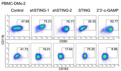
Corrected Figure 2B. Representative flow cytometric analysis of pro-inflammatory (CD11b+/CD80+) and anti-inflammatory macrophages (CD11b+/CD163+) in human PBMC-DMs from two healthy donors treated as indicated.
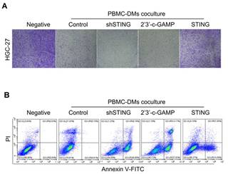
Corrected Figure 4A. Colony formation assay of human HGC-27 GC cells cocultured with human PBMC-DMs treated as indicated. (B) Left panel, representative flow cytometric plots of apoptosis markers (Annexin V+) in HGC-27 cells cocultured with human PBMC-DMs treated as indicated.
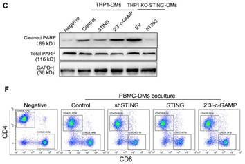
Corrected Figure S4. (C) Immunoblot analysis of cleaved-PARP and total PARP expression in human THP1-DMs treated as indicated. GAPDH was used as a loading control. (F) Representative flow cytometric plots of CD4/CD8 T cells in cocultures of human T cells with human PBMC-DMs treated as indicated.
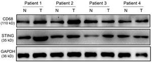
Corrected Figure S1. Immunoblot analysis showing CD68 and STING expression in paired normal mucosa and tumor samples from GC patients. GAPDH was used as a loading control.
Author contact
![]() Corresponding author: Rui-Hua Xu, Address: 651 Dongfeng East Road, Guangzhou 510060, China (e-mail: xurhorg.cn) or Hui-yan Luo, Address: 651 Dongfeng East Road, Guangzhou 510060, China (e-mail: luohyorg.cn)
Corresponding author: Rui-Hua Xu, Address: 651 Dongfeng East Road, Guangzhou 510060, China (e-mail: xurhorg.cn) or Hui-yan Luo, Address: 651 Dongfeng East Road, Guangzhou 510060, China (e-mail: luohyorg.cn)