Impact Factor
- Issue 14; 2026
- Issue 13; 2026
- Issue 12; 2026
- Issue 11; 2026
- Issue 10; 2026
- Volume 16; 2026
- Advance Articles
- Past Issues
- Cover Images
- Cover Suggestion
- Index & Coverage
- Special Issues
Introduction
1. Peptide-photosensitizer...
2. Peptide-drug assembly...
3. Multicomponent cooperative...
Conclusion
Acknowledgements
References
International Journal of Biological Sciences
International Journal of Medical Sciences
 Global reach, higher impact

Theranostics 2019; 9(11):3249-3261. doi:10.7150/thno.31814 This issue Cite
Review
Peptide-modulated self-assembly as a versatile strategy for tumor supramolecular nanotheranostics
Shukun Li1,3, Qianli Zou1, Ruirui Xing1, Thimmaiah Govindaraju4, Rawil Fakhrullin5, Xuehai Yan1,2,3 ![]()
1. State Key Laboratory of Biochemical Engineering, Institute of Process Engineering, Chinese Academy of Sciences, Beijing 100190, P. R. China.
2. Center for Mesoscience, Institute of Process Engineering, Chinese Academy of Sciences, Beijing 100190, P. R. China.
3. University of Chinese Academy of Sciences Beijing 100049, P. R. China.
4. Bioorganic Chemistry Laboratory, New Chemistry Unit, Jawaharlal Nehru Centre for Advanced Scientific Research, Jakkur P.O., Bengaluru 560064, Karnataka, India.
5. Bionanotechnology Lab, Institute of Fundamental Medicine and Biology, Kazan Federal University, Kazan 420008, Republic of Tatarstan, Russia
Received 2018-11-26; Accepted 2019-3-9; Published 2019-5-18
Abstract

Advances in supramolecular self-assembly have promoted the development of theranostics, the combination of both therapeutic and diagnostic functions in a single nanoplatform, which is closely associated with antitumor applications and has shown promising potential in personalized medicine. Peptide-modulated self-assembly serves as a versatile strategy for tumor supramolecular nanotheranostics possessing controllability, programmability, functionality and biosafety, thus promoting the translation of nanotheranostics from bench to bedside. In this review, we will focus on the self-assembly of peptide-photosensitizers and peptide-drugs as well as multicomponent cooperative self-assembly for the fabrication of nanotheranostics that integrate diagnosis and therapeutics for antitumor applications. Emphasis will be placed on building block design, interaction strategies and the potential relationships between their structures and properties, aiming to increase understanding of the critical role of peptide-modulated self-assembly in advancing antitumor supramolecular nanotheranostics.
Keywords: peptides, self-assembly, intermolecular interactions, nanotheranostics, cancer
Introduction
Nanotheranostics that combine therapeutic effects and diagnostic capabilities in a single nanoplatform have attracted increasing attention in recent years because of their potential for application in personalized medicine, including tumor-related treatments [1-6]. Such smart integrated protocols are advantageous over discrete steps of diagnosis and therapy because they can provide real-time readouts of lesion responses and thus, in turn, help to guide therapy in an adaptive, personal and precise manner. Nanotechnology, exploited as emerging techniques for the fabrication of nanomaterials, has produced a large arsenal of nanomaterials with diversified capabilities in diagnosis and therapy [7-11].
In nature, the self-assembly or organization of biomolecules into intricate and functional units in living organisms provides a rationale to construct supramolecular structures that harness the power of multiple noncovalent forces, including electrostatic forces, van der Waals forces, π interactions, hydrophobic interactions and coordination interactions [12-14]. These attractive forces are typically much weaker than covalent bonds, and these interactions occur in a dynamic and reversible manner, thus allowing self-assemblies to adapt to physiological conditions and maximize their biological functions [15-17]. In particular, peptides, as the fundamental components of proteins, show superior characteristics to those of existing assembly motifs as building blocks for the fabrication of nanotheranostics. These advantages include inherent biological origin, pharmacological safety, structural programmability, versatile functionality and easy availability. As a consequence, peptide self-assembly can be exploited as a fabrication strategy for creating various nanoscale theranostics [18-20]. Through peptide-regulated cooperative noncovalent interactions, diagnostic and/or therapeutic agents are combined to form an integrated nanoplatform. The unique significance of peptide modulation lies in three aspects. (i) With respect to structure, varying the sequences of peptides consisting of amino acids or modified peptides with regulatory groups can yield various nanostructures because of the diverse noncovalent interactions involved. For example, hydrophobic interactions play a predominant role in micelle formation [21], while hydrogen bonds can induce directional growth into nanofibers in a thermodynamically favorable pathway [22]. Alternatively, manipulating the kinetic parameters (pH, temperature, counter ions, concentrations, solvents) [23-25] can also trap assembly structures in a metastable state with different morphologies. Therefore, desired nanostructures can be obtained by thermodynamic control or kinetic control. (ii) With respect to function, the coassembly of peptides and other bioactive components is a facile and typical strategy, where two or more types of building blocks interact with each other through synergetic noncovalent interactions to form nanostructures. Simultaneously, the dynamic nature of noncovalent interactions can be easily exploited to achieve the targeted release of bioactive components in response to a specific lesion microenvironment. For example, coassembly involving metal coordination integrated the dual properties of robust stability in circulation and smart responsiveness to tumor lesions, thereby enhancing the therapeutic effect [26-29]. Furthermore, some metal ions with intrinsic magnetic or radioactive properties could also be incorporated into coassemblies to perform unique imaging functions [29-31]. Another strategy is to synthesize peptide conjugates due to the feasibility of conjugation chemistry [32-36], which allows the design of specific building blocks, including peptide-photosensitizer and peptide-chemodrug blocks, followed by self-assembly into nanostructures with relatively high covalent stability and high loading efficiency. In addition, stimuli-responsive [37-39], circulation-extending [40] and target-binding [41, 42] sites can be flexibly introduced to increase the target-to-background contrast in imaging application and simultaneously improve the local concentration of therapeutic compounds at the target of interest, with the purpose of improving the therapeutic index. (iii) Last but not least, structural regulation facilitates function amplification. In particular, the enhanced permeability and retention effect (EPR) occurs due to the increased permeability of the blood vessels and dysfunctional lymphatic drainage in solid tumors during the systemic circulation of the combined molecules [43], whereas this effect is not attainable by individual building blocks due to their quick excretion by the reticuloendothelial system (RES). In light of these advantages, peptide-modulated self-assembly serves as a flexible and versatile toolbox for the construction of tumor supramolecular nanotheranostics, highlighting the potential to predict the circulation and accumulation of agents at tumor sites, indicate uptake and achieve the sustainable release of drug molecules thereby improving therapeutic outcomes. However, elaboration of the structure-property relationship, including the manipulation of noncovalent interactions towards structural and functional regulation, is still a challenge [44, 45]. Therefore, a systematic summary of regulatory principles, properties and applications concerning peptide self-assembly is necessary for the future development of cancer nanotheranostics.
In this review, we mainly focus on three assembly strategies: (1) the self-assembly of peptide-photosensitizers, (2) the self-assembly of peptide-drugs and (3) multicomponent cooperative self-assembly for the fabrication of nanotheranostics that integrate diagnosis and therapeutics for antitumor application (Figure 1). Emphasis will be placed on building block design, intermolecular interactions and the potential relationships between their structures and properties, aiming to elucidate the critical role of peptide-modulated self-assembly in advancing antitumor supramolecular nanotheranostics. Representative studies based on above strategies are included, which will open promising prospects in the precise design of phototherapeutic agents, chemotherapeutic agents and multi-modal imaging-guided nanotheranositcs for highly efficient cancer therapy. Finally, we will outline the challenges and potential of the use of nanotheranostics to promote optimized treatment outcomes.
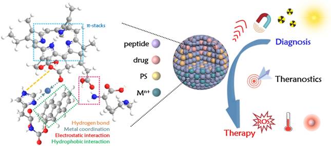
Schematic illustration of peptide-modulated self-assembly (peptide-photosensitizers; peptide-drugs; multicomponent cooperative self-assembly) as a versatile strategy for tumor supramolecular nanotheranostics.
1. Peptide-photosensitizer assembly strategies
Owing to their spatiotemporal selectivity and noninvasive nature, phototherapies, including photodynamic therapy (PDT) and photothermal therapy (PTT), have become promising approaches in the application of antitumor theranostics [46-48]. Overall, both modes rely on the specific delivery of photosensitizers to tumor sites, followed by photoirradiation to excite the photosensitizers. The excited photosensitizes decay backs to the ground state through three main pathways: photon emission (fluorescence), intersystem crossing (energy transfer to generate reactive oxygen species, PDT) and nonradiative relaxation (photothermal conversion, PTT). Based on these three pathways, the requirements of the photosensitizer state for PDT and PTT differ. For PDT, the encapsulated or coassembled photosensitizers should be quickly released as monomers at the site of interest to perform ROS generation [49, 50]. For PTT, however, the photosensitizer should be maintained in an aggregated state since close stacking efficiently quenches fluorescence and intersystem crossing, promoting the dissipation of the absorbed energy into the lesion through nonradiative relaxation, i.e., photothermal conversion [51, 52].
1.1 Peptide-modulated self-assemblies for PDT
The wide application of PDT in clinical use is barred by the high hydrophobicity of photosensitizers, as the resulting ordered or disordered aggregation reduces their bioavailability and accumulation at lesions, which has instigated the development of supramolecular materials that improve their pharmacokinetics and tumor targeting [32]. Peptide-modulated self-assembly nanotheranostics developed as a promising approach to increase the solubility of photosensitizers in a nanostructure, extend their blood circulation time, and thus improve their specific accumulation at tumor sites. Intriguingly, photosensitizer molecules sometimes participate in the process of peptide self-assembly for the formation of nanotheranostics [53]. For example, porphyrins are organic heterocyclic macrocycles with photophysical properties well suited for PDT and fluorescence imaging [52]. Importantly, their intrinsic hydrophobic properties and π groups can provide noncovalent interactions to promote coassembly with peptides and thereby afford the final nanostructures [54-56]. Our group [55] previously demonstrated that short peptides of a diphenylalanine derivative, H-Phe-Phe-NH2·HCl (CDP), and an amino acid derivative, 9-fluorenylmethoxycarbonyl-L-lysine (Fmoc-L-Lys), can both coassemble with chlorin e6 (Ce6) through π-stacks, hydrophobic effects and electrostatic interactions. The resulting nanoparticles with superior features of high loading efficiency and smart drug release can lead to fluorescence imaging-guided PDT.
To precisely release photosensitizers at the site of interest (tumor), a highly appreciated strategy has emerged: the incorporation of interaction sites or functional groups that can respond to the tumor microenvironment (low pH of 6.0-7.0 [57, 58], heightened glutathione (GSH) level [59, 61], overexpressed enzymes [62, 63] and biomarkers [62, 64]) to achieve controlled photosensitizer activation at tumor sites rather than normal tissues [65, 66]. Major mechanisms of stimuli sensing involve breaking the assembly-disassembly balance between noncovalent interactions. However, complex physiological components, dilution by body fluids and degradation by enzymes may cause the premature disassembly of nanotheranostics before they reach the tumor sites. To address this concern, a novel method has recently been suggested: coordination assembly. Natural self-organized proteins in living organisms, such as metalloproteins, are formed by metal ions and other organic cofactors through coordination interactions [67], where metal ions not only improve stability and mechanical strength but also play a significant role in regulating biological functions [68]. With natural examples as a guide, the metal-triggered self-assembly of peptides is a promising direction for constructing well-defined conformations or nanostructures [69-71]. Above all, the strategy meets the requirements of robust circulation in physiological environments and stimuli-responsive photosensitizer release, as metal ions can coordinate with competing ligands at high concentrations within tumor tissues.
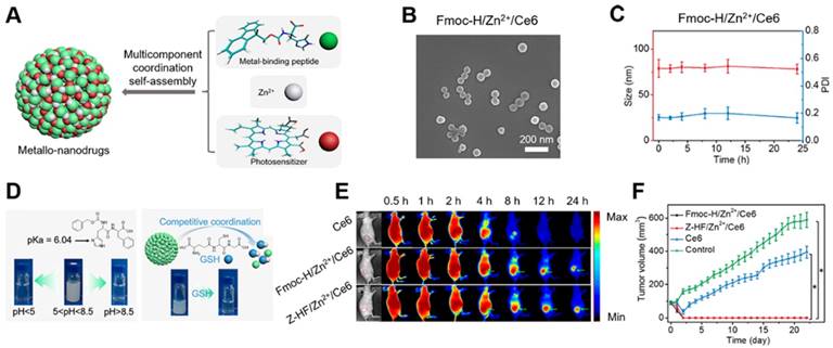
Hemoglobin organically integrates histidine residues and porphyrin derivatives through cooperative coordination. Natural examples inspired us to engineer metallo-nanodrug theranostics by a multicomponent coordination self-assembly fabrication method [26]. Histidine-containing dipeptides (N-benzyloxycarbonyl-L-histidine-L-phenylalanine, Z-HF) or amphiphilic histidine derivatives (fluorenylmethoxycarbonyl-L-histidine, Fmoc-H) are selected as starting building blocks for coordination self-assembly. Spherical metallo-nanodrugs (~ 80 nm) can be readily regulated by zinc ion coordination with peptides and chlorin e6 (Ce6) combined with other noncovalent interactions (Figure 2A, B). Such coordination interactions exhibit dual properties in theranostic applications. On the one hand, the intermolecular coordination forces are nearly strong as covalent bonds, and such stable interactions can resist dilution or decomposition under normal physiological conditions (Figure 2C), in which Ce6 molecules exist in an aggregated state with a self-quenching effect, protecting their photosensitization, and thus exhibit prolonged blood circulation and improved tumor accumulation. On the other hand, the dynamic nature of coordination forces and noncovalent interactions is susceptible to the tumor-related microenvironment. Decreased pH and increased GSH levels jointly promote the disassembly of metallo-nanodrugs due to the carboxyl protonation and competitive coordination of zinc ions in the presence of GSH (Figure 2D). Such ultrasensitive responsiveness can free Ce6 and activate both fluorescence feedback and ROS generation within tumor cells. The drug distribution can be traced by in vivo fluorescence imaging and provide therapeutic windows at 4 h post injection (Figure 2E). Under the guidance of the therapeutic window, effective molecular Ce6 drug concentration can promote PDT efficacy to eradicate tumors (Figure 2F). Recently, another assembly strategy involving metal coordination combined with other noncovalent forces was applied in host/guest chemistry by Granja and coworkers [72]. The host structure is composed of two self-complementary α,γ-cyclic peptides, which bear a Zn porphyrin cap for the selective recognition of the guest. The two components of the host structure are linked via two dynamic covalent bonds. Combined with hydrogen bonding, this linkage allows the two host molecules to self-assemble into a capsule structure. The metal ion coordination can recognize guest molecules (bipyridine) to form a sandwich complex structure. The hydrolysis of the hydrazones allows the reversible release of the encapsulated guest molecules due to the disruption of the capsule structure, suggesting the potential of these molecules in drug delivery vehicles, molecular machines and tumor theranostics.
(A) Construction of metallo-nanodrugs through multicomponent (small peptides, photosensitizers, zinc ions) coordination self-assembly. (B) SEM image of the metallo-nanodrugs Fmoc-H/Zn2+/Ce6. (C) Size and PDI of metallo-nanodrug under conditions mimicking physiological circulation. (D) Schematic illustration of ultrasensitive responsiveness to pH and GSH change. (E) Fluorescence images showing the preferential accumulation of metallo-nanodrugs at tumor sites in contrast to unencapsulated photosensitizers. (F) Tumor growth profiles after PDT. Adapted with permission from Ref [26]. Copyright 2018 American Chemical Society.
As a distinct alternative tool, aggregation-induced emission (AIE) based building blocks for fabrication of nanostructures has gained prominence in the field of cancer theranostics [73-75]. The building blocks are non-emissive in the molecularly dissolved state while induced to emit fluorescence in the aggregation state mainly due to the restriction of intramolecular rotation (RIR). In sharp contrast to conventional nanotheranostics based on photosensitizers with aggregation-caused quenching (ACQ) effect, the AIE-based nanotheranostics possess superb advantages, such as high stability and enhanced therapeutic effect, particularly when incorporation of smart activation strategy of photosensitizers [76-78]. For example, Liu and coworkers [76] designed a bioprobe for a target-specific light-up imaging and activatable photodynamic therapy. The bioprobe consists of four parts: Tetraphenylethene derivative (TPECM) functions as an imaging and photosensitizer reagent. Peptide sequence Gly-Phe-Leu-Gly (GFLG) can be responsive to cathepsin B. Tripeptide Asp-Asp-Asp (DDD) is a hydrophilic linker while increases the hydrophilicity of probe. A cyclic Arg-Gly-Asp (cRGD) moiety was incorporated for targeting tumor cells with overexpression of αvβ3 integrin. In aqueous media, the probe is nearly nonfluorescent and can rarely generate ROS owing to intramolecular motions. Once the bioprobe was selectively taken up by tumor cells, GFLG will be cleaved by cathepsin B and thus increase the hydrophobic interactions of TPECM for the formation of aggregation, leading to enhanced fluorescence emission and activated ROS generation for image-guided PDT. Furthermore, they developed another AIE-based probe, TPETP-SS-DEVD-TPS-cRGD [77], among which tetraphenylethenethiophene (TPETP) is a photosensitizer for PDT and peptide sequence Asp-Glu-Val-Asp (DEVD) is responsive to caspase-3/-7 for indicating cell apoptosis. In addition, cRGD and -S-S- are used as targeting ligand and linker, respectively, to realize dually-targeted activatable PDT. More importantly, when the probes were excited through 405 nm laser, the incorporated TPETP is red-emissive, while hydrophobic TPS showed intense green fluorescence after cleaved by caspases and therefore to report the therapeutic effect. Such a strategy based on distinct fluorescence turn-on signal is more favorable in complicated physiological conditions. Moreover, the activatable strategy can expand to other biological interactions, such as specific YSA peptide (YSAYPDSVPMMS)-EphA2 protein interaction [78].
1.2 Peptide-modulated self-assemblies for PTT
The use of peptide-photosensitizer conjugates as building blocks for nanotheranostic construction has attracted significant attention in recent years owing to their facile synthesis and their simple but compact structural architecture. Significantly, the unique photothermal efficiency can be enhanced by the high sensitizer loading density, which markedly reduces the intermolecular distance of the photosensitizers and thereby inhibits fluorescence and intersystem crossing pathways, opening an avenue for PTT. Light absorption by molecules can create a thermally induced pressure jump that produces ultrasonic waves, which are detected by acoustic detectors for imaging (photoacoustic imaging, PA) with deep tissue penetration and high spatial resolution [79, 80]. Therefore, PA imaging-guide PTT expanded the repertoire of imaging and therapeutic modalities in which porphyrin agents can engage [81]. In addition, compared to the modality of PDT, PTT shows advantages in the treatment of hypoxic tumors because of its oxygen independence.
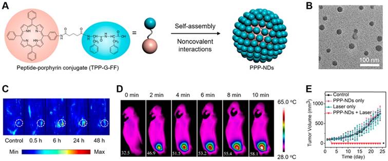
Zheng and coworkers [82] developed porphysomes, nanovesicles formed from self-assembled porphyrin bilayers that showed photothermal conversion property. Therefore, the obtained porphysomes were capable of visualization of lymphatic systems through PA imaging. After intravenous injection, such porphysomes accumulated in tumors and revealed laser irradiation-induced photothermal tumor ablation. However, lipid bilayers are generally considered unstable structures in blood circulation and in storage. In this regard, our group [83] designed and synthesized a diphenylalanine peptide-porphyrin conjugate (TPP-G-FF) (Figure 3A). Upon the incorporation of FF, which is capable of tuning self-assembly, the amphiphilic structures form strong π-stacking and hydrogen bonding interactions and thus assemble into regular nanodots (PPP-NDs) with a diameter of 25 ± 10 nm (Figure 3B). Supramolecular aggregation resulted in complete fluorescence quenching and the inhibition of ROS generation, leading to the absorbed light energy to dissipate thermally with high conversion efficiency (54.2%). Notably, the photothermal conversion of PPP-NDs can be visualized through PA imaging techniques in vivo. The imaging results demonstrated that the optimal tumoral accumulation and retention of PPP-NDs was 24 h (Figure 3C). In this therapeutic window, IR thermal imaging indicated that the mean temperature within 10 min at the tumor sites increased to 58.1 °C upon laser irradiation (Figure 3D). The elevated temperature induced tumor cell necrosis irreversibly, suggesting robust PTT potency towards antitumor therapy (Figure 3E).
(A) Construction of photothermal peptide-porphyrin nanodots (PPP-NDs) by peptide-porphyrin conjugate (TPP-G-FF) self-assembly. (B) TEM image of PPP-NDs. (C) PA images of mice over time after the intravenous injection of PPP-NDs (D) IR thermal images of intravenous PPP-NDs injected into mice under continuous irradiation. (E) Tumor growth profiles after PTT. Adapted with permission from Ref [83]. Copyright 2017 American Chemical Society.
In principle, such peptide-modulated porphyrin-based self-aggregates for nanotheranostics can also be applied to other photosensitizers, for example, phthalocyanine. Yoon and coworkers [84] designed a ''one-for-all'' nanomaterial, NanoPcTB, assembled from phthalocyanine conjugates, in which the photoactivity of phthalocyanine can be switched from PTT to PDT, and the imaging mode can simultaneously be changed from PA imaging to fluorescence imaging by biotin receptor-assisted partial disassembly at tumor sites. Spatiotemporally integrated phototherapy guided by the dual imaging mode can eradicate tumors thoroughly. Alternatively, a more contemporary approach focuses on tailoring peptide sequences to amplify the final therapeutic effect. The conjugation of functional sequences, including antibodies, receptors, and some chimeric stimuli-responsive groups, to peptides is being explored [85-89]. For example, a chimeric peptide sensitive to both pH and matrix metalloproteinase-2 (MMP-2), Fmoc-12-aminododecanoic acid-H8R8-PLGVR-PEG8 (Fmoc-ADDA-H8R8-PLGVR-PEG8), developed by Zhang and coworkers [90] can be used to codeliver the photosensitizer protoporphyrin IX (PpIX) and plasmid DNA to MMP-2-rich tumor cells simultaneously. The chimeric peptides undergo hydrolysis of the PLGVR peptide sequence and exfoliation of PEG, with increasing positive charges, leading to cationic R8 sequence exposure and preferential uptake by tumor cells. Furthermore, the decreased pH value in the endosomes leads to rapid protonation of the peptide H8 sequence and loss of its hydrophobic core, thereby releasing PpIX in the acidic endosomes. Moreover, a dual-stage light irradiation strategy was developed to realize the synergistic effect of drug and gene delivery. Upon short-term light irradiation, the ''proton sponge'' effect and the photochemical internalization effect can induce the endosomal escape of PpIX and DNA. Upon long-term irradiation, the synergistic efficacy of photodynamic and gene therapies was enhanced. As a result, chimeric peptide/PpIX/p53 complexes display significant inhibition of tumor growth.
2. Peptide-drug assembly strategies
Currently, chemotherapy is still one of the most effective ways for clinical treatment of cancer. Despite the clinical success of several chemotherapeutic drugs, it is still an unprecedented challenge to tailor the desired pharmacokinetics that allow chemotherapeutics to passively or actively target tumors while minimizing their adverse side effects. Over the past several decades, supramolecular nanotheranostics have emerged as an appealing approach to this goal, exhibiting improved solubility, prolonged circulation time and enhanced specific accumulation of chemodrugs in the tumor site owing to the EPR effect, thereby improving the overall therapeutic index [91]. Among a large number of research works, the chemodrug was passively encapsulated or absorbed in the nanostructure; this type of fixed “nanocarrier” protected the chemodrug molecules from degradative processes in the organism but resulted in limited, low drug payloads [92]. Peptides as building blocks possess multiple noncovalent interaction sites that can coassemble reciprocally with chemodrugs, presenting a more tunable and flexible way to increase drug loading. However, the intrinsic nature of noncovalent interactions could not resist harsh physiological conditions during in vivo application. Therefore, nanotheranostic designs that integrate flexible and high loading efficiencies as well as considerable stability are of interest. Two novel strategies aiming to solve the above problems are outlined below.
2.1 Metal coordination-driven self-assemblies for chemotherapy
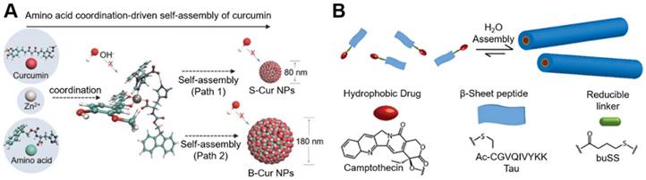
Metal coordination-driven multicomponent self-assembly is a versatile strategy that can improve the circulation stability of chemodrugs and achieve targeted release. Our group [27] suggested that curcumin-based nanostructures can be fabricated through the coordination interactions of metal ions with metal-binding peptides or amino acids. Curcumin nanoparticles with well-defined, uniform distributions were constructed from a histidine derivative (Fmoc-H), curcumin and zinc ions via cooperative coordination bonding together with other noncovalent interactions. Through control by kinetic parameters (tension coefficients of the solvent used to dissolve Fmoc-H) or dynamic parameters (concentration of curcumin), the size can be readily tuned from 80 nm to 180 nm (Figure 4A). The resultant nanodrugs possess high loading efficiency of curcumin (~52%) and protect it from degradation. Moreover, the dynamic ligand change or exchange in the tumor microenvironment conferred responsive release properties on the nanodrugs, therefore improving drug uptake by tumor cells. Fluorescence imaging indicated favorable circulation and accumulation in tumor sites. Collectively, these features increased the stability of curcumin and simultaneously improved tumor accumulation, resulting in enhanced antitumor therapy. The coordination-driven multicomponent self-assembly opens an avenue of chemodrug use for antitumor therapy.
(A) Curcumin nanoagents based on amino acid coordination-driven self-assembly. Adapted with permission from Ref [27]. Copyright 2018 Wiley VCH. (B) Illustration of a CPT-based drug amphiphile that can spontaneously self-assemble into nanofibers. Adapted with permission from Ref [110]. Copyright 2013 American Chemical Society.
2.2 Peptide-conjugate self-assemblies for chemotherapy
The covalent modification is an alternative clinically proven strategy to devise peptide-drug conjugates for self-assembly with enhanced treatment efficacy [93, 94]. Peptide sequences containing stimuli-responsive groups can be incorporated on demand, which not only plays a significant role in drug release but also promotes the strategy formation of intracellular (in vivo) self-assembly. Intracellular self-assembly based on biocompatible condensation reaction [95-97], endogenous stimuli reconstruction upon the control of either pH, disulfide reduction or enzymatic cleavage [17, 98] has shown great potential in improving targeting cellular uptake of the drug molecules in chemotherapy.
Xu and coworkers [99-103] performed pioneering works on designing and constructing drug-peptide amphiphilic hydrogelators that can self-assemble into supramolecular three-dimensional hydrogel networks. The dephosphorylation/phosphorylation cycle catalyzed by the alkaline phosphatase (ALP)/kinase switch has been applied to control self-assembly. For example [99], by introducing a phosphotyrosine into a Nap-Phe-Phe-Lys (PTX) hydrogelator, where Nap refers to a naphthalene moiety in order to increase the solubility or hydrophilicity but with no effect on the self-assembly property. Dephosphorylation occurred once the hydrogelator contacted the phosphatase enzyme; thus, the hydrophobicity was enhanced, further promoting spontaneous self-assembly via β-sheet interactions to form a hydrogel. Once the gel was formed, the PTX conjugates were sustainably released and exhibited effective cytotoxicity against HeLa cells. By comparison, Yang and coworkers [104, 105] recently provided a different approach to phosphorylation. The targeting tripeptide RGD with hydrophilic properties was conjugated to curcumin through a reducible disulfide bond linker to form a hydrogelator [104]. Due to the specific binding to integrin expressed on tumor cells, the localization of the hydrogelator was enhanced after systemic injection. After localization, the elevated intracellular GSH concentration reduced the disulfide bonds. Consequently, the RGD sequence was cleaved from the curcumin hydrogelator, increasing the hydrophobicity of building blocks and therefore allowing for their self-assembly spontaneously. The released curcumin showed obvious cytotoxic activity to tumor cells and inhibited tumor growth. Similarly, Ding and coworkers [106] designed another curcumin-peptide conjugate (Curcumin-FFE-CS-EE). By GSH reduction, the hydrophilic EE sequence was removed, leading to the formation of curcumin-based supramolecular nanofibers for use as radiosensitizers. Liang and coworkers [107, 108] design a precursor Cys(SEt)-Glu-Tyr(H2PO3)-Phe-Phe-Gly-CBT that is responsive to extracellular ALP and intracellular GSH in tumor site for yielding cyclic amphiphilic 2D building blocks. Interestingly, the precursor molecules cannot enter the cells, but the converted cyclic amphiphilic 2D molecules can be efficiently internalized by cells and locally self-assemble into the nanofibers with enhanced mechanical strength, highlighting great potential of smart nanotheranostics design.
Cui and coworkers [33, 53, 109, 110] demonstrated the rational design of an amphiphilic peptide-drug conjugate (“prodrug”) that can form a variety of nanostructures. Intriguingly, the drug itself constitutes one of these two domains, most commonly the hydrophobic, and serves a structural role in addition to eliciting a therapeutic effect. Such conjugated peptide-drug prodrugs with a suitable hydrophilic-hydrophobic balance can self-assemble into well-defined supramolecular nanostructures in aqueous solution. For example [110], the hydrophobic drug camptothecin (CPT) was conjugated to a β-sheet-forming peptide sequence derived from the Tau protein through a reducible disulfylbutyrate linker (buSS) (Figure 4B). In this design, the drug content can be precisely controlled by the number of CPT molecules, one, two or four, and the resultant drug loading can be 23%, 31%, and 38%, respectively. Moreover, the morphology can also be tuned by the number of branching points for attaching CPT molecules. Nanofilament to nanotube transitions can be tuned as a function of drug content since the hydrophobicity and possible π-π associative interactions among CPT inter- and intramolecular units were enhanced. This stable nanostructure sequestered CPT molecules in its core. Notably, biodegradable linkers offered responsiveness towards the external environment to release CPT sustainably and further increase the antitumor efficacy.
To maximize the effectiveness of chemotherapy, metal coordination and peptide conjugation can be flexibly combined. Cisplatin-based metal ions have been also reported to be capable of forming complexes with peptides containing carboxylic acid groups. Yang and coworkers [111] choose 10-hydroxycamptothecine (HCPT) and cisplatin to construct the dual anticancer drug assemblies. HCPT was firstly conjugated with peptide FFERGD to form amphiphilic building blocks, HCPT-peptide (HP). In order to assist HP entering into the cellular nucleus, cisplatin coordination was introduced to complex with HP. By tailoring the molar ratio of cisplatin/HP from 1 to 1.5, two different complexes were obtained and further self-assembled into long nanofibers (10-15 nm) and nanoparticles (20-25 nm), respectively. The nanomaterials play a role of “Trojan Horse” that transported dual anticancer drugs across the cell and nucleus membranes, showing synergistic inhibition effect to cancer cells.
Additionally, some active targeting groups, for example, surface markers (antigen or ligands), should be considered for peptide-drug conjugate design to enhance therapeutic results. The binding of ligands to receptors may cause receptor-mediated multivalent interactions to promote the internalization and release of drug molecules, alleviating the random diffusion of targeting drug molecules and thereby reversing multiple-drug resistance, which often causes chemotherapy treatments to fail [112, 113]. Moreover, conjugating peptides with functional groups targeting the tumor vasculature or tumor microenvironment as well as introducing imaging groups may also be necessary [114].
3. Multicomponent cooperative self-assembly strategies
Tumor heterogeneity and complex bionanostructure interactions have a considerable, underappreciated impact on treatment efficacy [57]. Consequently, it is reasonable to choose and explore a multicomponent cooperative self-assembly strategy to integrate manifold imaging techniques and multiple therapies into a single nanoplatform, thereby gaining mutually confirmative information on trafficking pathway and kinetics of delivery and accurately monitor therapeutic outcomes, an approach that shows great potential in clinical use [115].
Before initiating tumor treatment, it is essential to perform imaging to understand the tumoral biological information. In general, different imaging modalities have advantages and disadvantages that define their use in visualizing the genesis and development of lesions. Thus, the selected imaging modality should be suitable to reveal the dynamic features of tumors. Highly sensitive imaging techniques, including singlephoton emission computed tomography (SPECT), positron emission tomography (PET), and optical imaging, are being used to monitor tumor cell biology, tumor burden, tumor progression and metastasis, but they lack anatomical information to localize the signal, which is better provided by magnetic resonance imaging (MRI) and computed tomography (CT) [29, 30, 116]. Comprehensive imaging modalities can guide the field of nanotheranostics towards an era of more effective and personalized treatment.
3.1 Single mode imaging-guided therapy
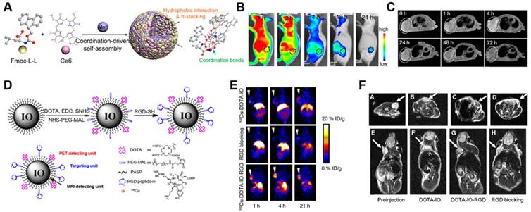
As an example, our group [28] has designed a multicomponent coordination nanotheranostic platform by mixing 9-fluorenylmethoxycarbonyl-L-histidine (Fmoc-L-L), a photosensitizer (Chlorin e6, Ce6), and Mn2+ in aqueous solution to form Fmoc-L-L/Mn2+ nanoparticles (FMNPs) driven by coordination and other noncovalent interactions (hydrophobic interaction, π-π stacking) (Figure 5A). Carboxyl groups involved in amino acid coordination with Mn2+ play a significant role in improving the stability of the final nanostructure. More importantly, introducing Mn2+ forced the FMNPs to act as an MRI agent. The resulting minimalist nanoplatform showed smart responsiveness within tumor tissue, restoring Ce6 photosensitization capability to generate ROS for PDT and enhancing Mn2+ retention for MRI due to stronger binding with GSH. This integrated fluorescence imaging with MRI combined the merits of the respective qualitative and quantitative information of both methods and provided feedback regarding the therapeutic efficacy. At 4 h post injection, PDT was performed under the guidance of fluorescence imaging (Figure 5B) and MRI (Figure 5C). In addition, therapeutic performance was observed in the process of tumor ablation within three days.
(A) Small amino acid-Mn2+ metal ion coordination-driven supramolecular self-assembly. (B) Fluorescence images of mice following the intravenous injection of FMCNPs. (C) T1-weighted MR images of mice following the intravenous injection of FMCNPs. Adapted with permission from Ref [28]. Copyright 2018 American Chemical Society. (D) Synthesis of PET/MRI dual-functional probe DOTA-IO-RGD. (E) PET images of mice after the injection of 64Cu-labeled nanoprobes. (F) T2-weighted MR images of mice before and after the injection of nanoparticles. Adapted with permission from Ref [117]. Copyright 2008 Society of Nuclear Medicine, Inc.
3.2 Dual/multiple mode imaging-guided therapy
Focusing on tumor-targeted drug delivery, PET/MRI dual-modality tumor imaging was achieved by using arginine-glycine-aspartic (RGD)-conjugated radiolabeled iron oxide (IO) nanoparticles fabricated by Chen and coworkers [117]. PET and MRI have already been used in clinical practice. Cyclic RGD peptide modification promotes the selective bonding of nanoparticles to tumor blood vessels (via avβ3, avβ5 and a5β1 integrin receptors overexpressed by activated endothelial cells), and the nanoparticles were further modified with DOTA to enable 64Cu complexation and simultaneous PET-MRI monitoring (Figure 5D). PASP-IO nanoparticles with a core size of 5 nm and a hydrodynamic diameter of approximately 45 nm were made by a single-step reaction. The saturation magnetization of PASP-IO nanoparticles is approximately 117 emu/g of iron, and the measured r2 and r2* are 105.5 and 165.5 (s mM)-1, respectively. The accumulation of 64Cu-DOTA-IO-RGD nanoprobes was found to start at 1 h post injection and reached a peak value at 4 h. In the case of nontargeted probes and in the blocking experiment, hardly any accumulation in tumors could be visualized (Figure 5E). T2-weighted MRI was conducted to confirm these findings (Figure 5F). As a complementary example [118], a new type of dual integrin αvβ3 and GRPR targeting radiotracer, 68Ga-BBN-RGD, was fabricated by their group and used for PET/CT imaging of breast cancer, showing great potential in discerning primary breast cancers, axillary lymph node metastasis and distant metastases.
Significantly, various imaging and therapeutic modalities can be integrated flexibly through peptide-modulated self-assembly to optimize final therapeutic outcomes [119, 120]. Wang and coworkers [119] introduced a paradigm of manipulating T1-T2 dual-modal MRI for tumor theranostics. A nanosystem of (Fe3O4@SiO2@mSiO2/DOX-(Gd-DTPA)-PEG-RGE NPs was presented, in which chemodrug DOX was loaded and the tumor-penetrating peptide RGERPPR (RGE) functioned to increase the cellular uptake and cytotoxicity of NPs, while Gd-DTPA served as the T1 contrast agent and Fe3O4 as the T2 contrast agent. In vitro and in vivo treatment results on U87MG cells showed that the NPs could be used as an effective platform for tumor theranostics. Chen and coworkers [120] conjugated a somatostatin receptor (SST) peptide derivative (DOTA-octreotate), to an Evans blue analog (EB) that can specifically bind to circulating serum albumin. The resulting molecule was used to chelate 86Y and 90Y, which play roles in PET imaging and radiotherapy, respectively. The imaging capability and the radiotherapeutic efficacy were demonstrated to be effective for the treatment of SSTR2-containing tumors.
Conclusion
In summary, we have provided an overview of recent fascinating developments in the area of peptide-modulated self-assembly as a versatile strategy for tumor supramolecular nanotheranostics. Peptides function as molecular building units or biologically active compounds to build supramolecular nanostructures owing to their molecular design versatility, tunable biocompatibility and biodegradability. The resultant peptide-photosensitizers, peptide-drugs and multicomponent cooperative self-assemblies can be regulated in terms of composition, noncovalent interactions and physiochemical properties. The obtained nanotheranostic platforms demonstrated precise selectivity, smart adaptability, and multifunctionality towards antitumor theranositcs, which amplified the therapeutic functions, providing prolonged blood circulation, enhanced tumor accumulation, and increased sensitivity to lesion response.
Although considerable success has been achieved in supramolecular nanotheranostics, the main challenge in cancer therapy stems from the heterogeneity and complexity of the tumor entity and microenvironment, which cause incomplete or unpredicted interactions (trafficking, internalization, penetration, distribution, fluid pressure) between nanotheranostics and tumor cells in vivo [121]. A deep understanding of tumor biology combined with noninvasive imaging techniques with fast response and ultrahigh resolution at the single-cell level will elucidate complex bionanotheranostic interactions. Moreover, most supramolecular nanotheranostics are delivered intravenously for systemic transport to tumors through defective tumor vessels and impaired lymphatics in the tissue: the enhanced permeability and retention (EPR effect). Such transportation is a fundamental principle but is substantially considered to be oversimplified [122] because it is increasingly clear that EPR varies among patients and tumor types, even within the same patient or the same tumor type over time [123]. Taking this point into consideration, the discovery of novel disease targets (overexpressed receptors) is necessary. Furthermore, even though satisfactory treatment outcomes have been achieved, the selection of animal models or tumor type models in research, cannot predict the clinical results; thus, the translation of most supramolecular nanotheranostics in humans remains largely unexplored. Therefore, a series of systematic, personalized studies in clinical use should be pursued. Last but not least, despite the promising therapeutic results of nanotheranostics covered in this review, two critical aspects on peptide-based supramolecular systems should be highlighted: (1) although peptides are biologically-originated building blocks, tedious chemical modification in order to meet desired demands for theranostics may provoke unwanted side effects such as inflammation and immunogenicity; (2) stability is another major concern. Peptide self-assembly is in harness of noncovalent interactions and thus prone to disassemble in complicated physiological environment, leading to premature drug release, side effects to normal tissue, and reduced drug efficacy in the site of interest. Therefore, ideal nanotheranostics should be stable enough in blood circulation while be unstable once they reach to the therapeutic site. In this regard, smart strategies that can enhance fabrication interactions and simultaneously manipulate nanodrugs to be smart responsiveness for tumor microenviornment will be highly appreciated. Insights into the mechanism for supramolecular nanotheranostics regarding materials design and therapeutic efficacy should be one of research focuses [51]. All in all, advancing supramolecular nanotheranostics may promote the clinical translation of nanodrugs and theranostics from bench to bedside.
Acknowledgements
We acknowledge financial support from the National Natural Science Foundation of China (Project Nos. 21773248 and 21802144), the National Natural Science Fund BRICS STI Framework Programme (No. 51861145304) and Innovation Research Community Science Fund (No.21821005) as well as the Key Research Program of Frontier Sciences of the Chinese Academy of Sciences (CAS, Grant No. QYZDB-SSW-JSC034).
Competing Interests
The authors have declared that no competing interest exists.
References
1. Chen HM, Zhang WZ, Zhu GZ, Xie J, Chen XY. Rethinking cancer nanotheranostics. Nat Rev Mater. 2017;2:17024
2. Shi JJ, Xiao ZY, Kamaly N, Farokhzad OC. Self-assembled targeted nanoparticles: evolution of technologies and bench to bedside translation. Acc Chem Res. 2011;44:1123-34
3. Kelkar SS, Reineke TM. Theranostics: combining imaging and therapy. Bioconjug Chem. 2011;22:1879-903
4. Kunjachan S, Ehling J, Storm G, Kiessling F, Lammers T. Noninvasive imaging of nanomedicines and nanotheranostics: principles, progress, and prospects. Chem Rev. 2015;115:10907-37
5. Huang P, Rong PF, Lin J, Li WW, Yan XF, Zhang MG. et al. Triphase interface synthesis of plasmonic gold bellflowers as near-infrared light mediated acoustic and thermal theranostics. J Am Chem Soc. 2014;136:8307-13
6. Cui HG, Wang J. Progress in the development of nanotheranostic systems. Theranostics. 2016;6:915-7
7. Webber MJ, Appel EA, Meijer EW, Langer R. Supramolecular biomaterials. Nat Mater. 2016;15:13-26
8. Fan WP, Yung B, Huang P, Chen XY. Nanotechnology for multimodal synergistic cancer therapy. Chem Rev. 2017;117:13566-638
9. Ariga K, Leong DT, Mori T. Nanoarchitectonics for hybrid and related materials for bio-oriented applications. Adv Funct Mater. 2018;28:1702905
10. Liu Y, Wang S, Ma Y, Lin J, Wang HY, Gu YQ. et al. Ratiometric photoacoustic molecular imaging for methylmercury detection in living subjects. Adv Mater. 2017;29:1606129
11. Sun TM, Zhang YS, Pang B, Hyun DC, Yang MX, Xia YN. Engineered nanoparticles for drug delivery in cancer therapy. Angew Chem Int Ed. 2014;53:12320-64
12. Zhang SG. Fabrication of novel biomaterials through molecular self-assembly. Nat Biotechnol. 2003;21:1171-8
13. Wang J, Liu K, Xing RR, Yan XH. Peptide self-assembly: thermodynamics and kinetics. Chem Soc Rev. 2016;45:5589-604
14. Omenetto FG, Kaplan DL. New opportunities for an ancient material. Science. 2010;329:528-31
15. Cui HG, Xu B. Supramolecular medicine. Chem Soc Rev. 2017;46:6430-2
16. Yang LL, Tan XX, Wang ZQ, Zhang X. Supramolecular polymers: historical development, preparation, characterization, and functions. Chem Rev. 2015;115:7196-239
17. Li LL, Qiao ZY, Wang L, Wang H. Programmable construction of peptide-based materials in living subjects: from modular sesign and morphological control to theranostics. Adv Mater. 2018. 1804 971
18. Xing PY, Zhao YL. Multifunctional nanoparticles self-assembled from small organic building blocks for biomedicine. Adv Mater. 2016;28:7304-39
19. Dou XQ, Feng CL. Amino acids and peptide-based supramolecular hydrogels for three-dimensional cell culture. Adv Mater. 2017;29:1605005
20. Rad-Malekshahi M, Lempsink L, Amidi M, Hennink WE, Mastrobattista E. Biomedical applications of self-assembling peptides. Bioconjug Chem. 2016;27:3-18
21. Tsonchev S, Niece KL, Schatz GC, Ratner MA, Stupp SI. Phase diagram for assembly of biologically-active peptide amphiphiles. J Phys Chem B. 2008;112:441-7
22. Krone MG, Hua L, Soto P, Zhou RH, Berne BJ, Shea JE. Role of water in mediating the assembly of Alzheimer amyloid-beta Abeta 16-22 protofilaments. J Am Chem Soc. 2008;130:11066-72
23. Lock LL, Reyes CD, Zhang P, Cui H. Tuning cellular uptake of molecular probes by rational design of their assembly into supramolecular nanoprobes. J Am Chem Soc. 2016;138:3533-40
24. Xing RR, Yuan CQ, Li SK, Song JW, Li JB, Yan XH. Charge-induced secondary structure transformation of Amyloid-derived dipeptide assemblies from beta-sheet to alpha-helix. Angew Chem Int Ed. 2018;57:1537-42
25. Kim J, Han TH, Kim YI, Park JS, Choi J, Churchill DC. et al. Role of water in directing diphenylalanine assembly into nanotubes and nanowires. Adv Mater. 2010;22:583-587
26. Li SK, Zou QL, Li YX, Yuan CQ, Xing RR, Yan XH. Smart peptide-based supramolecular photodynamic metallo-nanodrugs designed by multicomponent coordination self-assembly. J Am Chem Soc. 2018;140:10794-802
27. Li YX, Zou QL, Yuan CQ, Li SK, Xing RR, Yan XH. Amino acid coordination driven self-assembly for enhancing both the biological stability and tumor accumulation of curcumin. Angew Chem Int Ed. 2018;57:17084-8
28. Zhang H, Liu K, Li SK, Xin X, Yuan SL, Ma GH. et al. Self-assembled minimalist multifunctional theranostic nanoplatform for magnetic resonance imaging-guided tumor photodynamic therapy. ACS nano. 2018;12:8266-76
29. Lee N, Yoo D, Ling D, Cho MH, Hyeon T, Cheon J. Iron oxide based nanoparticles for multimodal imaging and magnetoresponsive therapy. Chem Rev. 2015;115:10637-89
30. de Jong M, Essers J, van Weerden WM. Imaging preclinical tumour models: improving translational power. Nat Rev Cancer. 2014;14:481-93
31. Price EW, Orvig C. Matching chelators to radiometals for radiopharmaceuticals. Chem Soc Rev. 2014;43:260-90
32. Li XS, Lee S, Yoon J. Supramolecular photosensitizers rejuvenate photodynamic therapy. Chem Soc Rev. 2018;47:1174-88
33. Cheetham AG, Chakroun RW, Ma W, Cui HG. Self-assembling prodrugs. Chem Soc Rev. 2017;46:6638-63
34. Li SK, Xing RR, Chang R, Zou QL, Yan XH. Nanodrugs based on peptide-modulated self-assembly: design, delivery and tumor therapy. Curr Opin Colloid Interface Sci. 2018;35:17-25
35. Zou QL, Liu K, Abbas M, Yan XH. Peptide-modulated self-assembly of chromophores toward biomimetic light-harvesting nanoarchitectonics. Adv Mater. 2016;28:1031-43
36. MacKay JA, Chen MN, McDaniel JR, Liu WG, Simnick AJ, Chilkoti A. Self-assembling chimeric polypeptide-doxorubicin conjugate nanoparticles that abolish tumours after a single injection. Nat Mater. 2009;8:993-9
37. Mu J, Lin J, Huang P, Chen XY. Development of endogenous enzyme-responsive nanomaterials for theranostics. Chem Soc Rev. 2018;47:5554-73
38. Lu Y, Aimetti AA, Langer R, Gu Z. Bioresponsive materials. Nat Rev Mater. 2016;2:16075
39. Torchilin VP. Multifunctional, stimuli-sensitive nanoparticulate systems for drug delivery. Nat Rev Drug Discov. 2014;13:813-27
40. Burdick JA, Anseth KS. Photoencapsulation of osteoblasts in injectable RGD-modified PEG hydrogels for bone tissue engineering. Biomaterials. 2002;23:4315-23
41. Srinivasarao M, Galliford CV, Low PS. Principles in the design of ligand-targeted cancer therapeutics and imaging agents. Nat Rev Drug Discov. 2015;14:203-19
42. Zhang P, Cui Y, Anderson CF, Zhang C, Li Y, Wang R. et al. Peptide-based nanoprobes for molecular imaging and disease diagnostics. Chem Soc Rev. 2018;47:3490-529
43. Matsumura Y, Maeda H. A New concept for macromolecular therapeutics in cancer chemotherapy: mechanism of tumoritropic accumulation of proteins and the antitumor agent smancs. Cancer Res. 1986;46:6387-92
44. Yan XH, Mohwald H. Organized peptidic nanostructures as functional materials. Biomacromolecules. 2017;18:3469-70
45. Adler-Abramovich L, Gazit E. The physical properties of supramolecular peptide assemblies: from building block association to technological applications. Chem Soc Rev. 2014;43:6881-93
46. Abbas M, Zou QL, Li SK, Yan XH. Self-assembled peptide- and protein-based nanomaterials for antitumor photodynamic and photothermal therapy. Adv Mater. 2017:29 1605021
47. Sun JJ, Guo Y, Xing RR, Jiao TF, Zou QL, Yan XH. Synergistic in vivo photodynamic and photothermal antitumor therapy based on collagen-gold hybrid hydrogels with inclusion of photosensitive drugs. Colloids Surf A Physicochem Eng Asp. 2017;514:155-60
48. Zhang PC, Hu CH, Ran W, Meng J, Yin Q, Li YP. Recent progress in light-triggered nanotheranostics for cancer treatment. Theranostics. 2016;6:948-68
49. Felsher DW. Cancer revoked: oncogenes as therapeutic targets. Nat Rev Cancer. 2003;3:375-80
50. Celli JP, Spring BQ, Rizvi I, Evans CL, Samkoe KS, Verma S. et al. Imaging and photodynamic therapy: mechanisms, monitoring, and optimization. Chem Rev. 2010;110:2795-838
51. Zhao LY, Liu YM, Chang R, Xing RR, Yan XH. Supramolecular photothermal nanomaterials as an emerging paradigm toward precision cancer therapy. Adv Funct Mater. 2019. 1806 877
52. Rajora MA, Lou JWH, Zheng G. Advancing porphyrin's biomedical utility via supramolecular chemistry. Chem Soc Rev. 2017;46:6433-69
53. Ma W, Cheetham AG, Cui HG. Building nanostructures with drugs. Nano Today. 2016;11:13-30
54. Zou QL, Zhang L, Yan XH, Wang AH, Ma GH, Li JB. et al. Multifunctional porous microspheres based on peptide-porphyrin hierarchical co-assembly. Angew Chem Int Ed. 2014;53:2366-70
55. Liu K, Xing RR, Zou QL, Ma GH, Mohwald H, Yan XH. Simple peptide-tuned self-assembly of photosensitizers towards anticancer photodynamic therapy. Angew Chem Int Ed. 2016;55:3036-9
56. Liu K, Xing RR, Li YX, Zou QL, Mohwald H, Yan XH. Mimicking primitive photobacteria: sustainable hydrogen evolution based on peptide-porphyrin co-assemblies with a self-mineralized reaction center. Angew Chem Int Ed. 2016;55:12503-7
57. Horsman MR, Vaupel P. Pathophysiological basis for the formation of the tumor microenvironment. Front Oncol. 2016;6:66
58. Sun HF, Li SK, Qi W, Xing RR, Zou QL, Yan XH. Stimuli-responsive nanoparticles based on co-assembly of naturally-occurring biomacromolecules for in vitro photodynamic therapy. Colloids Surf A Physicochem Eng Asp. 2018;538:795-801
59. Schafer FQ, Buettner GR. Redox environment of the cell as viewed through the redox state of the glutathione disulfide/glutathione couple. Free Radic Biol Med. 2001;30:1191-212
60. Chu CC, Lin HR, Liu H, Wang XY, Wang JQ, Zhang PF. et al. Tumor microenvironment-triggered supramolecular system as an in situ nanotheranostic generator for cancer phototherapy. Adv Mater. 2017;29:1605928
61. Huang P, Gao Y, Lin J, Hu H, Liao HS, Yan XF. et al. Tumor-specific formation of enzyme-instructed supramolecular self-assemblies as cancer theranostics. ACS nano. 2015;9:9517-27
62. Teng LS, Xie J, Teng LR, Lee RJ. Clinical translation of folate receptor-targeted therapeutics. Expert Opin Drug Deliv. 2012;9:901-8
63. Opoku-Damoah Y, Wang RN, Zhou JP, Ding Y. Versatile nanosystem-based cancer theranostics: design inspiration and predetermined routing. Theranostics. 2016;6:986-1003
64. Zhao X, Yang CX, Chen LG, Yan XP. Dual-stimuli responsive and reversibly activatable theranostic nanoprobe for precision tumor-targeting and fluorescence-guided photothermal therapy. Nat Commun. 2017;8:14998
65. Xing RR, Zou QL, Yuan CQ, Zhao LY, Chang R, Yan XH, Self-assembling endogenous biliverdin as a versatile near-infrared photothermal nanoagent for cancer theranostics. Adv Mater. 2019, 31. 1900 822
66. Mura S, Nicolas J, Couvreur P. Stimuli-responsive nanocarriers for drug delivery. Nat Mater. 2013;12:991-1003
67. Faller P, Hureau C, La Penna G. Metal ions and intrinsically disordered proteins and peptides: from Cu/Zn Amyloid-beta to general principles. Acc Chem Res. 2014;47:2252-9
68. Liu K, Yuan CQ, Zou QL, Xie ZC, Yan XH. Self-assembled zinc/cystine-based chloroplast mimics capable of photoenzymatic reactions for sustainable fuel synthesis. Angew Chem Int Ed. 2017;56:7876-80
69. Zou RF, Wang Q, Wu JC, Wu JX, Schmuck C, Tian H. Peptide self-assembly triggered by metal ions. Chem Soc Rev. 2015;44:5200-19
70. Novio F, Simmchen J, Vázquez-Mera N, Amorín-Ferré L, Ruiz-Molina D. Coordination polymer nanoparticles in medicine. Coord Chem Rev. 2013;257:2839-47
71. Zhang H, Kang L, Zou QL, Xin X, Yan XH. Coordination-assembled supramolecular nanoplatforms: structural modulation and theranostic applications. Curr Opin Biotechnol. 2018;58:45-52
72. Ozores HL, Amorin M, Granja JR. Self-assembling molecular capsules based on alpha, gamma-cyclic peptides. J Am Chem Soc. 2017;139:776-84
73. Hong YN, Lam JWY, Tang BZ. Aggregation-induced emission. Chem Soc Rev. 2011;40:5361-88
74. Mei J, Huang YH, Tian H. Progress and trends in AIE-based bioprobes: a brief overview. ACS Appl Mater Interfaces. 2018;10:12217-61
75. Ding D, Li K, Liu B, Tang BZ. Bioprobes based on AIE fluorogens. Acc Chem Res. 2013;46:2441-53
76. Yuan Y, Zhang CJ, Gao M, Zhang R, Tang BZ, Liu B. Specific light-up bioprobe with aggregation-induced emission and activatable photoactivity for the targeted and image-guided photodynamic ablation of cancer cells. Angew Chem Int Ed. 2015;54:1780-6
77. Yuan Y, Zhang CJ, Kwok RTK, Xu S, Zhang R, Wu J. et al. Light-up probe for targeted and activatable photodynamic therapy with real-time in situ reporting of sensitizer activation and therapeutic responses. Adv Funct Mater. 2015;25:6586-95
78. Chen C, Song ZG, Zheng XY, He ZK, Liu B, Huang XH. et al. AIEgen-based theranostic system: targeted imaging of cancer cells and adjuvant amplification of antitumor efficacy of paclitaxel. Chem Sci. 2017;8:2191-8
79. Wang LHV, Hu S. Photoacoustic tomography: in vivo imaging from organelles to organs. Science. 2012;335:1458-62
80. Cui LY, Rao JH. Semiconducting polymer nanoparticles as photoacoustic molecular imaging probes. Wiley Interdiscip Rev Nanomed Nanobiotechnol. 2017;9:e1418
81. Wang S, Lin J, Wang TF, Chen XY, Huang P. Recent advances in photoacoustic imaging for deep-tissue biomedical applications. Theranostics. 2016;6:2394-413
82. Lovell JF, Jin CS, Huynh E, Jin H, Kim C, Rubinstein JL. et al. Porphysome nanovesicles generated by porphyrin bilayers for use as multimodal biophotonic contrast agents. Nat Mater. 2011;10:324-32
83. Zou QL, Abbas M, Zhao LY, Li SK, Shen GZ, Yan XH. Biological photothermal nanodots based on self-assembly of peptide-porphyrin conjugates for antitumor therapy. J Am Chem Soc. 2017;139:1921-7
84. Li XS, Kim CY, Lee S, Lee D, Chung HM, Kim G. et al. Nanostructured phthalocyanine assemblies with protein-driven switchable photoactivities for biophotonic imaging and therapy. J Am Chem Soc. 2017;139:10880-6
85. Kumar R, Shin WS, Sunwoo K, Kim WY, Koo S, Bhuniya S. et al. Small conjugate-based theranostic agents: an encouraging approach for cancer therapy. Chem Soc Rev. 2015;44:6670-83
86. Sato K, Hendricks MP, Palmer LC, Stupp SI. Peptide supramolecular materials for therapeutics. Chem Soc Rev. 2018;47:7539-51
87. Chakrabortty S, Agrawalla BK, Stumper A, Veg NM, Fischer S, Reichardt C. et al. Mitochondria targeted protein-ruthenium photosensitizer for efficient photodynamic applications. J Am Chem Soc. 2017;139:2512-9
88. Piao W, Hanaoka K, Fujisawa T, Takeuchi S, Komatsu T, Ueno T. et al. Development of an azo-based photosensitizer activated under mild hypoxia for photodynamic therapy. J Am Chem Soc. 2017;139:13713-9
89. Zhang LQ, Wan S, Jiang Y, Wang YY, Fu T, Liu QL. et al. Molecular elucidation of disease biomarkers at the unterface of chemistry and biology. J Am Chem Soc. 2017;139:2532-40
90. Han K, Lei Q, Jia HZ, Wang SB, Yin WN, Chen WH. et al. A tumor targeted chimeric peptide for synergistic endosomal escape and therapy by dual-stage light manipulation. Adv Funct Mater. 2015;25:1248-57
91. Peer D, Karp JM, Hong S, FaroKhzad OC, Margalit R, Langer R. Nanocarriers as an emerging platform for cancer therapy. Nat Nanotechnol. 2007;2:751-60
92. Barreto JA, O'Malley W, Kubeil M, Graham B, Stephan H, Spiccia L. Nanomaterials: applications in cancer imaging and therapy. Adv Mater. 2011;23:18-40
93. Ekladious I, Colson YL, Grinstaff MW. Polymer-drug conjugate therapeutics: advances, insights and prospects. Nat Rev Drug Discov. 2018 doi:10.1038/s41573-018-0005-0
94. Cui HG, Chen XY. Peptides and peptide conjugates in medicine. Adv Drug Deliv Rev. 2017;110:1-2
95. Liang GL, Ren HJ, Rao JH. A biocompatible condensation reaction for controlled assembly of nanostructures in living cells. Nat Chem. 2010;2:54-60
96. Ye DJ, Shuhendler AJ, Cui LN, Tong L, Tee SS, Tikhomirov G. et al. Bioorthogonal cyclization-mediated in situ self-assembly of small-molecule probes for imaging caspase activity in vivo. Nat Chem. 2014;6:519-26
97. Yuan Y, Wang L, Du W, Ding ZL, Zhang J, Han T. et al. Intracellular self-assembly of taxol nanoparticles for overcoming multidrug resistance. Angew Chem Int Ed. 2015;54:9700-4
98. Qi GB, Gao YJ, Wang L, Wang H. Self-assembled peptide-based nanomaterials for biomedical imaging and therapy. Adv Mater. 2018;30:e1703444
99. Gao Y, Kuang Y, Guo ZF, Guo Z, Krauss IJ, Xu B. Enzyme-instructed molecular self-assembly confers nanofibers and a supramolecular hydrogel of taxol derivative. J Am Chem Soc. 2009;131:13576-7
100. Du XW, Zhou J, Shi JF, Xu B. Supramolecular hydrogelators and hydrogels: from soft matter to molecular biomaterials. Chem Rev. 2015;115:13165-307
101. Yang ZM, Liang GL, Wang L, Xu B. Using a kinase/phosphatase switch to regulate a supramolecular hydrogel and forming the supramoleclar hydrogel in vivo. J Am Chem Soc. 2006;128:3038-43
102. Zhou J, Du XW, Yamagata N, Xu B. Enzyme-instructed self-assembly of small D-peptides as a multiple-step process for selectively killing cancer cells. J Am Chem Soc. 2016;138:3813-23
103. Yang ZM, Xu KM, Guo ZF, Guo ZH, Xu B. Intracellular enzymatic formation of nanofibers results in hydrogelation and regulated cell death. Adv Mater. 2007;19:3152-56
104. Yang CB, Wang ZY, Ou CW, Chen MS, Wang L, Yang ZM. A supramolecular hydrogelator of curcumin. Chem Commun. 2014;50:9413-5
105. Zhan J, Cai YB, He SS, Wang L, Yang ZM. Tandem molecular self-assembly in liver cancer cells. Angew Chem Int Ed. 2018;57:1813-6
106. Xu HE, Wang TT, Yang CB, Li XL, Liu G, Yang ZM. et al. Supramolecular nanofibers of curcumin for highly amplified radiosensitization of colorectal cancers to ionizing radiation. Adv Funct Mater. 2018;28:1707140
107. Hai ZJ, Li JD, Wu JJ, Xu JC, Liang GL. Alkaline phosphatase-triggered simultaneous hydrogelation and chemiluminescence. J Am Chem Soc. 2017;139:1041-4
108. Zheng Z, Chen PY, Xie ML, Wu CF, Luo YF, Wang WT. et al. Cell environment-differentiated self-assembly of nanofibers. J Am Chem Soc. 2016;138:11128-31
109. Zhang PC, Cheetham AG, Lin YA, Cui HG. Self-assembled Tat nanofibers as effective drug carrier and transporter. ACS nano. 2013;7:5965-77
110. Cheetham AG, Zhang P, Lin YA, Lock LL, Cui HG. Supramolecular nanostructures formed by anticancer drug sssembly. J Am Chem Soc. 2013;135:2907-10
111. Cai YB, Shen HS, Zhan J, Lin ML, Dai LH, Ren CH. et al. Supramolecular “Trojan Horse” for nuclear delivery of dual anticancer drugs. J Am Chem Soc. 2017;139:2876-9
112. Wang FH, Huang Q, Wang Y, Zhang WJ, Lin R, Yu YN. et al. Rational design of multimodal therapeutic nanosystems for effective inhibition of tumor growth and metastasis. Acta Biomater. 2018;77:240-54
113. Shan LL, Zhuo X, Zhang FW, Dai YL, Zhu GZ, Yung BC. et al. A paclitaxel prodrug with bifunctional folate and albumin binding moieties for both passive and active targeted cancer therapy. Theranostics. 2018;8:2018-30
114. Kim I, Han EH, Ryu J, Min JY, Ahn H, Chung YH. et al. One-dimensional supramolecular nanoplatforms for theranostics based on co-assembly of peptide amphiphiles. Biomacromolecules. 2016;17:3234-43
115. Raymond DM, Nilsson BL. Multicomponent peptide assemblies. Chem Soc Rev. 2018;47:3659-720
116. Li C. A targeted approach to cancer imaging and therapy. Nat Mater. 2014;13:110-5
117. Lee HY, Li Z, Chen K, Hsu AR, Xu CJ, Xie J. et al. PET/MRI dual-modality tumor imaging using arginine-glycine-aspartic (RGD)-conjugated radiolabeled iron oxide nanoparticles. J Nucl Med. 2008;49:1371-9
118. Zhang JJ, Mao F, Niu G, Peng L, Lang LX, Li F. et al. Ga-68-BBN-RGD PET/CT for GRPR and integrin alpha(v)beta(3) imaging in patients with breast cancer. Theranostics. 2018;8:1121-30
119. Gao LP, Yu J, Liu Y, Zhou JE, Sun L, Wang J. et al. Tumor-penetrating peptide conjugated and doxorubicin loaded T1-T2 dual mode MRI contrast agents nanoparticles for tumor theranostics. Theranostics. 2018;8:92-108
120. Tian R, Jacobson O, Niu G, Kiesewetter DO, Wang ZT, Zhu GZ. et al. Evans blue attachment enhances somatostatin receptor subtype-2 imaging and radiotherapy. Theranostics. 2018;8:735-45
121. Heldin CH, Rubin K, Pietras K, Ostman A. High interstitial fluid pressure-an obstacle in cancer therapy. Nat Rev Cancer. 2004;4:806-13
122. Riehemann K, Schneider SW, Luger TA, Godin B, Ferrari M, Fuchs H. Nanomedicine-challenge and perspectives. Angew Chem Int Ed. 2009;48:872-97
123. Shi JJ, Kantoff PW, Wooster R, Farokhzad OC. Cancer nanomedicine: progress, challenges and opportunities. Nat Rev Cancer. 2017;17:20-37
Author contact
![]() Corresponding author: E-Mail: yanxhac.cn
Corresponding author: E-Mail: yanxhac.cn