13.3
Impact Factor
Theranostics 2018; 8(2):399-409. doi:10.7150/thno.21696 This issue Cite
Research Paper
A Versatile Nanowire Platform for Highly Efficient Isolation and Direct PCR-free Colorimetric Detection of Human Papillomavirus DNA from Unprocessed Urine
1. Biomarker Branch, National Cancer Center, 323 Ilsan-ro, Ilsan-dong-gu, Goyang, Gyeonggi. 10408, Korea;
2. Department of Cancer Biomedical Science, Graduate School of Cancer Science and Policy, 323 Ilsan-ro, Ilsan-dong-gu, Goyang, Gyeonggi. 10408, Korea;
3. Department of Medical Science, Yonsei University College of Medicine, 50 Yonsei-Ro, Seodaemun-Gu, Seoul 03722, South Korea;
4. Department of Laboratory Medicine, University of Ulsan College of Medicine and Asan Medical Center, Seoul, Republic of Korea;
5. Genopsy Inc., HongWoo BD 103-B, 373, Kangnamdaero, Seocho-Gu, Seoul 06621, Korea.
* These authors contributed equally to this work.
Received 2017-1-27; Accepted 2017-8-9; Published 2018-1-1
Abstract

Purpose: As human papillomavirus (HPV) is primarily responsible for the development of cervical cancer, significant efforts have been devoted to develop novel strategies for detecting and identifying HPV DNA in urine. The analysis of target DNA sequences in urine offers a potential alternative to conventional methods as a non-invasive clinical screening and diagnostic assessment tool for the detection of HPV. However, the lack of efficient approaches to isolate and directly detect HPV DNA in urine has restricted its potential clinical use. In this study, we demonstrated a novel approach of using polyethylenimine-conjugated magnetic polypyrrole nanowires (PEI-mPpy NWs) for the extraction, identification, and PCR-free colorimetric detection of high-risk strains of HPV DNA sequences, particularly HPV-16 and HPV-18, in urine specimens of cervical cancer patients. Materials and Methods: We fabricated and characterized polyethylenimine-conjugated magnetic nanowires (PEI/mPpy NWs). PEI/mPpy NWs-based HPV DNA isolation and detection strategy appears to be a cost-effective and practical technology with greater sensitivity and accuracy than other urine-based methods. Results: The analytical and clinical performance of PEI-mPpy NWs was evaluated and compared with those of cervical swabs, demonstrating a superior type-specific concordance rate of 100% between urine and cervical swabs, even when using a small volume of urine (300 µL). Conclusion: We envision that PEI-mPpy NWs provide substantive evidence for clinical diagnosis and management of HPV-associated disease with their excellent performance in the recovery and detection of HPV DNA from minimal amounts of urine samples.
Keywords: urine, cell-free DNA, HPV, cervical cancer, magnetic nanowires, colorimetric detection.
Introduction
Cervical cancer is the third most frequently diagnosed cancer and the fourth highest cause of cancer mortality among women globally [1, 2]. Cervical cancer exhibits an increased incidence in developing countries as a result of the absence of efficient and reliable screening services [3]. It is well established that persistent infection and progression of high-risk oncogenic types of human papillomavirus (HPV) initiates an abnormal dysplasia of the squamous cells on the surface of the cervix, which increases the likelihood of developing cervical cancer [4, 5]. According to the International Agency for Research on Cancer (IARC), ~25 strains of HPV are classified as high-risk oncogenic forms that are responsible for the incidence of cervical cell abnormalities or HPV-associated cancers. In such high-risk groups, HPV-16 and HPV-18 are the most common types and are found in ~70% of all cervical cancers [6]. Recently, the detection of urine-derived HPV DNA has opened up new clinical opportunities in developing applications for rapid detection and identification of HPV genotypes, offering great advantages over conventional methods such as the Pap smear test and liquid-based cytology [7-11]. In general, the standard screening method for cervical cancer involves invasive vaginal cytologic examinations that cause acute discomfort or pain to the patient. Moreover, such methods require specialized equipment and trained personnel, which is extremely difficult to establish and manage in resource-limited settings. However, urine as a potential alternative HPV source, allows for a simple, convenient, and efficient approach in collection and storage of the sample, as well as in the diagnosis of cervical cancer, which could contribute to maximizing the number of women receiving cervical screening tests. Recent studies provide the correlation results between urine and cervical samples by real-time PCR amplification, where the sensitivity and specificity of urine HPV testing were 68.6 % and 93.2 %, respectively, in comparison to those obtained in cervical samples [12].
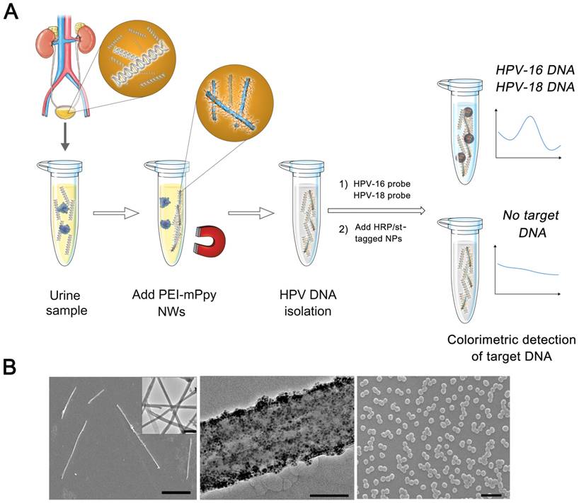
Since the discovery of elevated levels of cell-free DNA (cfDNA) in the bodily fluids of cancer patients (i.e., whole blood, plasma, sputum, and urine), there has been rapid progress in technological development of non-invasive diagnostic and prognostic applications that enable real-time monitoring of cancer progression and response to therapies [13-16]. Efficient recovery and analysis of circulating DNA can provide valuable clinical insights to evaluate carcinogenic HPV detection and genotyping in urine samples. In this study, we introduce a novel strategy of urine-based HPV screening using polyethylenimine-conjugated magnetic polypyrrole nanowires (PEI-mPpy NWs), which play a crucial role in both isolating and detecting cfDNA in urine (Figure 1). Indeed, it is necessary to establish a standard protocol for optimal extraction of cfDNA in urine, because it affords great opportunity to develop reliable and comprehensive urine-based HPV testing. By employing this approach, we developed a strategy for ultrasensitive PCR-free colorimetric analysis of urinary HPVs with genetic variations without the need for any specialized equipment, which is important for rapid, non-invasive, and inexpensive point-of-care (POC) testing in resource-poor areas.
Results and Discussion
Magnetic nanowires can serve as a versatile platform for extracting large quantities of cfDNA from urine as well as detecting target HPV genes in colorimetric recognition with great accuracy and sensitivity. The polycation polyethylenimine (PEI) offers the advantage of having very high affinity for negatively-charged DNA by readily forming nanowire-DNA complexes through electrostatic interactions [17-21]. We applied this cationic nature of PEI for the rapid and efficient isolation of cfDNA from urine with high yield and purity, and for direct PCR-free detection of target HPVs via the addition of multiple horseradish peroxidase (HRP)- and streptavidin-labelled polypyrrole (Ppy) nanoparticles (HRP/st-tagged NPs) to greatly magnify colorimetric signals (Figure 1A; PEI-mPpy NWs). Initially, Ppy nanowires were electrochemically deposited within the pores of the anodic aluminum oxide (AAO) template in which biotin and Fe3O4 magnetic nanoparticles (MNPs; 10 nm in diameter) were employed as co-dopants. Using an AAO membrane as a sacrificing layer offers particular advantages in that it allows not only precise manipulation of the diameter and length of the nanowires, but also ensures sufficient accumulation of a high density of biotin and magnetic nanoparticles in the channels of the template. Field emission scanning and transmission electron microscopy (SEM and TEM, respectively) images provided detailed characterization of PEI-mPpy NWs showing an average diameter of 200 nm and a mean length of 18 µm (Figure 1B, Left). High-magnification TEM imaging confirmed the presence of ~10 nm Fe3O4 MNPs in the nanowires with random orientation and densely packed distribution (Figure 1B, Middle). In addition, the morphology of HRP/st-tagged NPs was observed by SEM (Figure 1B, Right). On the other hand, PEI-conjugated magnetic Ppy nanowires (PEI-mPpy NWs) displayed a relatively high saturation magnification of 87 emu/g, whereas no magnetization saturation and no hysteresis features were observed in PEI-conjugated Ppy nanowires (PEI-Ppy NWs) (Supporting Figure S1).
It is likely that the incorporation of high amounts of magnetic nanoparticles in the nanowires is strongly correlated with the enhanced magnetization behavior of the nanowires. Recently, urine-based HPV screening approaches have been widely investigated as a potential alternative to conventional cytology tests to detect cervical cancer [22-24]. Indeed, as urine represents an easily accessible specimen, it readily allows simple and non-invasive self-collection of large amounts with repeated sampling. We therefore anticipate that, with advances in isolating and detecting urine-derived cfDNA implicated in the development of cervical carcinogenesis, urine testing could be a more reliable and effective tool for identifying and monitoring women who are at risk for cervical cancer. However, HPV detection by urine is particularly difficult to implement because HPV DNA is found at extremely low levels in urine and inappropriate extraction protocols are often employed. The lack of adequate strategies in isolating cfDNA from urine renders the urinary HPV detection rate relatively low and sometimes unsuccessful compared to that from conventional cervical swab samples [25, 26]. The development of DNA extraction protocols yielding sufficiently high quality and quantity of DNA is the most crucial step in the process of the molecular analyses that subsequently ensures high-performance PCR-free colorimetric detection of HPVs with diverse genetic variations. Accordingly, the first step in enhancing the accuracy and reliability of urine-based HPV DNA testing is to maximize the isolation efficiency of cfDNA from urine. Taking this consideration into account, the thin and extended features of the nanowires are likely to improve recovery efficiency by greatly increasing the interaction frequency and duration of HPV DNA present in urine. We expect that the recovery of elevated levels of cfDNA from urine directly contributes to a high HPV type-specific concordance between urine and cervical swab samples. Therefore, we validated the capability of PEI-mPpy NWs that were conjugated with different molecular weights of branched PEI (800 Da and 25 kDa) through ex vivo spiking with a known concentration of genomic DNA (gDNA) from HPV-positive cell lines (HeLa: HPV-18-positive, and SiHa: HPV-16-positive cells) into the HPV-negative urine pool (Supporting Figure S2A-B).
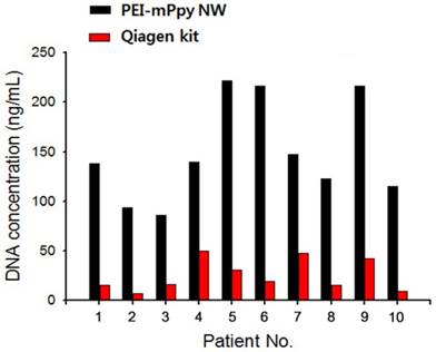
Higher DNA recovery was achieved with >90% isolation efficiency by using PEI-mPpy NWs labeled with 25 kDa PEI across a range of concentrations of input gDNA. Meanwhile, the efficacy of PEI-mPpy NWs conjugated with low molecular weight PEI (800 Da) was markedly decreased with increasing concentrations of gDNA. We found that PEIs with a higher molecular weight (25 kDa) exhibited better binding and condensation ability with DNA molecules than low-molecular-weight PEIs (800 Da), likely due to the fact that complex formation by electrostatic interactions is strongly dependent on the molecular weight of PEIs, which is consistent with previous studies [19, 21]. Using quantitative real time polymerase chain reaction (qPCR), we subsequently analyzed the limit of detection (LOD) of various concentrations of genomic DNA from HPV-positive cell lines (HeLa: HPV-18-positive, and SiHa: HPV-16-positive cells) that were ex vivo spiked into the HPV-negative urine pool. Generally, the LOD was defined as the lowest concentration of HPV DNA detected with positive test results of at least 95% based on threshold cycle (Ct) values, which were compared with those from the Roche Cobas 4800 HPV test. The analytical sensitivity (LOD) of PEI-mPpy NWs in the cfDNA isolated from urine was 0.5 pg/µL for HPV-16-positive SiHa cells and 1.2 pg/µL for HPV-18-positive HeLa cells, whereas the LOD for the Roche Cobas HPV test was 5.2 pg/µL for HPV-16-positive SiHa cells and 1.6 pg/µL for HPV-18-positive HeLa cells, indicating that the nanowires showed substantially improved analytical performance compared to the values reported in previous studies (Supporting Figure S2C) [2]. The feasibility of PEI-mPpy NWs in HPV DNA isolation was further examined using urine samples of ten representative patients with cervical cancer. Initially, we attempted to measure and compare the concentration of cfDNA isolated by using PEI-mPpy NWs (black bars) and a Qiagen DNA extraction kit (red bars) (Figure 2).
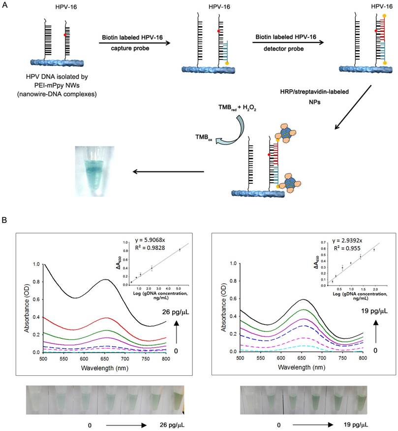
Interestingly, the total amount of DNA obtained from urine using PEI/mPpy NWs was approximately 4-fold higher than that obtained using the Qiagen kit. Most importantly, an elevated level of eluted cfDNA appears to be closely relevant to the accuracy of HPV DNA detection and genotyping, as shown in Table 1. For this study, HPV genotyping profiles of cervical cancer patients were established from direct swab samples of cervical tissue and confirmed by the Roche Cobas 4800 HPV Test with high-risk HPV types (HPV-16, -18, and -others). Paired urine samples were applied to PEI-mPpy NWs and Qiagen reagents to efficiently isolate HPV DNA and elucidate their genotype profiles by qPCR. Of the 15 HPV-positive samples, HPV DNA was identified in all 15 (100%) when isolated by using PEI-mPpy NWs, whereas HPV DNA was detected in 5 urine samples (33.3%) when extracted with the Qiagen kit. Indeed, HPV DNA recovered by the PEI-mPpy NWs exhibited obviously lower cycle threshold (Ct) values compared to that from the Qiagen kit, which strongly suggests higher performance of the nanowires in DNA recovery yield and integrity. Significant enhancement in the overall yield of target HPV in urine samples could be attributed to the elongated shape of the nanowires, which allows a large number of interactions with HPV DNA while retaining bioactivity. Indeed, it is likely that the pencil-like shape of the nanowires not only gives them a large surface-to-volume ratio, but also enables them to move freely through the complex urinary constituents, and thus these nanowires are capable of binding preferentially to cfDNA due to increased duration and frequency of exposure. Next, we attempted to isolate genomic DNA (HPV-positive SiHa cells (HPV-16) and HeLa cells (HPV-18); 250 ng/mL) ex vivo spiked into the HPV-negative urine pool using the PEI-mPpy NWs and utilized them (i.e., nanowire-DNA complexes) as a template for the PCR-free detection of HPVs via a novel colorimetric signal amplification strategy. Without the PCR amplification step, detection occurs following hybridization of biotin-labelled capture probe (CP) and detector probe (DP) with its corresponding target HPV DNA in the nanowire-DNA complexes, as shown in Figure 3A.
Indeed, both probes sequentially hybridize to their complementary HPV sequences on the nanowire-DNA template. After hybridization of the probe to its target sequence, multiple horseradish peroxidase (HRP)- and streptavidin-labeled polypyrrole nanoparticles (HRP/st-tagged NPs) were added for the specific recognition, with capture and detector probes linked with biotin in a solution containing 1 mM colorimetric TMB substrate and 0.5 mM H2O2. Ultimately, we were able to visualize the colorimetric signals that were derived from the specific interaction of the probes with their respective target HPV sequences [27, 28]. The addition of HRP/st-tagged NPs obviously facilitates H2O2-mediated catalytic oxidation of TMB, which triggers substantial color change in the presence of target DNA [26]. Under optimal experimental conditions, we evaluated the dynamic range of colorimetric UV signals using the calibration plots that were strongly correlated with the concentration of the target DNA, exhibiting a linear relationship between the absorbance value of the oxidized TMB at 650 nm and the concentration of the target DNA, with detection limits of 0.052 pg/µL (HPV-16-positive SiHa cells) and 0.12 pg/µL (HPV-18-positive HeLa) based on a signal-to-noise ratio of 3. The magnetic nanowires are optimally suitable for the isolation of cfDNA from urine at high efficiency, and thus can be a key component in improving the workflow of routine urine-based HPV DNA analysis. The clinical utility of the PCR-free colorimetric assay was further evaluated by testing urine samples that were collected from cervical cancer patients and healthy controls (Figure 4).
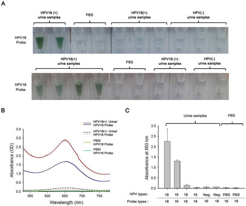
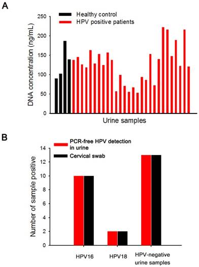
Firstly, cfDNA was extracted from urine samples with a volume <300 µL from HPV18-positive cancer patients, HPV16-positive cancer patients, and HPV-negative healthy controls by using PEI-mPpy NWs, where HPV genotyping had already been confirmed by standard cervical swab clinical examinations. PCR-free optimized procedures were applied for in situ detection of HPVs in the nanowire-DNA complexes following these protocols: i) the denaturation of nanowire-DNA complexes at 95 oC for 1 min, ii) the introduction of complementary CP/DP to nanowire-DNA complexes for target-induced hybridization, and iii) the addition of HRP/st-tagged NPs to dramatically amplify the colorimetric signals. As shown in Figure 4A, two HPV18-positive urine samples displayed strong visible colorimetric response through the specific hybridization with HPV18 probes, whereas PBS, HPV16-positive, and HPV-negative urine samples showed no color change, confirming specific binding of their complementary probes with high sensitivity and specificity. Similarly, four HPV16-positive urine samples yielded a significant increase in colorimetric response upon exposure to HPV16 probes. However, all HPV16-negative samples including PBS, HPV18-positive, and HPV-negative urine samples did not respond to HPV16 probes. As expected, these results were in good agreement with absorbance values obtained by UV-Vis absorption spectroscopy (Figure 4B). Indeed, HPV18-positive urine samples were found to be highly specific and hybridized only in the presence of HPV18 probes, exhibiting strong absorption peaks at 650 nm even without PCR amplification. The quantitative colorimetric measurements showed that HPV16- and HPV18-positive urine samples yielded appreciably higher absorbance with their corresponding probes, but exhibited significantly low cross-reactivity with other HPV genotypes. On the other hand, HPV16-/18-negative urine samples and PBS resulted in negligible colorimetric signals in both probe types. We further evaluated the proposed nanowire-based strategy for ultrasensitive colorimetric determination of the HPV genotype distribution patterns using urine samples of cervical cancer patients and healthy controls, and ultimately compared the results with those from cervical swab samples. Initially, we compared the amount of the cfDNA extracted from a total of 28 urine samples (24 HPV-positive and 4 HPV-negative samples) of healthy controls and patients with cervical cancer by using PEI-mPpy NWs (Figure 5A).
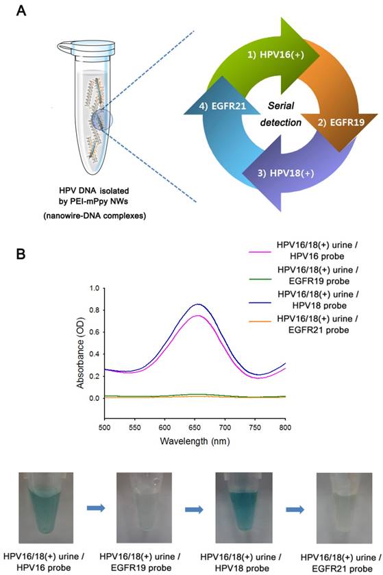
We found that there were no apparent differences in the concentration of the eluted cfDNA between healthy individuals and HPV-positive cancer patients. We also investigated type-specific HPV detection in paired conventional swab sampling and urine testing (300 µL). Surprisingly, PEI-mPpy NWs greatly improved extraction and detection efficiency of urinary cfDNA with an excellent genotyping concordance rate (100%) between cervical swabs and urine-based colorimetric detection (Figure 5B, Table 2). Continually, we identified multiple serial HPV genotypes in the same nanowire-DNA complexes. The cfDNA extracted by PEI-mPpy NWs can serve as a versatile sensing platform for inducing specific recognition and binding of the probe to complementary DNA based on the colorimetric assay (Figure 6A). With the addition of a series of type-specific CP/DP and HRP/st-tagged NPs continuously in the same nanowire-DNA complexes that were generated from the urine of patients with both HPV16 and 18, we identified and discriminated multiple HPV targets with genetic diversity using one nanowire-DNA template. As shown in Figure 6B, we observed obvious color change and UV-Vis absorbance at 650 nm of the nanowire-DNA complexes after adding the first target probe (i.e., HPV16 probes) and HRP/st-tagged NPs. However, as expected, no response was observed with the addition of EGFR19 probe to the same template. Immediately, a second target probe (i.e., HPV18 probes) and HRP/st-tagged NPs were added to the same nanowire-DNA complexes that confirmed the successful colorimetric response of specific HPV DNA segments with excellent sensitivity and specificity, even in repeated detection. Finally, the addition of the EGFR21 probe confirmed that no reaction occurred involving the nanowire-DNA template.
(a) Schematic diagram of highly efficient isolation of urinary cfDNA using polyethyleneimine-conjugated magnetic nanowires (PEI-mPpy NWs) and direct PCR-free colorimetric detection of target HPVs via sequential addition of complementary probes and multiple horseradish peroxidase (HRP)-/streptavidin-labelled polypyrrole nanoparticles (HRP/st-tagged NPs) to dramatically amplify colorimetric signals. (b) SEM image of PEI-mPpy NWs (Left, Scale bar = 10 µm). Inset is a TEM image of the nanowires (scale bar = 1 µm). TEM image at high magnification showing the presence of a large quantity of magnetic nanoparticles (MNPs; 10 nm) doped within the nanowire (Middle, Scale bar = 50 nm). SEM image of HRP/streptavidin-conjugated nanoparticles (Right, scale bar = 200 µm)
Validation of PEI-mPpy NWs in the extraction of cfDNA in urine samples of cervical cancer patients. Comparisons of the concentration of cfDNA isolated from urine samples of ten representative cervical cancer patients by using PEI-mPpy NWs and a Qiagen DNA extraction kit. PEI-mPpy NWs and a Qiagen kit were employed for the extraction of cfDNA from urine.
The detection and genotyping of HPV DNA in cervical swabs and urine samples of cervical cancer patients.
| Patient No. | Cervical swab HPV type | Urine HPV type | ||
|---|---|---|---|---|
| PEI-mPpy NW (Ct) | Qiagen kit (Ct) | ΔCt | ||
| P1 | HPV16 | HPV16 (34.2) | N/A | - |
| P2 | HPV16 | HPV16 (37.2) | N/A | - |
| P3 | 12 other types | 12 other types (29.6) | 12 other types (33.9) | 4.3 |
| P4 | HPV16 | HPV16 (34.4) | N/A | - |
| P5 | 12 other types | 12 other types (32.8) | 12 other types (36.8) | 4.0 |
| P6 | 12 other types | 12 other types (32.7) | 12 other types (35.2) | 2.5 |
| P7 | 12 other types | 12 other types (37.2) | N/A | - |
| P8 | 12 other types | 12 other types (34.6) | 12 other types (40.0) | 5.4 |
| P9 | 12 other types | 12 other types (28.2) | 12 other types (29.7) | 1.5 |
| P10 | HPV16 | HPV16 (33.9) | N/A | - |
| P11 | 12 other types | 12 other types (30.7) | N/A | - |
| P12 | 12 other types | 12 other types (31.0) | 12 other types (40.0) | 9.0 |
| P13 | HPV16 | HPV16 (33.4) | HPV16 (38.3) | 4.9 |
| P14 | HPV16 | HPV16 (34.2) | N/A | - |
| P15 | HPV18 | HPV18 (31.6) | N/A | - |
Conclusions
We have successfully evaluated the capabilities of PEI-mPpy NWs as a versatile platform for the isolation of HPV cfDNA from urine of cervical cancer patients, and for the identification and detection of multiple type-specific HPVs via direct PCR-free colorimetric signal amplification. The elongated features of the nanowires offer great advantages in enhancing the extraction yield of cfDNA by significantly increasing the interactions with DNA molecules in urine, thus promoting the ability for directly detecting the target HPV DNA from a small volume of urine (300 µL) even without PCR amplification step. Indeed, colorimetric results can even be discriminated by the naked eye. Our PEI-mPpy NWs-based HPV DNA isolation and detection strategy appears to be a cost-effective and practical technology with greater sensitivity and accuracy than other urine-based methods. These findings suggest that the use of PEI/mPpy NWs in urine-based HPV screening shows significant advancement in the isolation, identification, and analysis of multiple high-risk genotypes of HPV DNA, which provides a promising direction in non-invasive screening and detection of multiple genotypes in HPV-related cancers.
Experimental Section
Chemicals and reagents
Polyethyleneimine (PEI, branched, average MW ~25,000), iron oxide (II, III), magnetic nanoparticles solution (MNP, 10 nm average particle size, 5 mg/mL in H2O), N-(3-dimethylaminopropyl)-N′-ethylcarbodiimide hydrochloride (EDC), N-hydroxy succinimide (NHS), pyrrole, biotin, and streptavidin were purchased from Sigma-Aldrich (St. Louis, MO, USA). An anodized aluminum oxide (AAO) membrane filter (200 nm pore diameter) was obtained from Whatman (Pittsburgh, PA, USA). For real-time PCR analysis, the Cobas 4800 HPV controls and amplification/detection kits were purchased from Roche Molecular Diagnosis (Pleasanton, CA, USA).
Preparation and characterization of PEI/mPpy NWs
Polyethylenimine-conjugated magnetic nanowires (PEI/mPpy NWs) were fabricated as previously described [17]. Briefly, 30 µL of magnetic nanoparticles (MNPs, 5 µg/mL; ~10 nm in diameter) were deposited on top of the Au-coated Anodic Aluminum Oxide (AAO) membrane (~150 nm thick) and infiltration into the AAO nanopores was promoted by moderate aspiration at room temperature (RT). All electrochemical techniques were conducted using a potentiostat/galvanostat (SP-150, BioLogic, USA). Ppy was electrochemically polymerized in the MNP-loaded AAO template nanochannels in a solution of 0.01 M poly(sodium 4-styrenesulfate) containing 0.1 M pyrrole and 1 mM biotin by applying chronoamperometry at 1.0 V (vs. Ag/AgCl) for 7 min. The resulting AAO templates were rinsed several times with distilled water, immersed in 2 M NaOH for 3 h, and then placed in a sonication bath (Bioruptor® UCD-200, Diagenode Inc, USA) to obtain individual, free-standing Ppy NWs. Then, 30 mM N-(3-dimethylaminopropyl)-N-ethylcarbodiimide hydrochloride (EDC) and 6 mM N-hydroxysuccinimide (NHS) were added to the resulting Ppy NWs to activate carboxylic acid groups. Subsequently, the resulting nanowires were incubated with streptavidin (10 µg/mL) for 45 min and washed with distilled water. Then, biotinylated PEI (800 or 25000 Da) was added to the streptavidin-labeled nanowires at RT for 1 h. The morphologies of the PEI/mPpy NWs were analyzed by SEM (JSM-7800F, JEOL, USA) and TEM (G2 F30ST, Tecnai, USA). Magnetic field measurements were investigated by a SQUID-VSM magnetometer (MPMS 3, Quantum Design, USA).
(A) Magnetic nanowire-based colorimetric assay for HPV DNA detection and genotyping in urine samples. Colorimetric detection was performed on the nanowire-DNA complexes that contained cfDNA isolated from the urine samples using PEI-mPpy NWs. The biotin-labeled capture and detector probes were specifically designed to recognize the corresponding target HPV DNAs attached to the nanowire, even without a PCR amplification step. After hybridization of the target with type-specific probes, multiple horseradish peroxidase (HRP)- and streptavidin-labeled polypyrrole nanoparticles (HRP/st-tagged NPs) were added to produce amplified colorimetric signals that are even visible to the naked eye. (B) UV-Vis absorption spectra of nanowire-DNA complexes that are hybridized specifically with their complementary capture/detector probes followed by the addition of HRP/st-tagged NPs. Known concentrations of genomic DNA from HPV-positive SiHa cells (Left; HPV-16; 0, 0.16, 0.52, 1.3, 2.6, 5.2, 26 pg/µL) and that from HPV-positive HeLa cells (Right; HPV-18; 0, 0.12, 0.4, 1.8, 3.9, 19 pg/µL) were spiked into HPV-negative urine pool ex vivo, for HPV DNA isolation and colorimetric detection. The insets show plots of the corresponding absorbance at 650 nm versus various concentrations of genomic DNA from HPV-positive SiHa cells (Left; HPV-16) and HeLa cells (Right; HPV-18) that were extracted by PEI-mPpy NWs. The error bars represent the standard deviations from five independent measurements.
(a) Assessment of clinical performance of the proposed PCR-free colorimetric assay by evaluating urine samples of HPV-positive cervical cancer patients (HPV16(+) and HPV18(+)), HPV-negative healthy controls (HPV(-)), and PBS. The photographs display the color change of nanowire-DNA complexes after hybridization with biotin-labelled capture and detector probes with the corresponding target HPV DNA isolated using the nanowire, followed by the addition of HRP/st-tagged NPs. (b) The UV-Vis spectra of nanowire-DNA complexes that contain cfDNA isolated from HPV18-positive urine by PEI-mPpy NWs. With the addition of HPV16/HPV18 probes and HRP/st-tagged NPs, type-specific HPVs can be specifically detected. (c) Average absorbance values of circulating cfDNA isolated from urine samples of HPV-positive cervical cancer patients (HPV16(+) and HPV18(+)), HPV-negative healthy controls (HPV(-)), and PBS after the reaction with different probe types specific for HPV16 or HPV18. A total of 24 HPV-positive and HPV-negative urine samples were collected and tested. The error bars represent the standard deviations from five independent measurements.
Urine Samples
Urine samples were obtained from thirty HPV-positive patients and five healthy volunteers at the National Cancer Center Hospital (Goyang, Korea) according to procedures approved by the NCC Institutional Review Board (NCC2016-0282). The collected urine samples were immediately aliquoted and stored at -20 °C until use.
Evaluation of genomic DNA capture and release efficiency by using PEI-mPpy NWs
Defined quantities of genomic DNA from HPV-positive cell lines (HeLa: HPV-18-positive and SiHa: HPV-16-positive cells) were ex vivo spiked into a HPV-negative urine pool at concentrations ranging from 0.01 to 1,000 ng/mL. Subsequently, DNA capture was carried out by adding PEI/mPpy NWs (0.5 mg/mL) to the urine samples (300 µL) containing the genomic DNA with gentle shaking (120 × g) for various time durations (10, 30, 60, and 90 min) at RT. Then, the resulting solutions were placed onto a magnetic rack for 30 min to separate unbound or non-specifically bound constituents and capture DNA-bound magnetic nanowires. The captured DNA was then eluted in 10 mM Tris-HCl buffer (pH 10.0) by maximum speed shaking (750 × g) for various times (10, 30, 60, and 90 min) at RT. The captured and released DNA was quantified by PicoGreen assay according to the manufacturer's instructions.
Limit of detection
To evaluate the LOD, known concentrations of genomic DNAs from HeLa (HPV-18, 10-50 copies/cell) and SiHa (HPV-16, 1-2 copies/cell) cell lines were ex vivo spiked into the HPV-negative urine pools (n = 5). The PEI-Ppy MNWs (0.5 mg/mL) were added to each sample, followed by shaking (120 × g) at RT for 1 h. Then, HPV DNA was eluted into nuclease-free water. Finally, the isolated DNA was mixed with an equal volume of Cobas HPV master mix and amplified with the Roche LC480 instrument.
(a) The concentration of cfDNA isolated from urine samples of HPV-negative healthy controls and HPV-positive cervical cancer patients using PEI-mPpy NWs. (b) Type-specific concordance of HPV DNA genotypes of the results from cervical swabs and PCR-free urinary HPV detection by PEI-mPpy NWs.
Comparison of HPV detection in cervical swabs and PCR-free urinary HPV detection by PEI-mPpy NWs. A total of 25 HPV-positive and HPV-negative urine samples were collected and tested.
| Urine (PCR-free colorimetric HPV detection) | Cervical swab | ||
|---|---|---|---|
| HPV positive | HPV negative | Sum | |
| HPV positive | 12 | 0 | 12 |
| HPV negative | 0 | 13 | 13 |
| Sum | 12 | 13 | 25 |
cfDNA isolation from patient's urine samples
cfDNA was isolated from HPV-positive and -negative urine samples (300 µL) by using PEI/mPpy NWs (0.5 mg/mL) and QIAamp circulating nucleic acid kit (Qiagen, USA) according to the manufacturer's protocol. The yield of cfDNA eluted from urine was routinely analyzed by PicoGreen fluorescence assay and gel electrophoresis for direct quantification and visualization. Subsequently, the detection and genotyping of the eluted DNA were performed with the Roche Cobas 4800 HPV Test by adding an equal volume of Cobas HPV master mix reagent and amplifying the HPV target.
Preparation and characterization of HRP/streptavidin-tagged polypyrrole (Ppy) nanoparticles
HRP/streptavidin-tagged PPy nanoparticles (HRP/st-tagged NPs) were prepared as previously described [25]. Briefly, PVP was dissolved in distilled water and then, pyrrole and HA were added to polymerize. Subsequently, the resulting HA-PPy NPs were dialyzed against distilled water (DW) for 2 days to remove unreacted chemicals and lyophilized. Then, ~2 mg of HA-PPy NPs were dispersed in 1 mL of 0.4 M EDC/0.1 M NHS solution and incubated for 45 min. After removing excess chemicals, 1 mg of HRP and 25 µg of streptavidin were added to the carboxyl-activated HA-PPy NPs and mixed overnight at 4°C. The resulting HRP/streptavidin-tagged Ppy NPs were precipitated with DW several times and incubated at 4℃ until use.
Colorimetric detection of HPV DNA
To isolate cell-free HPV DNA, PEI-mPpy NWs (10 µg/mL) were added to 200 µL of HPV-positive patient urines and mixed for 30 min at RT. After precipitation with PBS, the resulting captured DNA was denatured at 95℃ for 1 min. Immediately, 1 pM of biotin-terminated CP and DP were added to the resulting solution and incubated for 1 h at 37℃. Then, the HRP/st-tagged NPs were added to the sample and incubated for 30 min at 37℃. Subsequently, 25 µL of 10 mM 3,3′,5,5′-Tetramethylbenzidine (TMB), 25 µL of 0.1 M H2O2, and 200 µL of 0.2 M sodium acetate trihydrate buffer (pH 5.0) were added to the captured HPV DNA sample and incubated for 3 min at RT in the dark. To evaluate any correlation between the concentration of HPV DNA and the absorbance, UV-Vis detection was performed at a wavelength of 652 nm using a DU 730 UV-Vis spectrophotometer (Beckman Coulter, USA). For the serial detection of HPV-16 and HPV-18 DNA, PEI-mPpy NWs (10 µg/mL) were added to 200 µL of the urine samples taken from patients who were HPV-positive, and mixed for 30 min to capture cfDNA. After being precipitated in PBS, the resulting cfDNA-magnetic nanowires were denatured at 95℃ for 1 min and hybridized with 1 pM biotin-terminated HPV-16-CP and HPV-16-DP for 1 h at 37℃. Next, the HRP/st-tagged NPs were added to the samples and incubated for 30 min at 37℃. To the resulting solution, 25 µL of 10 mM 3,3′,5,5′-Tetramethylbenzidine (TMB), 25 µL of 0.1 M H2O2, and 200 µL of 0.2 M sodium acetate trihydrate buffer (pH 5.0) were added to observe the colorimetric signals. After that, the DNA captured by the nanowires were consecutively incubated in a solution containing biotin-terminated EGFR 19-, HPV-18-, or EGFR 21- probes, followed by the addition of the HRP/st-tagged NPs and 25 µL of 10 mM 3,3′,5,5′-Tetramethylbenzidine (TMB) in 200 µL of 0.2 M sodium acetate trihydrate buffer (pH 5.0) containing 25 µL of 0.1 M H2O2, and ultimately the colorimetric signals were detected using the same procedure as described previously.
(a) A novel approach of PEI-mPpy NWs in the extraction, identification, and PCR-free sequential detection of multiple HPV genotypes from urine specimens of cervical cancer patients. (b) The UV-Vis colorimetric results of cfDNA isolated by PEI-mPpy NWs from urine of cervical cancer patients who were found to be positive for both HPV16 and HPV18, demonstrating multiple uses of the same nanowire-DNA complexes for the detection of HPV with different genetic variations. However, no response was observed for non-HPV probes (EGFR19 and EGFR21). The photographs indicate the color change as a result of type-specific hybridization between target HPVs and their complementary probes.
HPV capture and detector probe sequences
| Probe label | Sequences |
|---|---|
| HPV 16 - CP | 5' biotin - GAG GAG GAG GAT GAA ATA GAT GGT - 3' |
| HPV 16 - DP | 5' - TTG GAA GAC CTG TTA ATG GGC - biotin 3' |
| HPV 18 - CP | 5' biotin - CAC ATT GTG GCA CAA TCT TTT A - 3' |
| HPV 18 - DP | 5' - GCC ATA TCG CTT TCA TCT GT - biotin 3' |
| EGFR 19 - CP | 5' biotin - GGAATTAAGAGAAGCAACATCTCC -3' |
| EGFR 19 - DP | 5' - AACCTCAGGCCCACCTTTT - biotin 3' |
| EGFR 21 - CP | 5' biotin - CCAGGAACGTACTGGTGAAAA - 3' |
| EGFR 21 - DP | 5' - GGAAGAGAAAGAATACCATGCA - biotin 3' |
Abbreviations
HPV: human papillomavirus; EGFR: epidermal growth factor receptor; PEI-mPpy NWs: polyethylenimine-conjugated magnetic polypyrrole nanowires; cfDNA: cell-free DNA; POC: point-of-care; PCR: polymerase Chain Reaction; HRP: horseradish peroxidase; AAO: Anodic Aluminum Oxide; Ppy: polypyrrole; CP: capture probe; DP: detector probe; TMB: 3,3′,5,5′-Tetramethylbenzidine.
Supplementary Material
Supplementary figures.
Acknowledgements
This work was supported by a National Cancer Center grant from the Republic of Korea (1510070-3, 1611170-2) and the National Research Foundation of Korea (NRF) grant funded by the Korean government (MEST) (NRF-2017R1A2B4007800).
Author contributions
S.J., H.L., and Y.C. designed the study and analyzed the data. S.J. and H.L. fabricated the platforms and performed the DNA capture and release experiments. K.B. and K.Y. collected and analyzed the digital PCR data. K.Y. and E.L. prepared the clinical samples. S.J., H.L., K.Y., and Y.C. co-wrote the manuscript. All authors discussed the results and the comments on the manuscript.
Competing Interests
The authors have declared that no competing interest exists.
References
1. Steenbergen RD, Snijders PJ, Heideman DA, Meijer CJ. Clinical implications of (epi) genetic changes in HPV-induced cervical precancerous lesions. Nature Reviews Cancer. 2014;14:395-405
2. Rao A, Young S, Erlich H, Boyle S, Krevolin M, Sun R. et al. Development and characterization of the cobas human papillomavirus test. Journal of clinical microbiology. 2013;51:1478-84
3. Schiffman M, Castle PE, Jeronimo J, Rodriguez AC, Wacholder S. Human papillomavirus and cervical cancer. The Lancet. 2007;370:890-907
4. Bosch F, Lorincz A, Munoz N, Meijer C, Shah K. The causal relation between human papillomavirus and cervical cancer. J Clin Pathol. 2002;55:244-65
5. Clifford G, Gallus S, Herrero R, Munoz N, Snijders P, Vaccarella S. et al. Worldwide distribution of human papillomavirus types in cytologically normal women in the International Agency for Research on Cancer HPV prevalence surveys: a pooled analysis. The Lancet. 2005;366:991-8
6. Walboomers JM, Jacobs MV, Manos MM, Bosch FX, Kummer JA, Shah KV, Snijders PJ, Peto J, Meijer CJ, Muñoz N. Human papillomavirus is a necessary cause of invasive cervical cancer worldwide. J Pathol. 1999;189:12-9
7. Bryzgunova O, Laktionov P. Extracellular Nucleic Acids in Urine: Sources, Structure, Diagnostic Potential. Acta Naturae (англоязычная версия). 2015;7:48-54
8. Mendez K, Romaguera J, Ortiz AP, López M, Steinau M, Unger ER. Urine-based human papillomavirus DNA testing as a screening tool for cervical cancer in high-risk women. International J Gynecol Obstet. 2014;124:151-155
9. Sahasrabuddhe VV, Gravitt PE Dunn ST, Brown D Allen RA, Eby YJ et al. Comparison of human papillomavirus detections in urine, vulvar, and cervical samples from women attending a colposcopy clinic. J Clin Microbiol. 2014;52:187-192
10. Song ES, Lee HJ, Hwang TS. Clinical efficacy of human papillomavirus DNA detection in urine from patients with various cervical lesions. J Korean Med Sci. 2007;22:99-104
11. Stanczuk GA, Kay P, Allan B, Chirara M, Tswana SA, Bergstrom S. et al. Detection of human papillomavirus in urine and cervical swabs from patients with invasive cervical cancer. J Med Virol. 2003;71:110-114
12. Khunamornpong S, Settakorn J, Sukpan K, Lekawanvijit S, Katruang N, Siriaunkgul S. Comparison of human papillomavirus detection in urine and cervical samples using high-risk HPV DNA testing in northern thailand. Obstet Gynecol Int. 2016;2016:8
13. Karachaliou N, Mayo-de-las-Casas C, Molina-Vila MA, Rosell R. Real-time liquid biopsies become a reality in cancer treatment. Ann Transl Med. 2015;3:36
14. Isobe K, Hata Y, Kobayashi K, Hirota N, Sato K, Sano G. et al. Clinical significance of circulating tumor cells and free DNA in non-small cell lung cancer. Anticancer Res. 2012;32:3339-3344
15. Breitbach S, Tug S, Helmig S, Zahn D, Kubiak T, Michal M. et al. Direct quantification of cell-free, circulating DNA from unpurified plasma. PLoS One. 2014;9:e87838
16. Schmidt B, Weickmann S, Witt C, Fleischhacker M. Improved method for isolating cell-free DNA. Clin Chem. 2005;51:1561-1563
17. Lee HJ, Hwang NR, Hwang SH, Cho Y. Magnetic nanowires for rapid and ultrasensitive isolation of DNA from cervical specimens for the detection of multiple human papillomaviruses genotypes. Biosen Bioelectron. 2016;86:864-870
18. Zeng X, Sun Y-X, Zhang X-Z, Zhuo R-X. Biotinylated disulfide containing PEI/avidin bioconjugate shows specific enhanced transfection efficiency in HepG2 cells. Org Biomol Chem. 2009;7:4201-10
19. Mady M, Mohammed W, El-Guendy NM, Elsayed A. Effect of polymer molecular weight on the DNA/PEI polyplexes properties. Rom J Biophys. 2011;21:151-65
20. Bolhassani A, Javanzad S, Saleh T, Hashemi M, Aghasadeghi MR, Sadat SM. Polymeric nanoparticles: potent vectors for vaccine delivery targeting cancer and infectious diseases. Hum Vaccines Immunother. 2014;10:321-32
21. Vancha AR, Govindaraju S, Parsa KV, Jasti M, González-García M, Ballestero RP. Use of polyethyleneimine polymer in cell culture as attachment factor and lipofection enhancer. BMC Biotechnol. 2004;4:1
22. Hauwers Kd, Depuydt C, Bogers J-P, Stalpaert M, Vereecken A, Wyndaele J-J. et al. Urine versus brushed samples in human papillomavirus screening: study in both genders. 2007.
23. Shan Z, Zhou Z, Chen H, Zhang Z, Zhou Y, Wen A. et al. PCR-ready human DNA extraction from urine samples using magnetic nanoparticles. J Chromatogr B. 2012;881:63-8
24. Siddiqui H, Nederbragt AJ, Jakobsen KS. A solid-phase method for preparing human DNA from urine for diagnostic purposes. Clin Biochem. 2009;42:1128-35
25. Xue X, Teare MD, Holen I, Zhu YM, Woll PJ. Optimizing the yield and utility of circulating cell-free DNA from plasma and serum. Clin Chim Acta. 2009;404:100-4
26. Board RE, Williams VS, Knight L, Shaw J, Greystoke A, Ranson M. et al. Isolation and extraction of circulating tumor DNA from patients with small cell lung cancer. Annals of the New York Academy of Sciences. 2008;1137:98-107
27. Song Y, Wei W, Qu X. Colorimetric Biosensing Using Smart Materials. Adv Mater. 2011;23:4215-4236
28. Tao Y, Lin Y, Ren J. Self-assembled, functionalized graphene and DNA as a universal platform for colorimetric assays. Biomaterials. 2013;34:4810-4817
Author contact
![]() Corresponding author: E-mail: ynchore.kr
Corresponding author: E-mail: ynchore.kr
 Global reach, higher impact